Vega preclinical ultrasound
- Hands-free, automated transducer positioning and movement
- High speed, high-throughput performance with 3 mice scanning in just a few minutes
- 3D Widefield acquisitions enabling whole subject imaging
- Standard B-mode and M-mode capability
- Shear Wave Elastography (SWE) mode for quantifying and evaluation tissue stiffness
- Acoustic angiography mode for visualizing microvasculature
- Fits on the benchtop
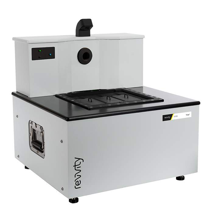
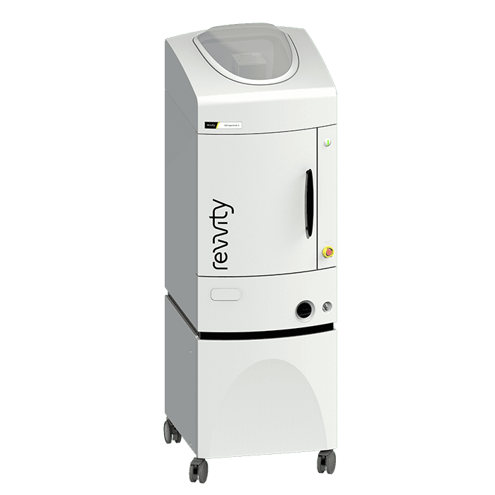
Vega Preclinical Ultrasound System
Designed with the researcher in mind, the Vega removes the challenges associated with traditional hand-held ultrasound, and uses a bottom-up imaging approach through the use of automated hands-free transducers located under the imaging stage. This unique design requires minimal training with no dedicated sonographer needed, enables high-throughput imaging, and produces more consistent results than conventional hand-held ultrasound systems.
This powerful ultrasound system gives you:
- Hands-free - Automated transducer positioning and movement
- Easy-to-use requiring minimal training
- High-speed, high-throughput performance with 3 mice scanning in just a few minutes
- 3D widefield acquisitions enabling whole subject imaging
- Standard B-Mode and M-Mode capability
- Shear Wave Elastography (SWE) mode for quantifying tissue stiffness
- Acoustic Angiography (AA) mode for visualization of microvasculature
- Flexible visualization and analysis software
- Fits on the benchtop
IVIS Lumina S5 & X5
- High-sensitivity 2D optical imaging (bioluminescence and fluorescence)
- High-throughput format with a 20 x 20 FOV sufficient for imaging 5 mice simultaneously
- High resolution, low dose X-ray with optical overlay (IVIS Lumina X5 only)
- Compact design that fits on your benchtop
- Unique animal handling accessories and software tools to streamline throughput
- Complimentary Living Image™ software licenses are provided with the IVIS systems and upon request.
IVIS Lumina S5 Imaging System
The IVIS Lumina S5 in vivo imaging system has all the capabilities of the current IVIS Lumina platform with improved throughput and accessories to streamline imaging workflow, data acquisition and analysis, ideal for accelerating your research.
High-throughput High-Sensitivity Optical Imaging
The IVIS Lumina S5 integrates a next generation 1 inch CCD camera into our benchtop instrument providing a high throughput 20 x 20 cm Field of View (FOV) sufficient for imaging 5 animals at a time for bioluminescence and fluorescence in vivo imaging.
As with other IVIS Lumina in vivo optical imaging systems, the IVIS Lumina S5 is equipped with 26 filters tunable to image fluorescent sources that emit from green to near-infrared. Novel illumination technology effectively increases fluorescent transmission through 900 nm. Moreover, the IVIS Lumina S5 incorporate Revvity's patented Compute Pure Spectrum (CPS) algorithm for spectral library generation software tools to ensure accurate autofluorescence removal, unmixing and fluorophore quantitation. Standard on all IVIS instruments, absolute calibration affords consistent and reproducible results independent of magnification, filter selection from one instrument to any another IVIS instrument within an organization or around the world.
IVIS Lumina S5 – A High Throughput Solution
Not only does the IVIS Lumina S5 offer higher throughput via the 1 inch CCD, but it is also compatible with a set of smart animal handling accessories (purchased separately) designed with throughput and safety in mind.
Smart loading trays allows users to pose animals on the benchtop before placing the tray into the IVIS. Fiducials built into the tray enable the software to automatically recognize and draw ROIs providing automated animal identification.
Animal trays are designed with ease of use and user safety in mind. No nose cones are required thus minimizing cleanup. When used with the next generation anesthesia unit (RAS-4), strong vacuum capabilities minimize excess gas from escaping thus preventing exposure of users to anesthetic gas.
Finally, Living Image™ software brings IVIS technology to life by facilitating an intuitive workflow for in vivo optical image acquisition, analysis and data organization. The software’s design creates an intuitive, seamless workflow for researchers of all skill levels.
Key Features:
- High throughput (5 mice) optical imaging
- Increased throughput (10 mice) using optional manifold
- Supports mouse and rat imaging
- Compute Pure Spectrum (CPS) spectral unmixing
- Full fluorescence tunability through the NIR spectrum
- Unique accessories to speed acquisition and analysis
- Small footprint–sits on your benchtop
- Complimentary Living Image software licenses are provided with the IVIS systems and upon request.
IVIS Lumina X5 Imaging System
The IVIS Lumina X5 has all the capabilities of the IVIS Lumina S5 imaging system with integrated industry leading high-resolution x-ray for greater detail. The IVIS Lumina X5 also includes state of the art spectral unmixing features for sensitive multispectral imaging to monitor multiple biological events in the same animal.
High-throughput Optical and X-ray Imaging – No Compromise
The IVIS Lumina X5 integrates a next generation 1 inch CCD camera into our benchtop instrument providing a high throughput 20 x 20 cm FOV sufficient for imaging 5 mice at a time with bioluminescence and fluorescence. Moreover, the large, independently deployed scintillator facilitates X-ray acquisitions of 5 mice and larger rodents up to 500-600 grams with seamless, accurate overlay onto the optical image at any field of view.
As with other IVIS Lumina systems, this instrument is equipped with 26 filters tunable to image fluorescent sources that emit from green to near-infrared. Novel illumination technology effectively increases fluorescent transmission through 900 nm. Additionally, the IVIS Lumina X5 incorporates Revvity's patented Compute Pure Spectrum (CPS) algorithm for spectral library generation software tools to ensure accurate autofluorescence removal, unmixing and fluorophore quantitation.
Standard on all IVIS instruments, absolute calibration affords consistent and reproducible results independent of magnification, filter selection from one instrument to any another IVIS instrument within an organization or around the world.
Industry Leading X-ray Resolution
The IVIS Lumina X5 is equipped with a microfocus X-ray source and geometric magnification and when combined, achieve industry leading X-ray resolution in a 2D optical/X-ray system setting a high standard in multimodal 2D imaging resolution. With optical image overlays at every X-ray resolution, never miss underlying anatomical and structural changes. Get more from your data and explore new applications.
IVIS Lumina X5 – A High Throughput Solution
Not only does the IVIS Lumina X5 offer higher throughput via the next generation 1 inch CCD, but it is also compatible with a set of smart animal handling accessories (purchased separately) designed with throughput and safety in mind.
Smart loading trays enable users to pose animals on the benchtop before placing the tray into the IVIS. Fiducials built into the tray allows the software to automatically recognize and draw ROIs providing automated animal identification.
Animal trays are designed with ease of use and user safety in mind. No nose cones are required thus minimizing cleanup. When used with the next generation anesthesia unit (RAS-4), strong vacuum capabilities minimize excess gas from escaping thus preventing exposure of users to anesthetic gas.
Finally, Living Image™ software brings IVIS technology to life by facilitating an intuitive workflow for in vivo optical, X-ray image acquisition, analysis and data organization. The software’s design creates an intuitive, seamless workflow for researchers of all skill levels.
Key Features:
- High throughput (5 mice) optical and X-ray
- Increased throughput (10 mice) using optional manifold
- High resolution, low dose X-ray with optical overlay
- Supports mouse and rat imaging
- Compute Pure Spectrum (CPS) spectral unmixing
- Full fluorescence tunability through the NIR spectrum
- Unique accessories to streamline workflow, data acquisition and analysis
- Complimentary Living Image software licenses are provided with the IVIS systems and upon request
IVIS Lumina III series
- 2D optical imaging (bioluminescence and fluorescence)
- Low-dose X-ray with optical overlay (IVIS Lumina XRMS only)
- Images up to 3 mice simultaneously
- Compact design that fits on your benchtop
- Complimentary Living Image™ software licenses are provided with the IVIS systems and upon request.
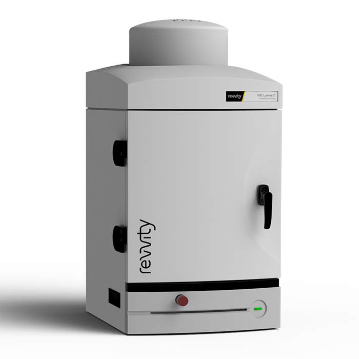
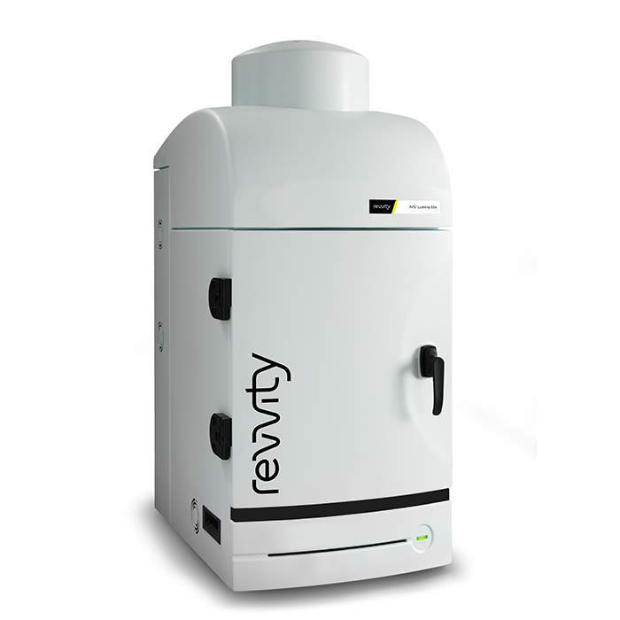
IVIS Lumina LT In Vivo Imaging System
The IVIS Lumina LT optical imaging system is equipped with 2D bioluminescence, fluorescence, and radioisoptic (Cerenkov) imaging capabilities. For more sophisticated fluorescent models, the IVIS Lumina LT can be easily upgraded to an IVIS Lumina III system offering users enhanced fluorescence imaging tunability for improved sensitivity from the visible to near infrared imaging spectrum.
Features/Benefits:
- 2D Bioluminescence
- 2D Fluorescence
- Radioisotopic Cerenkov Imaging
- Compute Pure Spectrum Spectral Unmixing
- DyCE Imaging (Optional Upgrade)
- Extended NIR Range 150W Tungsten EKE
- Absolute Calibration to NIST® Standards
- Complimentary Living Image™ software licenses are provided with the IVIS systems and upon request.
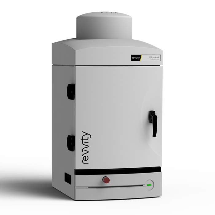
IVIS Lumina III In Vivo Imaging System
The IVIS Lumina III imaging system is capable of imaging both fluorescent and bioluminescent reporters. The system is equipped with up to 26 filter sets that can be used to image reporters that emit from green to near-infrared. Superior spectral unmixing can be achieved with the IVIS Lumina III system's optional high resolution short cut off filters.
Features and Benefits
- Market trusted technology offering the fullest suite of leading imaging technologies, reagents and support
- Exquisite sensitivity in bioluminescence
- Full fluorescence tunability through the NIR spectrum
- Compute Pure Spectrum spectral umixing for ultimate fluorescence sensitivity
- Expandable system tailored to your workflow
- Complimentary Living Image™ software licenses are provided with the IVIS systems and upon request.
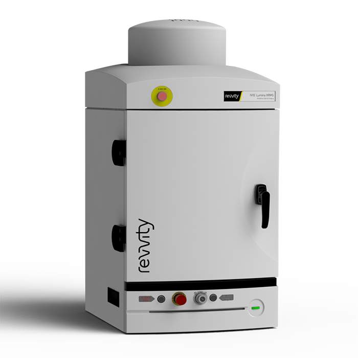
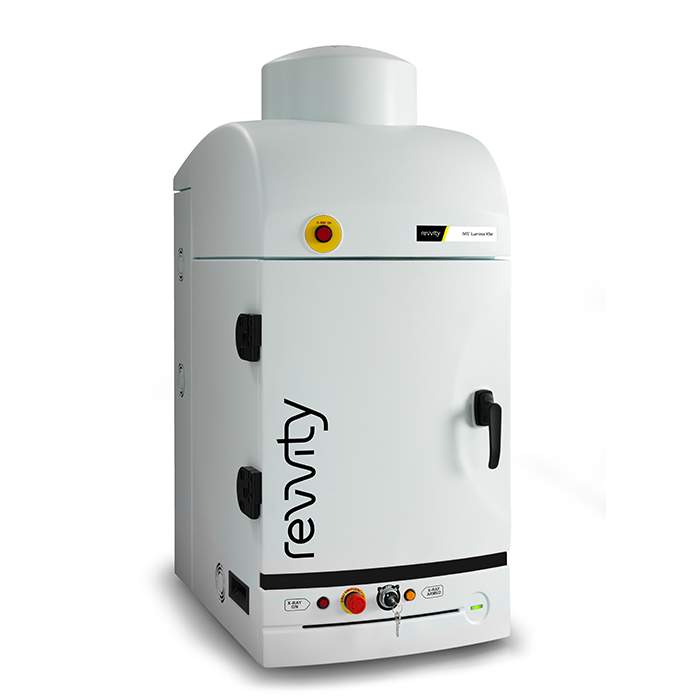
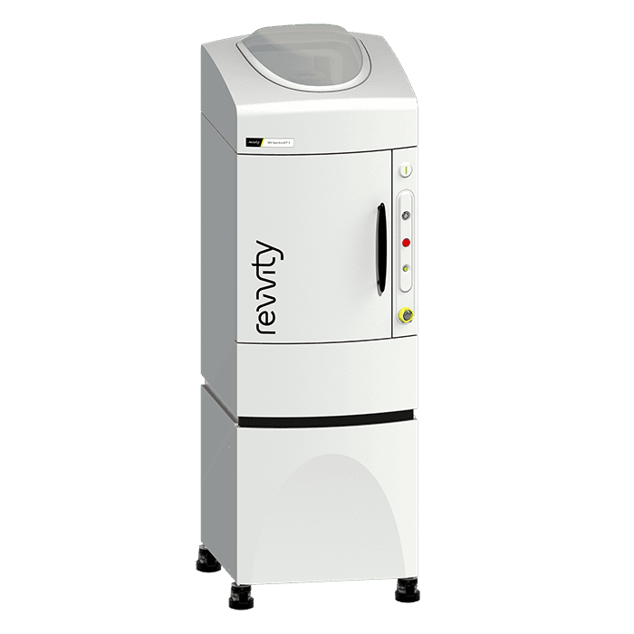
IVIS Lumina XRMS In Vivo Imaging System
The Lumina XRMS includes state of the art spectral unmixing for sensitive multispectral imaging to monitor multiple biological events in the same animal. Use our Living Image® software to automate all the controls and settings required for seamless image acquisition and processing. Typical X-ray image acquisitions take only 10 seconds and can be overlaid with both optical and photographic images.
Superior Optical Imaging with Spectral Unmixing
The IVIS Lumina XRMS Series III is capable of imaging all common fluorescent and bioluminescent reporters or dyes. The system is equipped with up to 21 filter sets to image reporters that emit from green to near-infrared. High resolution, sharp cut-off filters are simply interchangeable to achieve the highest performance, sensitivity and spectral unmixing. The Lumina XRMS imaging system also accommodates Petri dishes or micro-titer plates for in vitro imaging.
The system can incorporate premium animal handling features such as a heated stage, gas anesthesia connections and a syringe injection system for simultaneous compound administration. Living Image software yields high-quality, reproducible, quantitative results incorporating instrument calibration, background subtraction and the image algorithms. Simple user guided spectral unmixing allows detection and separation of multiple reporters, and Living Image provides the precise overlay to see your optical reporters together with anatomical surface or X-ray features.
Features and Benefits:
- Multispecies optical and X-ray imaging
- Image mice, rats and other large animals
- High resolution, low dose digital X-Ray
- Exquisite sensitivity in bioluminescence
- Compute Pure Spectrum (CPS) spectral unmixing for ultimate fluorescence sensitivity
- Full fluorescence tunability through the NIR Spectrum
- Complimentary Living Image™ software licenses are provided with the IVIS systems and upon request.
IVIS Spectrum 2 series
- High sensitivity 2D and 3D bioluminescence & fluorescence imaging
- High throughput simultaneous imaging of up to 10 mice
- Fast data acquisition for rapid visualization of images in real-time
- Two powerful modes of fluorescence excitation - epi-fluorescence and trans-illumination
- Proprietary spectral unmixing algorithms for autofluorescence removal and multiplex imaging
- Easy, one-click co-registration with the Quantum GX3 microCT system
- Broadly adopted, easy to use, and intuitive, Living Image™ visualization and analysis software
- Integrated low-dose, ultra-fast microCT (IVIS SpectrumCT 2 only)
- Complimentary Living Image™ software licenses are provided with the IVIS systems and upon request.
IVIS Spectrum 2 In Vivo Imaging System
Building upon the IVIS Spectrum in vivo optical system with proven 2D bioluminescence and 2D fluorescence imaging and 3D optical tomography capabilities combined in a single system, the IVIS Spectrum 2 is our next generation in optical imaging. This advanced imaging system incorporates a CCD camera with eXcelon® coating that enables detection of more signal at higher efficiency across a broader spectrum of wavelengths. With exclusivity to this innovative camera for in vivo imaging, the IVIS Spectrum 2 preclinical optical imaging system delivers the sensitivity you demand for non-invasive longitudinal imaging to better understand early disease-related biological changes, track disease progression, and help guide the drug development process.
IVIS SpectrumCT 2 In Vivo Imaging System
The IVIS SpectrumCT 2 in vivo optical imaging system is an integrated platform that combines the full suite of IVIS optical features including; spectral unmixing and 2D and 3D quantitative bioluminescence & fluorescence imaging, with fast, low dose microCT imaging.
Similar to the IVIS Spectrum 2 (part number CLS158738) optical imaging system, the IVIS SpectrumCT 2 incorporates a CCD camera with eXcelon® coating that enables detection of more signal at higher efficiency across a broader spectrum of wavelengths.
The simple user interface along with advanced visualization and analysis tools are driven by our market leading, easy to use Living Image® software. The IVIS SpectrumCT 2 enables longitudinal workflows to characterize disease progression and therapeutic effect throughout the complete experimental time frame with both quantitative CT and optical reconstructions. Fast imaging and the ability to image multiple animals offers the throughput required to scan large cohorts of animals quickly and draw sound conclusions from your experimental data.
Quantum GX3 microCT imaging system
- Superior spatial resolution of 5 microns
- Wide FOV range from 8 mm to 86 mm
- Improved image-based respiratory gating
- Proprietary active ring reduction
- Continuous and step scanning modes
- Seamless co-registration with the IVIS 3D optical imaging system
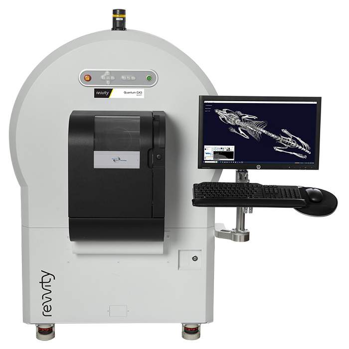
Quantum GX3 microCT System
The Quantum GX3 system represents the latest advances in microCT imaging with high image resolution, high speed, low-dose, and flexibility.
With the combination of higher resolution, increased field of view (FOV) range, and enhanced image-based respiratory and cardiac gating, the Quantum GX3 low-dose microCT system enables researchers to gain a better understanding of healthy and diseased tissue in a broad range of areas including bone, respiratory, cardiovascular, liver/kidney, brain, and oncology research.
Featured resources