US
Revvity Sites Globally
Select your location.
*e-commerce not available for this region.


Automated ultrasound for non-invasive in vivo evaluation of liver disease progression in mice
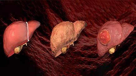
Being able to characterize and monitor the extent of fibrosis, steatosis, and inflammation in the liver non-invasively is valuable to clinicians and researchers to comprehensively stage disease progression and evaluate therapeutic response.
Read this review to learn how researchers used the Vega® automated, hands-free, high-throughput ultrasound system as a turnkey solution to evaluate, monitor, and quantify liver disease progression in murine models.
Case studies include:
- Evaluation of NAFLD using a mouse western diet mouse model
- Disease progression in a chemically induced mouse fibrosis model
- Monitoring treatment response in Mdr2 knockout mice
- Multimodal imaging of NAFLD/NASH progression in mouse model
For research use only. Not for use in diagnostic procedures.
To view the full content please answer a few questions
Download Resource
Automated ultrasound for non-invasive in vivo evaluation of liver disease progression in mice