US
Revvity Sites Globally
Select your location.
*e-commerce not available for this region.


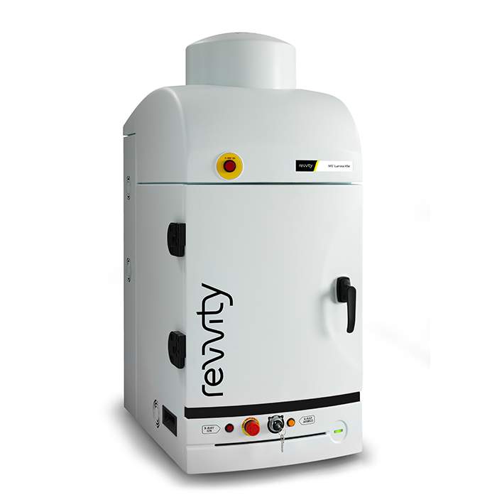
IVIS Lumina X5 Imaging System
High-Sensitivity Optical Meets High-Resolution X-ray
The IVIS™ Lumina X5 high-throughput 2D optical imaging system combines high-sensitivity bioluminescence and fluorescence with high-resolution x-ray into a compact system that fits on your benchtop. With an expanded 5 mouse field of view for 2D optical imaging plus our unique line of accessories to accelerate setup and labeling, it has never been easier or faster to get robust data- and answers- on anatomical and molecular aspects of disease.
For research use only. Not for use in diagnostic procedures.

IVIS Lumina X5 Imaging System
IVIS® Lumina S5 high throughput, high sensitivity optical in vivo imaging system with integrated x-ray.
Part #:
CLS148590
Imaging Modality:
2D Bioluminescence, 2D Fluorescence, X-ray
Loading...
Product information
Overview
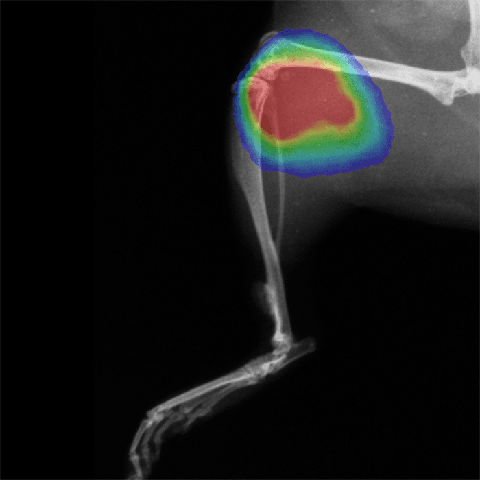
The IVIS Lumina X5 has all the capabilities of the IVIS Lumina S5 imaging system with integrated industry leading high-resolution x-ray for greater detail. The IVIS Lumina X5 also includes state of the art spectral unmixing features for sensitive multispectral imaging to monitor multiple biological events in the same animal.
High-throughput Optical and X-ray Imaging – No Compromise
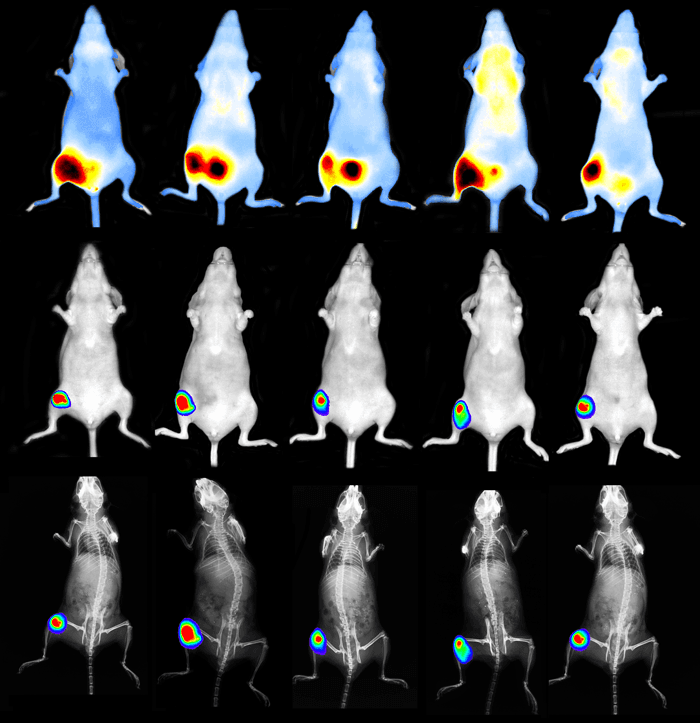
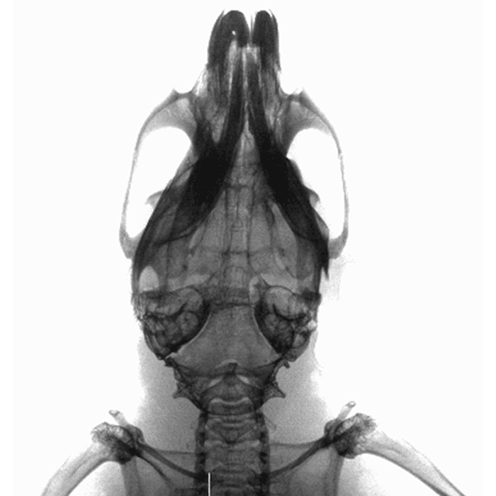
The IVIS Lumina X5 integrates a next generation 1 inch CCD camera into our benchtop instrument providing a high throughput 20 x 20 cm FOV sufficient for imaging 5 mice at a time with bioluminescence and fluorescence. Moreover, the large, independently deployed scintillator facilitates X-ray acquisitions of 5 mice and larger rodents up to 500-600 grams with seamless, accurate overlay onto the optical image at any field of view.
As with other IVIS Lumina systems, this instrument is equipped with 26 filters tunable to image fluorescent sources that emit from green to near-infrared. Novel illumination technology effectively increases fluorescent transmission through 900 nm. Additionally, the IVIS Lumina X5 incorporates Revvity's patented Compute Pure Spectrum (CPS) algorithm for spectral library generation software tools to ensure accurate autofluorescence removal, unmixing and fluorophore quantitation.
Standard on all IVIS instruments, absolute calibration affords consistent and reproducible results independent of magnification, filter selection from one instrument to any another IVIS instrument within an organization or around the world.
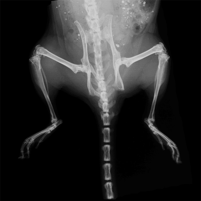
Industry Leading X-ray Resolution
The IVIS Lumina X5 is equipped with a microfocus X-ray source and geometric magnification and when combined, achieve industry leading X-ray resolution in a 2D optical/X-ray system setting a high standard in multimodal 2D imaging resolution. With optical image overlays at every X-ray resolution, never miss underlying anatomical and structural changes. Get more from your data and explore new applications.
IVIS Lumina X5 – A High Throughput Solution
Not only does the IVIS Lumina X5 offer higher throughput via the next generation 1 inch CCD, but it is also compatible with a set of smart animal handling accessories (purchased separately) designed with throughput and safety in mind.
Smart loading trays enable users to pose animals on the benchtop before placing the tray into the IVIS. Fiducials built into the tray allows the software to automatically recognize and draw ROIs providing automated animal identification.
Animal trays are designed with ease of use and user safety in mind. No nose cones are required thus minimizing cleanup. When used with the next generation anesthesia unit (RAS-4), strong vacuum capabilities minimize excess gas from escaping thus preventing exposure of users to anesthetic gas.
Finally, Living Image™ software brings IVIS technology to life by facilitating an intuitive workflow for in vivo optical, X-ray image acquisition, analysis and data organization. The software’s design creates an intuitive, seamless workflow for researchers of all skill levels.
Key Features:
- High throughput (5 mice) optical and X-ray
- Increased throughput (10 mice) using optional manifold
- High resolution, low dose X-ray with optical overlay
- Supports mouse and rat imaging
- Compute Pure Spectrum (CPS) spectral unmixing
- Full fluorescence tunability through the NIR spectrum
- Unique accessories to streamline workflow, data acquisition and analysis
- Complimentary Living Image software licenses are provided with the IVIS systems and upon request
Specifications
| Dimensions | 48.26 cm (W) x 116.84 cm (H) |
|---|
| Brand |
IVIS
|
|---|---|
| Imaging Modality |
2D Bioluminescence
2D Fluorescence
X-ray
|
| Unit Size |
1 unit
|
Image gallery
IVIS Lumina X5 Imaging System
References
- Zhou et al (2021). Dipeptidyl Peptidase-4 modulates Long-chain Acyl-CoA synthetase 4 to Promote Lipid Peroxidation and Regulate Ferroptosis in Pancreatic Cancer. Res Square. https://doi.org/10.21203/rs.3.rs-173980/v1
- Rupp et al (2021). Therapeutic potential of Fingolimod in triple negative breast cancer preclinical models. Translational Oncology. 14(1): 100926. https://doi.org/10.1016/j.tranon.2020.100926
- Jiang et al (2020). Combined Treatment With CCR1-Overexpressing Mesenchymal Stem Cells and CCL7 Enhances Engraftment and Promotes the Recovery of Simulated Birth Injury-Induced Stress Urinary Incontinence in Rats. Fontiers in Surgery. 7:40. https://dx.doi.org/10.3389%2Ffsurg.2020.00040
- Trikha et al (2020). Inhibition of Angiotensin Converting Enzyme Impairs Anti-staphylococcal Immune Function in a Preclinical Model of Implant Infection. Front. Immunol. https://doi.org/10.3389/fimmu.2020.01919
- Brandon et al (2020). Identification of ovarian high-grade serous carcinoma cell lines that show estrogen-sensitive growth as xenografts in immunocompromised mice. Sci Reports. 10: 10799. https://doi.org/10.1038/s41598-020-67533-1
- Kelley et al (2020). In vivo Mouse Model of Spinal Implant Infection. Medicine. https://dx.doi.org/10.3791/60560
- Lefeuvre et al (2020). Effects of topical corticosteroids and lidocaine on Borrelia burgdorferi sensu lato in mouse skin: potential impact to human clinical trials. Sci Reports. 10: 10552. https://doi.org/10.1038/s41598-020-67440-5
- Liang et al (2019). Idarubicin-loaded methoxy poly(ethylene glycol)-b-poly(l-lactide-co-glycolide) nanoparticles for enhancing cellular uptake and promoting antileukemia activity. Int J Nanomedicine. 14: 543–556. https://www.ncbi.nlm.nih.gov/pmc/articles/PMC6333394/
- Karches et al (2019). Bispecific Antibodies Enable Synthetic Agonistic Receptor-Transduced T Cells for Tumor Immunotherapy. Translational Cancer Mechanisms and Therapy. https://doi.org/10.1158/1078-0432.CCR-18-3927
Citations
Resources
Are you looking for resources, click on the resource type to explore further.
Case Study
A novel non-Invasive in vivo tool for the assessment of NASH
Non-alcoholic fatty liver disease (NAFLD) describes a progressive pathology that affects the liver. Fat accumulation causes fatty...
Technical Note
Adaptive Fluorescence Background Subtraction for IVIS systems
Instrument background occurs when excitation light leaks through the emission filter. This occurs more frequently when the...
Application Note
AI-assisted high-throughput analysis of multimodal in vivo imaging data for cancer research
Preclinical cancer research faces significant workflow bottlenecks due to the manual, time-intensive nature of analyzing...
Literature - Publication Review
Applications of In Vivo bioluminescence imaging for SARS-CoV-2
Scientists continue to explore several options to treat SARS-CoV-2 infection with hundreds of therapeutics at various phases of...
Technical Note
AutoExposure
This tech note outlines procedures on using auto-exposure on the IVIS® preclinical optical imaging platform using Living Image®...
Loading...


How can we help you?
We are here to answer your questions.

