MicroCT Imaging
Biology doesn't happen in 2-dimensions
Being able to study 3-dimensional (3D) structure and morphology of tissues with high resolution imaging is becoming increasingly important in biomedical research. Micro-computed tomography is a unique imaging modality that enables you to image 3D structure rapidly and non-invasively at high resolution providing valuable anatomical information for your basic research or drug discovery and development programs.
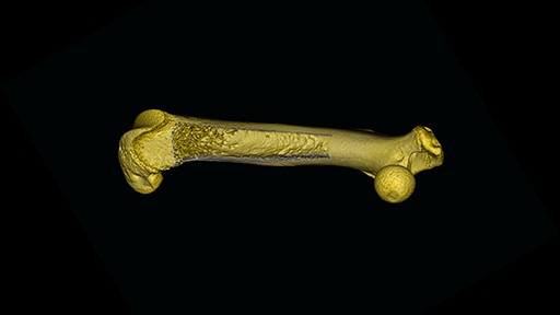
Micro-CT offers excellent cross-sectional images of skeletal structures and has long been an established tool for evaluating bone structure. It is often used to study bone diseases such as osteoporosis as well as evaluating the efficacy of therapeutics such as bisphosphates. In addition to bone research, microCT is commonly used to study cancer biology. With the development of new contrast agents, microCT scanning of soft tissue is an emerging area for this modality particularly in the area of cardiovascular and pulmonary research.
Whether you are studying tumor biology, bone, cardiovascular or pulmonary disease research, or evaluating drug efficacy, our microCT imaging solutions can help you get the answers you need.
Micro-computed tomography uses x-rays to create a 3D image composed of 2D planar images that are reconstructed and processed into 3D models.
Often referred to as 3D x-ray, microtomography, or microCT imaging, it's similar to CT scans performed in hospitals but on a smaller scale and offers greatly enhanced resolution.
For research use only. Not for use in diagnostic procedures.