


PhenoVue cell painting kits
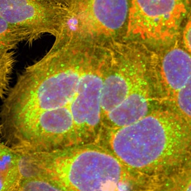
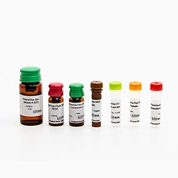
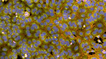
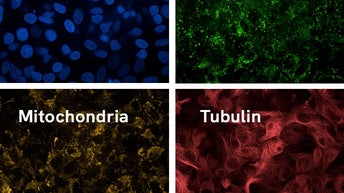
PhenoVue™ cell painting kits comprise 6 validated, pre-optimized fluorescent probes to stain the actin cytoskeleton, nucleoli, nucleus, plasma membranes, ER and Golgi and apparatus, together with a stain diluent, to streamline the cell painting workflow. We are proud to be supporting partners of the Joint Undertaking in Morphological Profiling-Cell Painting (JUMP-CP) consortium, providing our PhenoVue Cell Painting JUMP kits to the Consortium to create the world's largest, public cell painting dataset.
PhenoVue™ cell painting kits comprise 6 validated, pre-optimized fluorescent probes to stain the actin cytoskeleton, nucleoli, nucleus, plasma membranes, ER and Golgi and apparatus, together with a stain diluent, to streamline the cell painting workflow. We are proud to be supporting partners of the Joint Undertaking in Morphological Profiling-Cell Painting (JUMP-CP) consortium, providing our PhenoVue Cell Painting JUMP kits to the Consortium to create the world's largest, public cell painting dataset.
HCS/HCA instruments
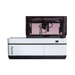
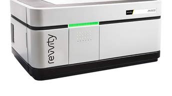
Opera Phenix OptIQ™ high-content screening system offers fast acquisition for multiplexing experiments by combining multi-camera technology with our proprietary automated high NA water immersion objectives that allow acquisition of images with shorter exposure times and higher resolution. Synchrony™ Optics combine a pinhole disk with a microlens-enhanced Nipkow disk to capture the illuminating light for a strong excitation of fluorophores and delivers high quality confocality. Optional high-power lasers minimize exposure times further and help to increase throughput of your cell painting screen.
Opera Phenix OptIQ™ high-content screening system offers fast acquisition for multiplexing experiments by combining multi-camera technology with our proprietary automated high NA water immersion objectives that allow acquisition of images with shorter exposure times and higher resolution. Synchrony™ Optics combine a pinhole disk with a microlens-enhanced Nipkow disk to capture the illuminating light for a strong excitation of fluorophores and delivers high quality confocality. Optional high-power lasers minimize exposure times further and help to increase throughput of your cell painting screen.
Operetta CLS™ high-content analysis system offers a medium throughput multiplexing imaging solution with flexible labelling due to its 8-wavelength excitation source and accessible 8 position emission filter wheel. Direct optical coupling of the powerful solid-state LED light source provides increased excitation performance offering attractive exposures times. Read times can be decreased with our optionally available proprietary automated high NA water immersion objectives providing better light efficiency. For efficient background rejection a spinning disk is available to acquire beautiful confocal images.
PhenoPlate microplates
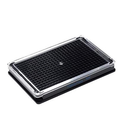
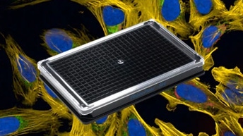
PhenoPlate™ (formerly CellCarrier™ Ultra) microplates have been engineered to deliver superior images and the highest quality data for high-content applications. The excellent flatness of the plate bottom provides optimal clarity and fast autofocusing, while the high optical quality of the cyclic olefin imaging surface ensures superior image quality.
PhenoPlate™ (formerly CellCarrier™ Ultra) microplates have been engineered to deliver superior images and the highest quality data for high-content applications. The excellent flatness of the plate bottom provides optimal clarity and fast autofocusing, while the high optical quality of the cyclic olefin imaging surface ensures superior image quality.
Powerful software
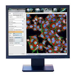
Harmony® imaging and analysis software offers all the tools needed to extract the multitude of cellular parameters in an easy and straightforward way. With its building block approach to image analysis, as few as four building blocks are needed to set up the analysis sequence.
Image Artist™ image analysis and management platform for high-content screening and cell imaging data allows you to quickly process, analyze, share, and store the vast volumes of data generated by high-content screening and cellular imaging, including 3D imaging, phenotypic screening, and cell painting – so you get answers sooner.
Signals One data analysis and management software offers tools for quality control, data visualization and hit detection.
explorer G3 customized cell painting workstation
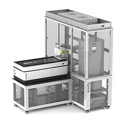
explorer™ G3 customized cell painting workstation provides an integrated, turn-key solution to automate cell painting assays using our PhenoVue Cell Painting Kits with image acquisition on our Opera Phenix OptIQ high content imager.
explorer™ G3 customized cell painting workstation provides an integrated, turn-key solution to automate cell painting assays using our PhenoVue Cell Painting Kits with image acquisition on our Opera Phenix OptIQ high content imager.
Featured products


Featured resources
