Study dynamic cellular processes with live-cell imaging
Live-cell imaging is the study of living cells using images acquired by time-lapse microscopy. It is becoming a requisite technique in many fields of life science and biomedical research, such as cell biology, developmental biology and cancer biology, as well as in drug discovery.
Live-cell imaging assays can be broadly divided into three categories:
- Live-cell end-point assays – cells are incubated for a specific period of time followed by acquisition of a single image
- Live-cell kinetic assays – cells are incubated and images acquired periodically over a time-course
- Live-cell fast response assay - cells are incubated and a compound or modulator is dispensed into the wells shortly before imaging.
Advantages of live-cell imaging
Live-cell imaging enables researchers to study dynamic cellular processes, behavior and function in real time and over time, thereby giving a more realistic view of biological function.
Live-cell imaging is also helpful when the endpoint of the assay is unknown, e.g. when analyzing long-term toxic drug effects. Depending on the type of question being asked, it may also be beneficial to avoid fixation reagents, as they influence the sample in an uncontrolled way and may change protein localization, e.g. when studying protein trafficking.
Kinetic live-cell imaging avoids the need to prepare a separate sample for every time point to be analyzed - a single sample can be analyzed over time.
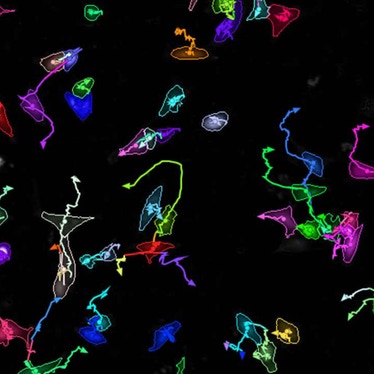
Furthermore, single-cell tracking software allows scientists to follow individual cells over time even as they divide. As a result, dynamic processes or effects that might have been overlooked when studying the whole cell population, can be seen and studied at a single cell level.
Challenges of kinetic live-cell imaging
A major challenge of kinetic live-cell imaging is keeping cells alive and functioning as naturally as possible for the duration of the experiment – usually days or weeks. Cells require incubation under controlled environmental conditions, such as temperature, CO₂ and humidity, with minimal disruption throughout the experiment to avoid inducing stress responses that might alter the cellular processes being studied.
Also, as cells are typically not exposed to light during their natural life cycle, it's important in imaging applications to minimize light exposure to reduce phototoxicity and photobleaching.
With well-designed systems, live-cell assays allow research biologists and drug discovery scientists to discover more, achieve greater understanding and provide the confidence and reassurance that their results are true to life.
Solutions for live-cell imaging
Our high-content screening systems, Opera Phenix® Plus and Operetta® CLS™ have the flexibility to run live-cell assays, combining the benefits of observing live-cells over time with the quantitative, multiparametric data analysis required in drug discovery programs. In addition, the Opera Phenix Plus system is equipped with fast frame rate imaging and has the option of on-board liquid handling to enable “dispense and read” live-cell fast response assays.