US
Revvity Sites Globally
Select your location.
*e-commerce not available for this region.


Image Artist
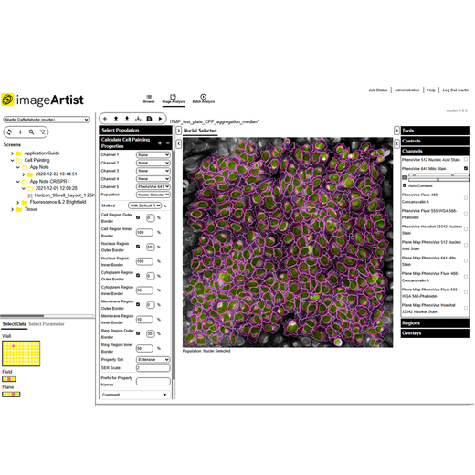

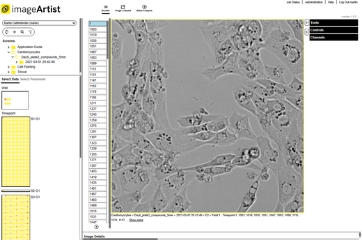
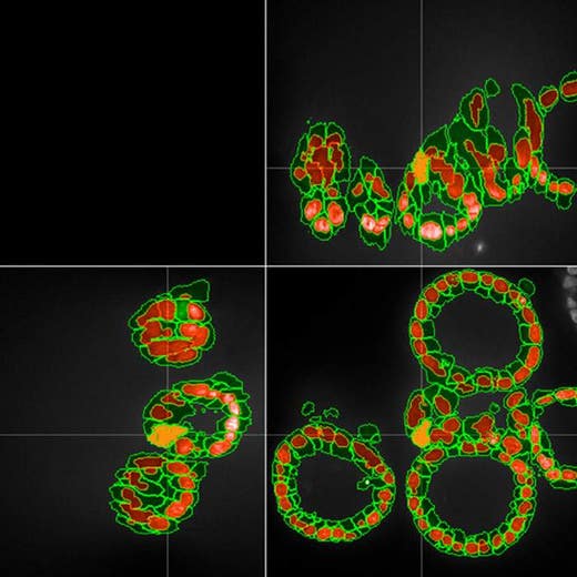
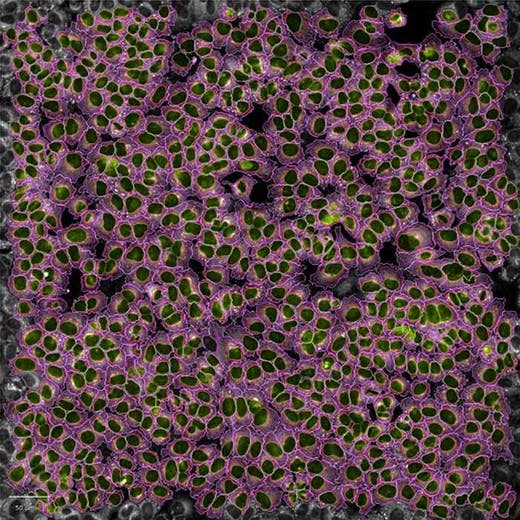

Image Artist
Signals Image Artist Image Data Analysis and Management Platform
Image Artist™ is our next generation image analysis and management platform for high-content screening and cell imaging data. Quickly process, analyze, share, and store the vast volumes of data generated by high-content screening and cellular imaging, including live cell imaging, 3D imaging, phenotypic screening, and cell painting – so you can get your answers sooner. Reduce time to results from days and weeks to hours!
The only commercially available platform that provides universal high-volume image data storage and analysis, Image Artist supports image data from all major high-content screening and cellular imaging systems. It uses high performance computing and an industry standard object store to provide a scalable, cost-effective, and multi-user solution for image analysis and management that can expand with your labs evolving needs.
Product variant
Unit Size: 1 each
Part #:
INF02358
For research use only. Not for use in diagnostic procedures.
Loading...
Product information
Overview
Next generation image analysis and management
Image Artist has powerful capabilities for processing, analyzing, storing, and sharing all your high-content screening and cell imaging data – so you can significantly cut your time to results.
- Fast image data processing and image analysis even for complex assays powered by high performance computing
- Easy-to-use assay building blocks with integrated Artificial Intelligence (AI) that make advanced image analysis
straightforward – now with improved segmentation and analysis capabilities for 3D cell applications - A central location to store all your image data with associated instrument metadata for an enduring picture of
experiments - Compatible with all major high-content screening and cell imaging systems
- Multi-user solution that can support your entire lab without compromising performance
- Scalable data storage to expand with your labs needs over time
- Seamless integration with Revvity Opera Phenix OptIQ™ and Operetta CLS™ high-content screening systems operated
by Harmony™ software, as well as Signals One
for profiling image data, hit selection, and more - NEW! The latest version is compatible with data from a broader range of systems including the Celigo™ image
cytometer - NEW! Wider range of on-premise and cloud options, with AWS S3 support. Now featuring flexible tier-based storage
options, and Intelligent-Tiering for automatic cost-savings. - NEW! Phenologic.AI is an optional module that provides an efficient and reliable method for identifying cells
and cellular nuclei within fluorescent and brightfield images by harnessing the power of pre-trained deep neural
networks (DNNs).
It is also part of our complete workflow solution for high-content screening that includes PhenoVue™ cellular imaging reagents, PhenoPlate™ imaging microplates, HCS instruments, image cytometers, software, and automation.
For all your high-content screening and cellular imaging data
High-content screening and cellular imaging experiments generate huge amounts of image data. With the latest applications such as phenotypic screening, cell painting, and 3D imaging which use more complex,
physiologically-relevant disease models, the volume of data continues to grow.
To maximize all this valuable data, you need a powerful solution for image processing, management and storage, and for sharing data with colleagues.
Image Artist allows you to bring together cell imaging data from a wide range of sources on a single software platform so that you can store, share, analyze, and re-analyze it seamlessly.
Not only does it make managing image data easier for research scientists but also for your IT team. With cloud deployment options (such as AWS S3), your IT team have just one platform and installation to manage and store research image data which can easily be scaled. They can also have peace of mind with enhanced security for cloud deployments. What's more, with AWS S3, you can choose from a range of storage classes based on your cost and data access requirements, and optional Intelligent-Tiering allows automatic transfer of data to more cost-effective tiers, based on access frequency.
Image analysis designed for biologists
Whether you are performing phenotypic screening, cell painting, 3D imaging, live cell imaging, or more routine assays, Image Artist is designed to make it easy for biologists to perform sophisticated image analysis without any coding experience.
The software platform’s image analysis building blocks encapsulate many years of knowledge and expertise in cellular imaging and analysis - so that you can focus on the biology. By simply adding together building blocks such as “Find Nuclei” and “Calculate Cell Painting Properties”, users can quickly create image analysis protocols in just a few simple steps.
With 3D cell models bridging the gap between 2D cell cultures and in vivo animal models, Image
Artist has been further optimized for 3D cell applications, with improved segmentation and analysis capabilities – so you can generate more accurate results than ever.
Built-in Artificial Intelligence (AI) and machine learning technologies allow users to train the software to develop image analysis algorithms. While other systems may require an image analysis expert to create an algorithm, Image Artist uses proprietary machine-learning technology to make it easy for you to do it on your own. Using a learn-by-example approach, in just a few clicks segmented images can be classified quickly and easily.
This is all powered by high performance computing, so you can get your answers faster.
- Get started without extensive training
- Measure complex and subtle phenotypic responses
- Compare multiple samples, plates, or batches to quality check your results
- Kinetic measurement capabilities to track cells, reveal changes in cell properties, and provide information on
cell movement - Use with Signals One to perform screening data analysis and validation, QC analyses,
calculate reliable normalization, multivariate hit stratification, dose response curves, and drug response
profiling
Specifications
| Brand |
Signals Image Artist
|
|---|---|
| Unit Size |
1 each
|
Image gallery
Image Artist
Resources
Are you looking for resources, click on the resource type to explore further.
Brochure
3D cell culture workflow solutions
More than ever, researchers are turning to 3D cell cultures, microtissues and organoids to bridge the gap between 2D cell cultures...
FAQ
Cell painting: your questions answered
With powerful potential for phenotypic drug discovery, cell painting is being rapidly adopted by leading scientists across the...
Scientific Poster
Enhancing brightfield high-content analysis: AI-driven segmentation and advanced organoid detection
Improve brightfield imaging accuracy for complex cell and organoid analysis.
Brightfield imaging can provide valuable morphological...
Whitepaper
High content screening in three dimensions
Researchers are increasingly looking to 3D cell cultures, microtissues, and organoids to bridge the gap between 2D cell cultures...
Case Study
Phenotypic characterization of mitochondria in breast cancer cells using morphology and texture properties
Mitochondria are complex networks that are highly variable, both between cell lines and within cell populations. Reliably...
Loading...


How can we help you?
We are here to answer your questions.

