3D Cell Model Imaging & Analysis
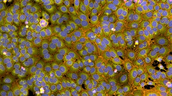
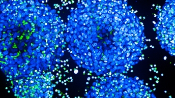
3D cell culture models, such as spheroids, organoids, and microtissues, have gained popularity as tools for research, drug discovery and toxicity studies. 3D cell models are more physiologically relevant than biochemical assays and 2D cell cultures, as they more closely represent the microenvironments, cell-to-cell interactions, and biological processes that occur in vivo.
We provide a portfolio of products for high-content imaging and analysis of 3D cell cultures to help you generate more physiologically relevant data that can power better-informed decisions.
Our high-content analysis systems, plus innovative reagents, provide you with highly sensitive solutions for 3D cell imaging, and our intuitive software platforms give you the tools to optimize imaging and analyze the most complex 3D cellular models.
For research use only. Not for use in diagnostic procedures.


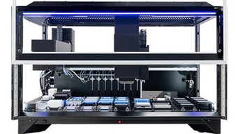
Imaging systems designed with 3D cell culture models in mind
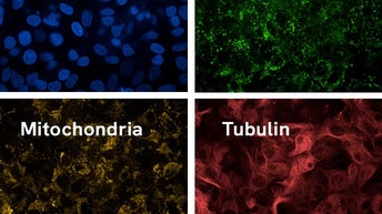
Due to their size, 3D cell models and tissues pose characteristic challenges to any imaging system but benefit greatly from confocal imaging. Our spinning disk confocal high-content imaging systems with automated water-immersion objectives let you quickly and easily generate content-rich, physiologically relevant data from 3D samples.

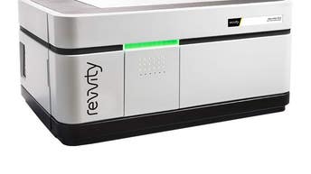
The Operetta CLS™ system's unique combination of technologies provides flexibility, sensitivity, and resolution for 3D cell culture imaging, whilst the Opera Phenix OptIQ™ system is our premier confocal solution for 3D high-content screening. It features a microlens-enhanced dual-disk design with pinhole distances optimized for thick samples such as 3D cell models. With 3D cells models in mind, both systems utilize automated water-immersion objectives and confocal spinning-disk technology, together with the optional PreciScan™ intelligent acquisition software plug-in.

Automated water-immersion objectives
Captures up to four times more light than air objectives and provides higher resolution in X,Y, and Z, so you can get more detail faster, and image deeper into 3D structures.
Captures up to four times more light than air objectives and provides higher resolution in X,Y, and Z, so you can get more detail faster, and image deeper into 3D structures.

Confocal spinning-disk technology
Ideal for imaging 3D spheroids, this technology allows you to acquire image stacks with improved signal-to-noise ratios, high X, Y, and Z resolution, high frame rates and minimal sample illumination.
Ideal for imaging 3D spheroids, this technology allows you to acquire image stacks with improved signal-to-noise ratios, high X, Y, and Z resolution, high frame rates and minimal sample illumination.

Save time with pre-scan intelligent acquisition
Pre-scan at low magnification to locate spheroids, then automatically rescan at higher magnification with the spheroid centered in the image.
Pre-scan at low magnification to locate spheroids, then automatically rescan at higher magnification with the spheroid centered in the image.
Intuitive software to analyze the most complex 3D cellular models
Our software solutions give you the tools to optimize imaging of 3D cell models and their automated workflow-centric interfaces, with integrated artificial intelligence, make advanced 3D image analysis straightforward. Turn data into understanding with the intuitive Harmony® imaging and analysis software for the Opera Phenix OptIQ and Operetta CLS systems. You can:
- Speed up 3D image acquisition by targeted imaging of microtissues
- Better understand your cell models by exploring them in the 3D viewer and XYZ viewer
- Create Z stack overviews (or gallery)
- Produce 3D renderings and movies
- Analyze morphologies and volumes in 3D
- Calculate Maximum Intensity Projections containing height profiles
As datasets for 3D cell models are often huge, data from Harmony software and all major high-content screening and cell imaging systems can seamlessly integrate with Image Artist™, our next generation image analysis and management platform.

Harmony™ high-content imaging and analysis software
Drives the Operetta CLS and Opera Phenix systems and includes everything you need to analyze the most complex cellular models in 3D, reliably discriminate phenotypes, and turn your biological data into knowledge.
Drives the Operetta CLS and Opera Phenix systems and includes everything you need to analyze the most complex cellular models in 3D, reliably discriminate phenotypes, and turn your biological data into knowledge.

Image artist™ high-content image analysis and management platform
Quickly process, analyze, share and store the vast volumes of data generated by high-content screening and cellular imaging of 3D objects, and get answers sooner.
Quickly process, analyze, share and store the vast volumes of data generated by high-content screening and cellular imaging of 3D objects, and get answers sooner.
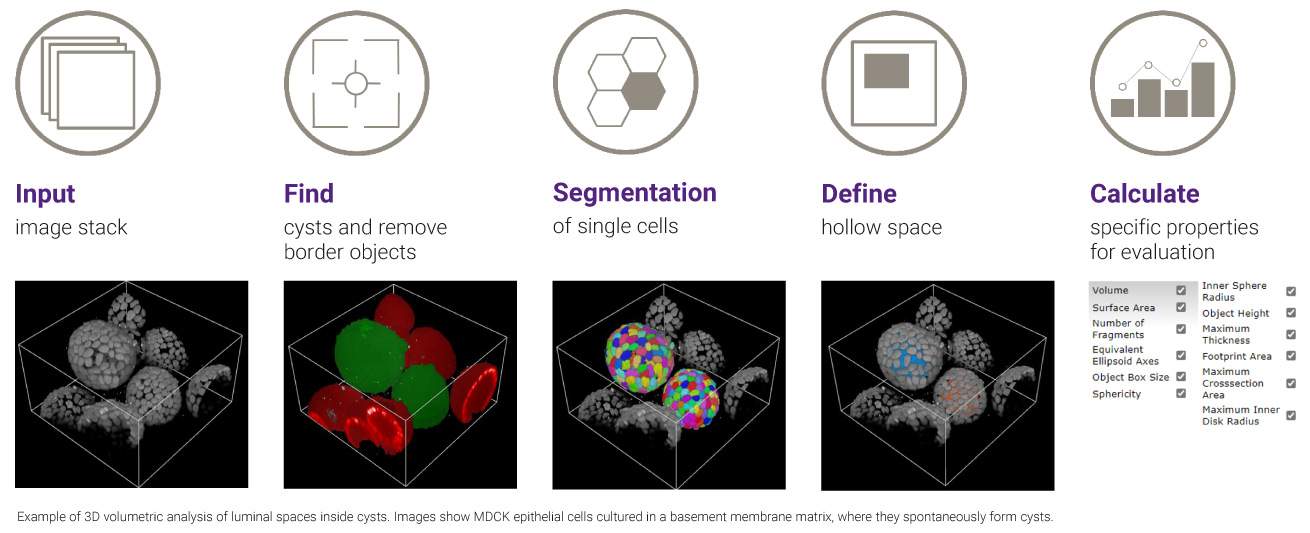
Workflow-centric software for simplified 3D image analysis
Explore our resources


Explore our solutions


Cellular Imaging Software

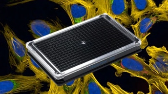
Microplates for Cell imaging


Featured products