


US
Revvity Sites Globally
Select your location.
*e-commerce not available for this region.

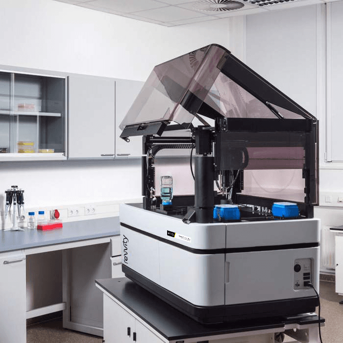
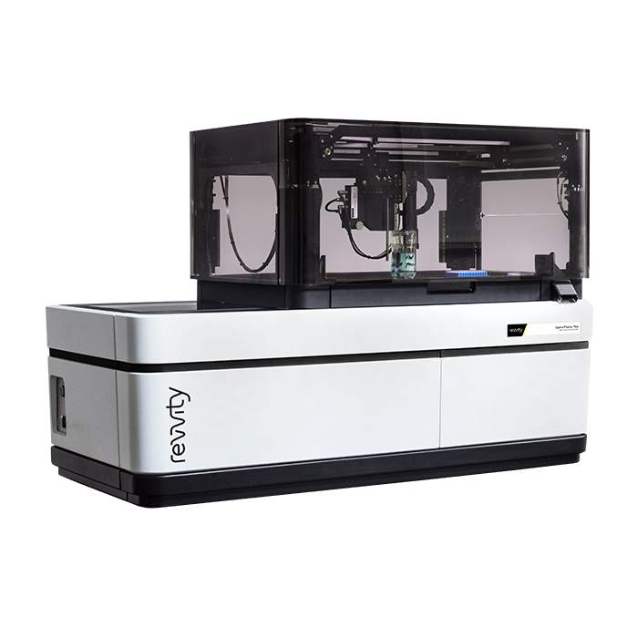
Opera Phenix Plus High-Content Screening System
The Opera Phenix™ Plus high-content imaging system is a premier confocal solution for your most demanding high content applications. Drawing on over 20 years of experience, the Opera Phenix Plus is designed for high-throughput imaging assays, phenotypic screening, assays using complex disease models, such as live cells, primary cells and microtissues, and fast-response assays, such as Ca2+ flux.
For research use only. Not for use in diagnostic procedures.
/poster.jpg?format=auto&width=40&height=40)
/poster.jpg?format=auto&width=40&height=40)


Opera Phenix Plus High-Content Screening System
Opera Phenix Plus High-Content Screening System
Part #:
HH14001000
Imaging Modality:
Brightfield, Confocal, Digital phase contrast, Fluorescence
Loading...
Unlock deeper insights with the Opera Phenix Plus high-content imaging system
Make meaningful discoveries faster with our advanced high-content imaging solutions.
Discover more
High content imaging provides a greater depth of information than other approaches, allowing you to explore cellular processes in detail.
Quantify in detail
Analyze phenotypic changes at scale, from single cells to entire populations. High-content imaging empowers detailed quantification.
Enhance 3D imaging
Water immersion objectives elevate 3D image quality, revealing intricate details within complex biological structures.
Boost throughput
Add more cameras to increase speed and efficiency. High-content imaging accelerates your research pipeline.
Video overview
Key features
Multiple cameras
Increase imaging speed by using multiple cameras and simultaneous acquisition, especially for extensive stacks for 3D models.
Microlens-enhanced pinhole
Microlenses increase the excitation efficiency allowing for a bigger pinhole to pinhole distance thereby reducing out of focus light in confocal imaging.
Automated water-immersion objectives
Improve image quality and get better data by enhancing the signal and improving the z resolution while capturing more light.
Proprietary Synchrony™ optics
Combine a microlens-enhanced pinhole disk with dual-view confocal optics. This minimizes spectral crosstalk during simultaneous acquisition by separating fluorescence excitation and emission. Provide greater speed and higher sensitivity.
Machine learning
Easily create algorithms without being an image analysis expert using our PhenoLOGIC proprietary machine-learning technology.
Intelligent image acquisition
Image only the objects you are interested in, centered at high magnification, thereby reducing imaging time and data size.
Powerfully simple analysis
Harmony™ high-content imaging and analysis software can make you more productive faster with ready made templates and simple steps to a custom analysis.
3D analysis
Explore your cell models by visualizing them in a 3D- and an XYZ-viewer and quantify volumetric and other 3D related phenotypic readouts.
Applications
Micro Physiological Systems (MPS)
Deepen understanding of 3D cell models with clearer images that reveal critical features and processes.
Functional genomic screening
Create genetic evidence with ease by phenotyping cellular response to thousands of gene manipulations in a single experiment.
Cell painting
Simplify multiparametric screening with out of the box reagents and analysis to reduce complexity (and create more time for you to focus on the science).
What our clients say about us
Opera Phenix Plus family: typical configurations
Opera Phenix Plus Single
The same sensitivity and resolution as the rest of the Phenix family, with the ability to upgrade later with additional cameras.
Opera Phenix Plus Simultaneous
Higher speed, dual-camera system for multi-color, simultaneous confocal image acquisition and fast multiplexing.
Opera Phenix Plus FRET
With its five lasers and four-camera setup, it supports CFP/YFP FRET applications to map protein-protein interactions.
Opera Phenix Plus Screener
The ultimate in throughput and performance, it delivers four cameras and four higher powered lasers - supporting screening of large libraries.
Configuration details
| Single | Simultaneous | FRET | Screener | ||
|---|---|---|---|---|---|
| System options | Number of cameras | 1 | 2 | 4 | 4 |
| Camera upgrade path | 2 or 4 cameras | 4 cameras | - | - | |
| Automated water immersion lenses | ✔ | ✔ | ✔ | ✔ | |
| Transmitted light | ✔ | ✔ | ✔ | ✔ | |
| Environmental control | ✔ | optional | ✔ | optional | |
| On-board liquid handling | optional | optional | optional | optional | |
| Robotics/automation compatible | ✔ | ✔ | ✔ | ✔ | |
| Emission filters | 8 | up to 16 | up to 14 | up to 14 | |
| Acquisition speed | 2D/3D | +/+ | ++/++ | +++/++++ | +++/++++ |
| Lasers | 375/425 nm | - | - | ✔ | - |
| 405 nm | ✔ | ✔ | - | ✔ | |
| 488 nm | ✔ | ✔ | ✔ | ✔ | |
| 561 nm | ✔ | ✔ | ✔ | ✔ | |
| 640 nm | ✔ | ✔ | ✔ | ✔ | |
| Imaging modes | Synchrony optics for minimized simultaneous imaging cross talk | - | ✔ | ✔ | ✔ |
| Multi-color fluorescence imaging | ✔ | ✔ | ✔ | ✔ | |
| Multi-color simultaneous confocal imaging | - | ✔ | ✔ | ✔ | |
| 3D imaging | ✔ | ✔ | ✔ | ✔ | |
| Brightfield and digital phase contrast | ✔ | ✔ | ✔ | ✔ | |
| Fluorescence widefield imaging | ✔ | ✔ | ✔ | ✔ | |
| Ratiometric FRET* | + | + | ++ | + | |
| Fast frame rate imaging | ✔ | ✔ | ✔ | ✔ |
*CFP/YFP or spectrally similar pairs
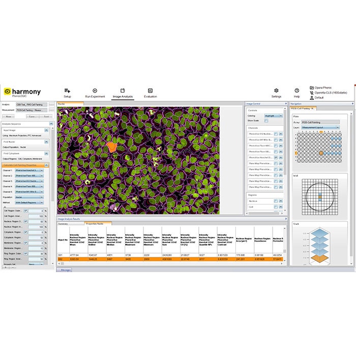
Image acquisition and analysis simplified with Harmony software
Intuitive workflow
Harmony™ software offers an intuitive user interface that guides you from image acquisition to analysis and evaluation.
Templates for quick set-up
Templates allow you to set up acquisition channels and parameters efficiently.
Ready-made solutions
Choose from pre-built solutions for common image analysis tasks, simplifying your workflow.
Customizable building blocks
Create, configure, and customize your own high-content analysis applications using image analysis building blocks.
Advanced features
Harmony™ includes advanced analysis capabilities, such as texture and STAR morphology analysis, providing detailed descriptions of cellular morphology and robust differentiation of phenotypes.
Data management
The software automatically stores analysis results and metadata, including assay layout, instrument settings, and user-defined keywords and annotations.
Image gallery


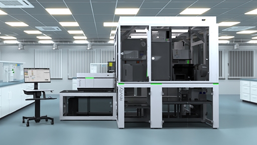
Opera Phenix Plus liquid handling module
Opera Phenix Plus system with liquid handling module
Cell painting assay imaged on the Opera Phenix Plus system
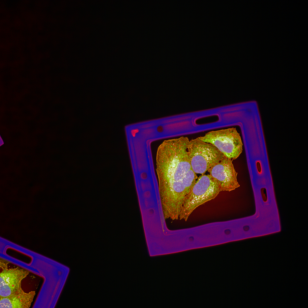
U2OS cell imaging on the Opera Phenix Plus system
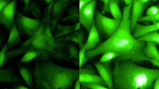
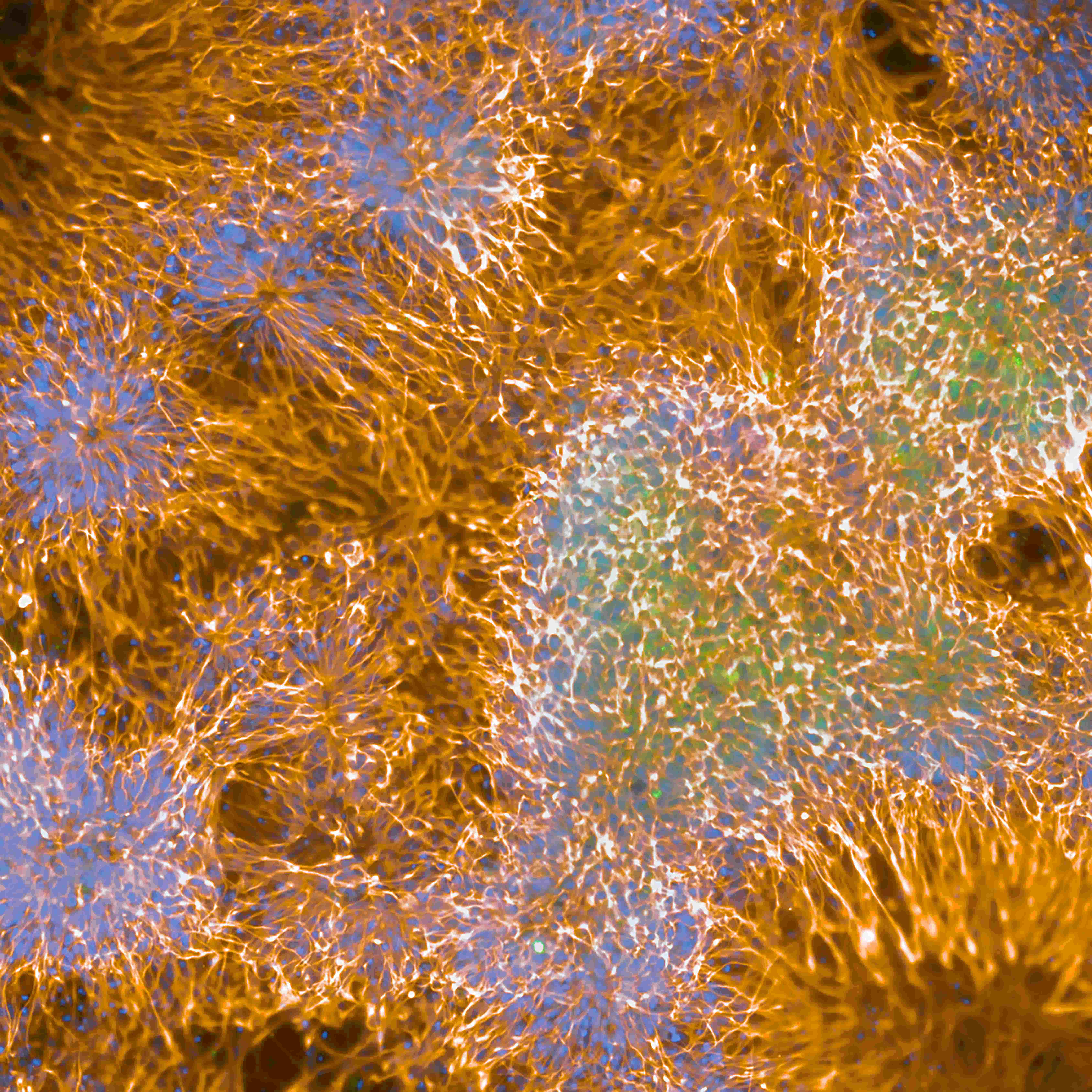
Live cell staining on the Opera Phenix Plus system
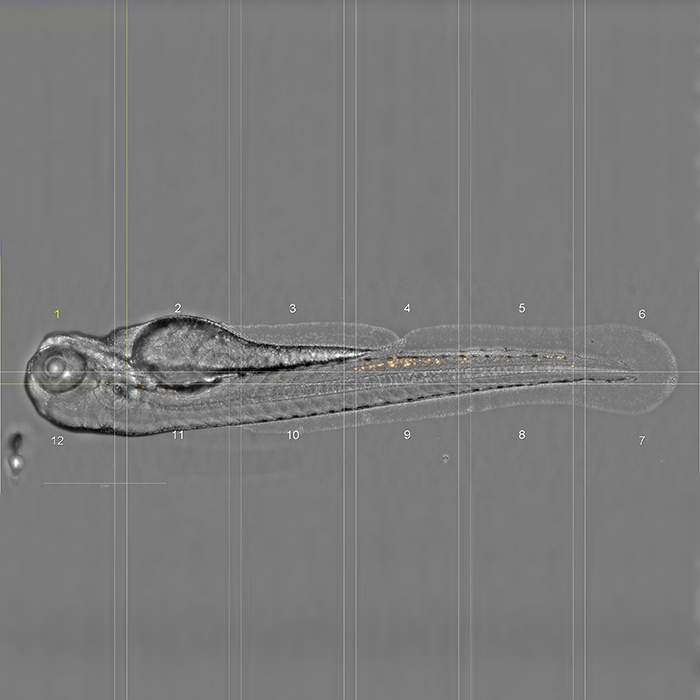
Zebra fish imaging on the Opera Phenix Plus system
Cell painting assay imaged on the Opera Phenix Plus system
Calcium flux assay imaged on the Opera Phenix Plus system
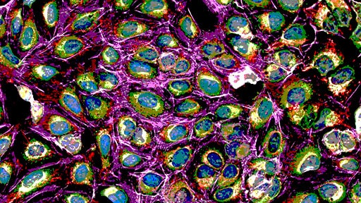
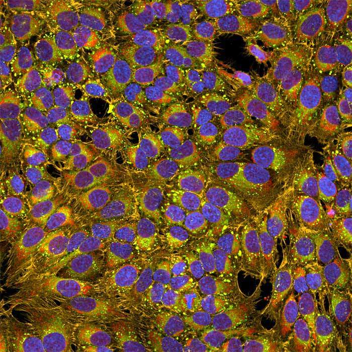
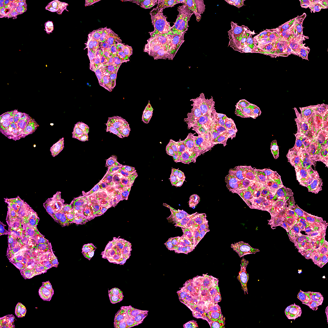
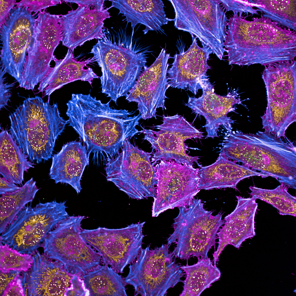
Cell painting assay imaged on the Opera Phenix Plus system
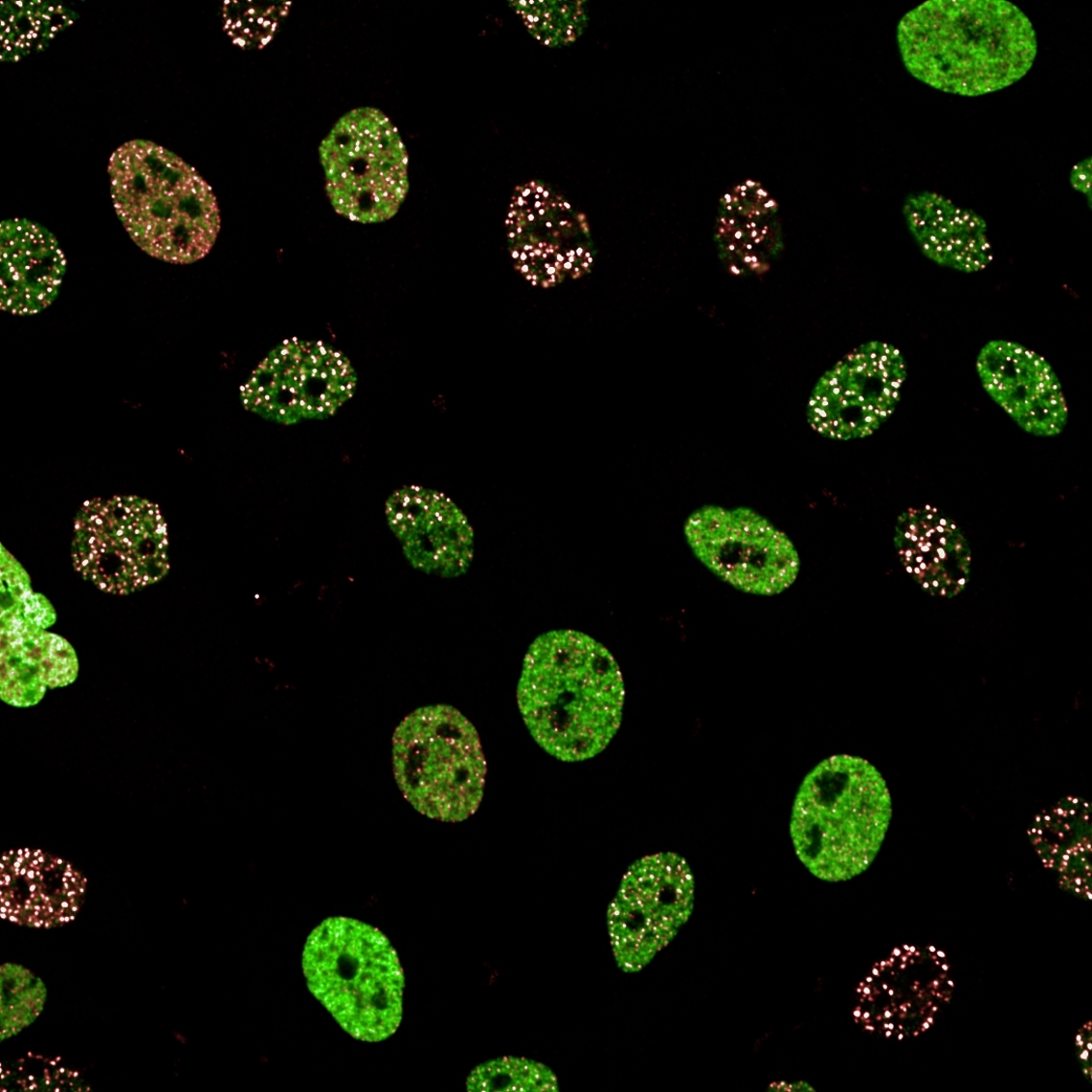
U2OS cell imaging on the Opera Phenix Plus system
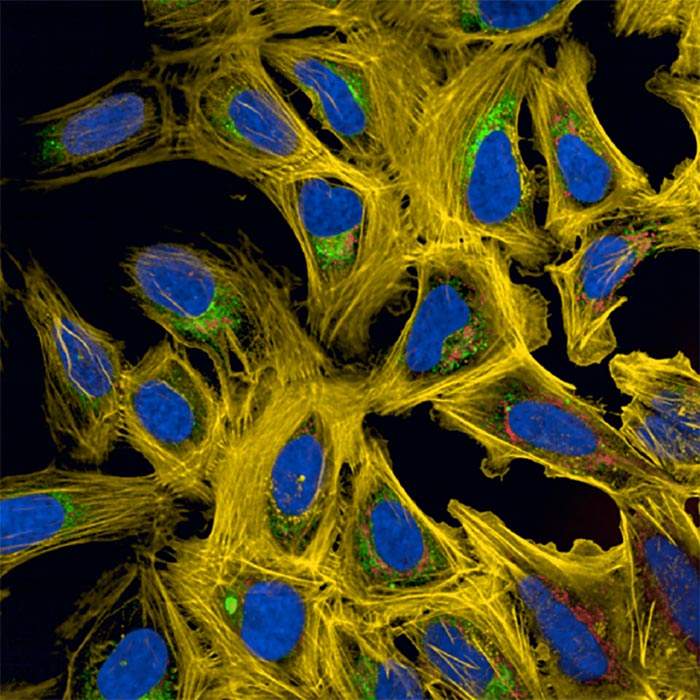
5-plex staining on the Opera Phenix Plus system
Cell painting assay imaged on the Opera Phenix Plus system
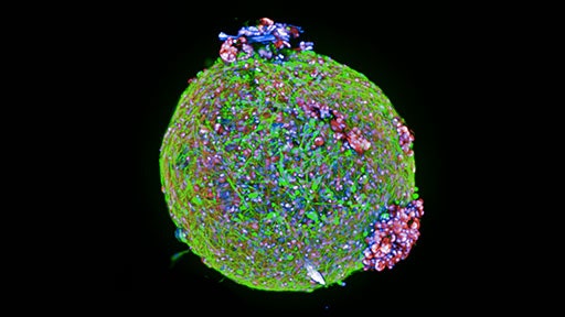
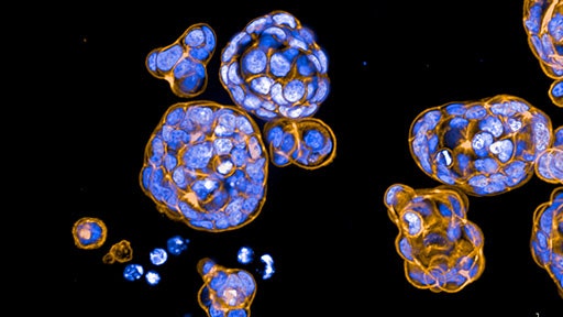
Organoids from pediatric brain tumor imaged on the Opera Phenix Plus high-content screening system
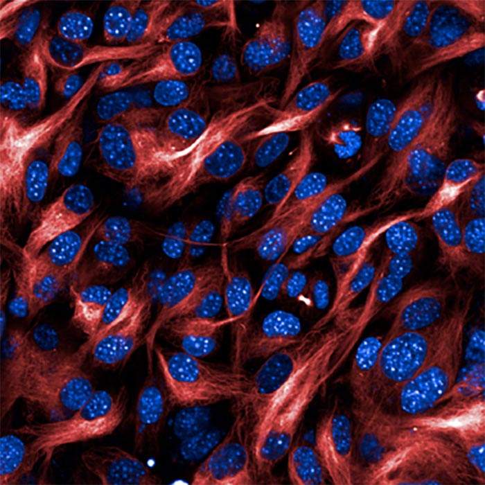
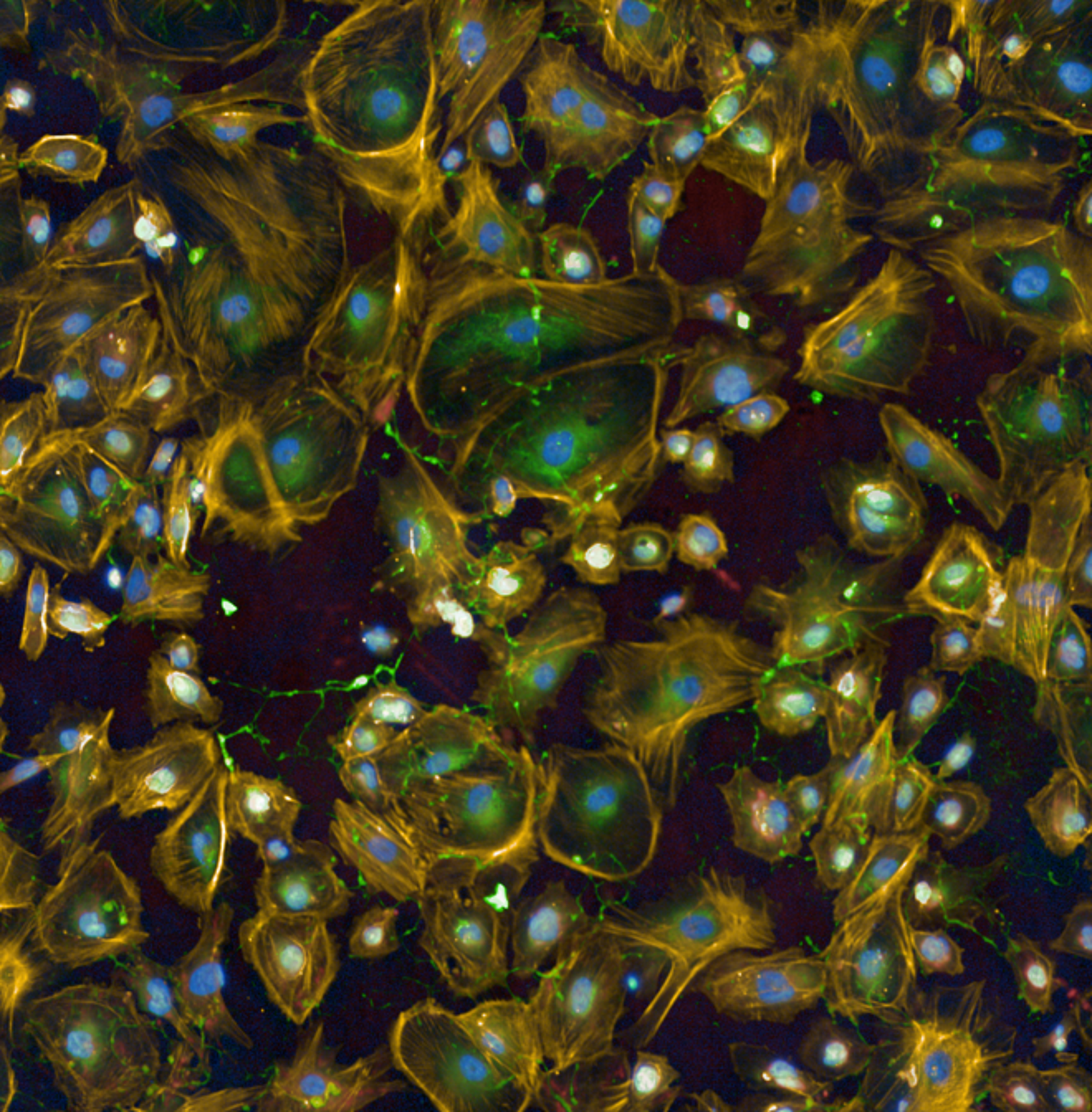
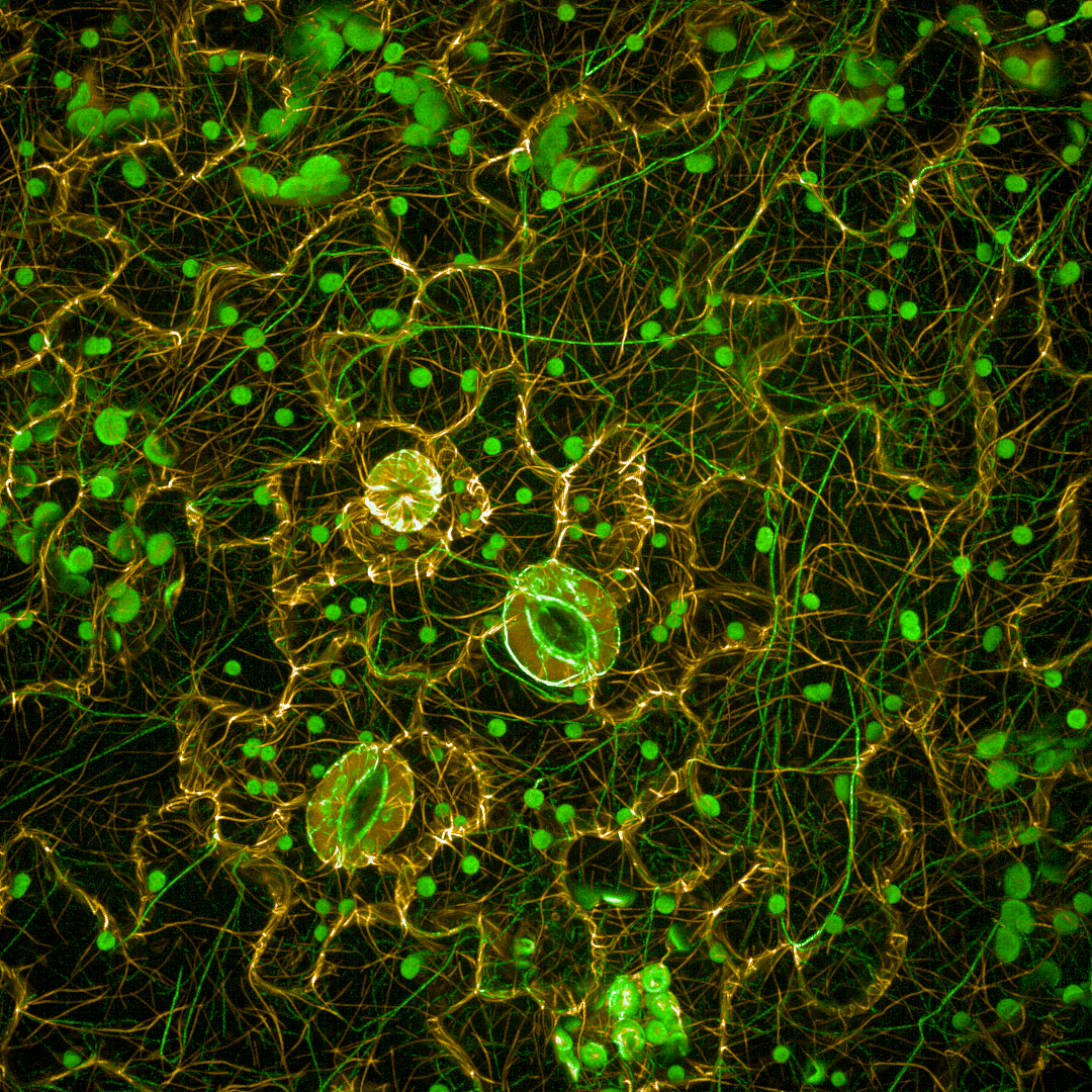
Immortalized human podocytes imaged on the Opera Phenix Plus high-content screening system
Arabidopsis cells imaged on the Opera Phenix high-content screening system
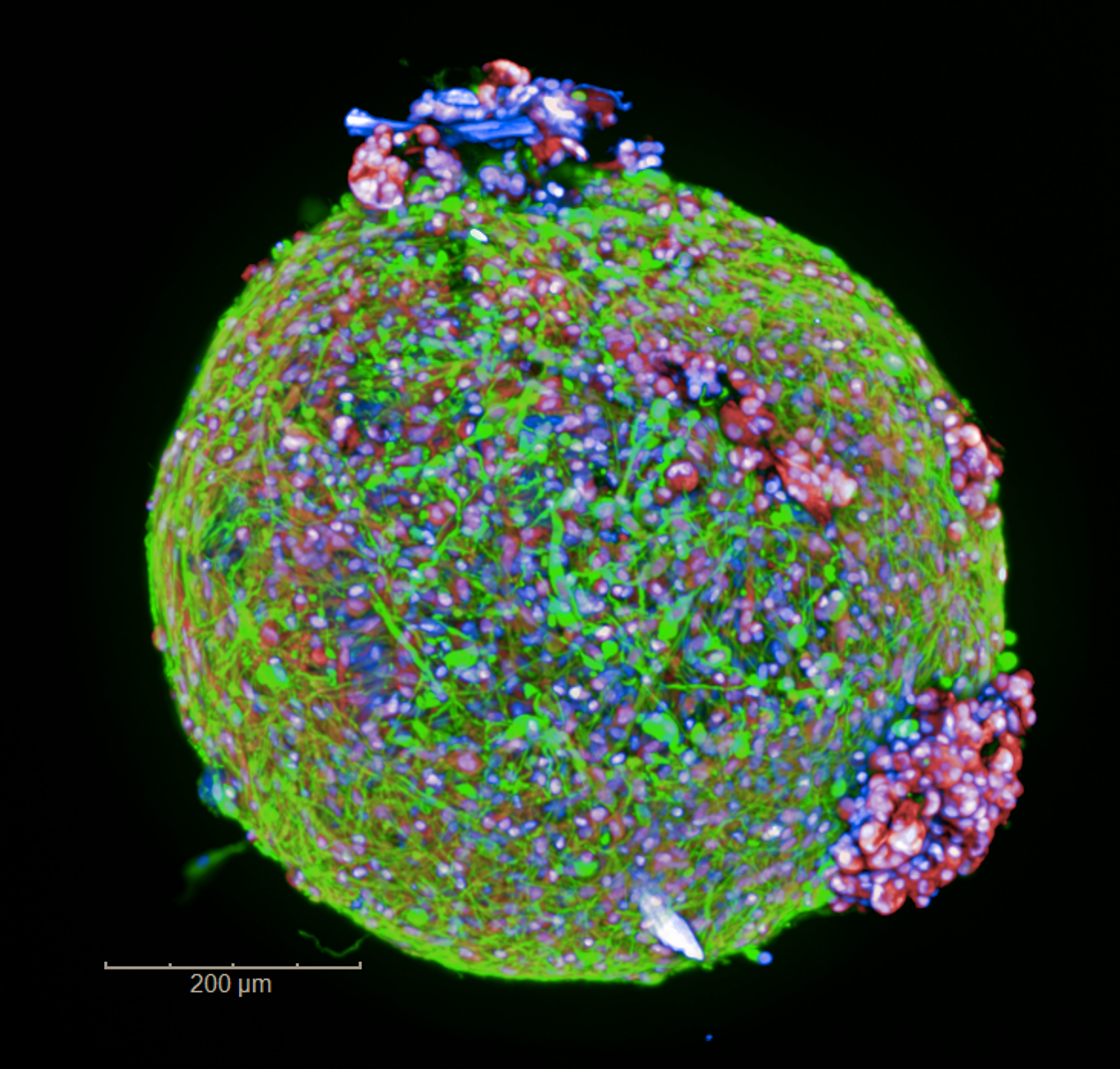
HepG2 spheroids imaged on the Opera Phenix Plus high-content screening system.
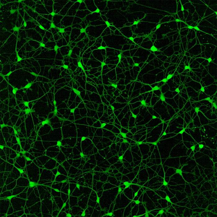
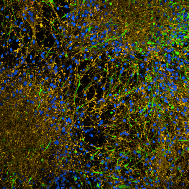
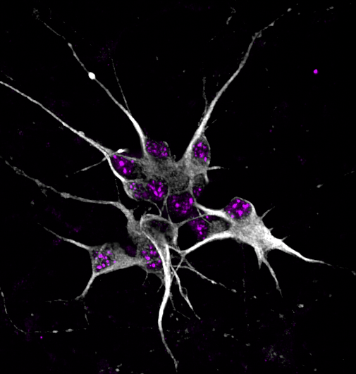
Human iPSC-derived neurons imaged on the Opera Phenix Plus high-content screening system.
Neuroblastoma SHSY5Y cells imaged on the Opera Phenix Plus high-content screening system.
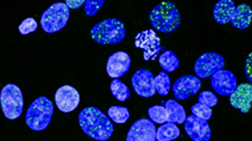
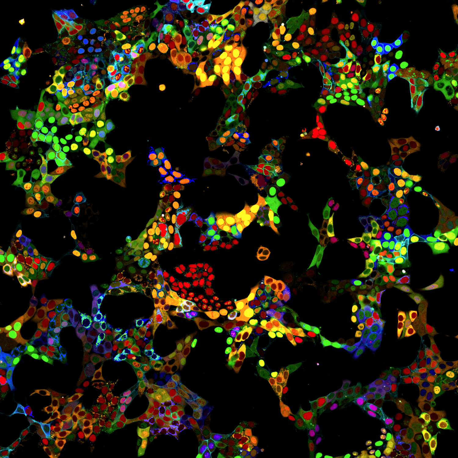
HAP1 cell pool imaged on the Opera Phenix Plus high-content screening system.
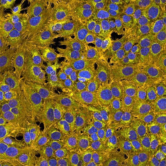
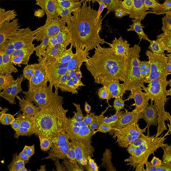
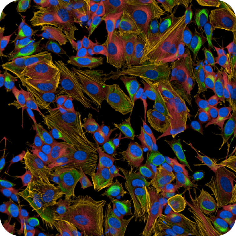
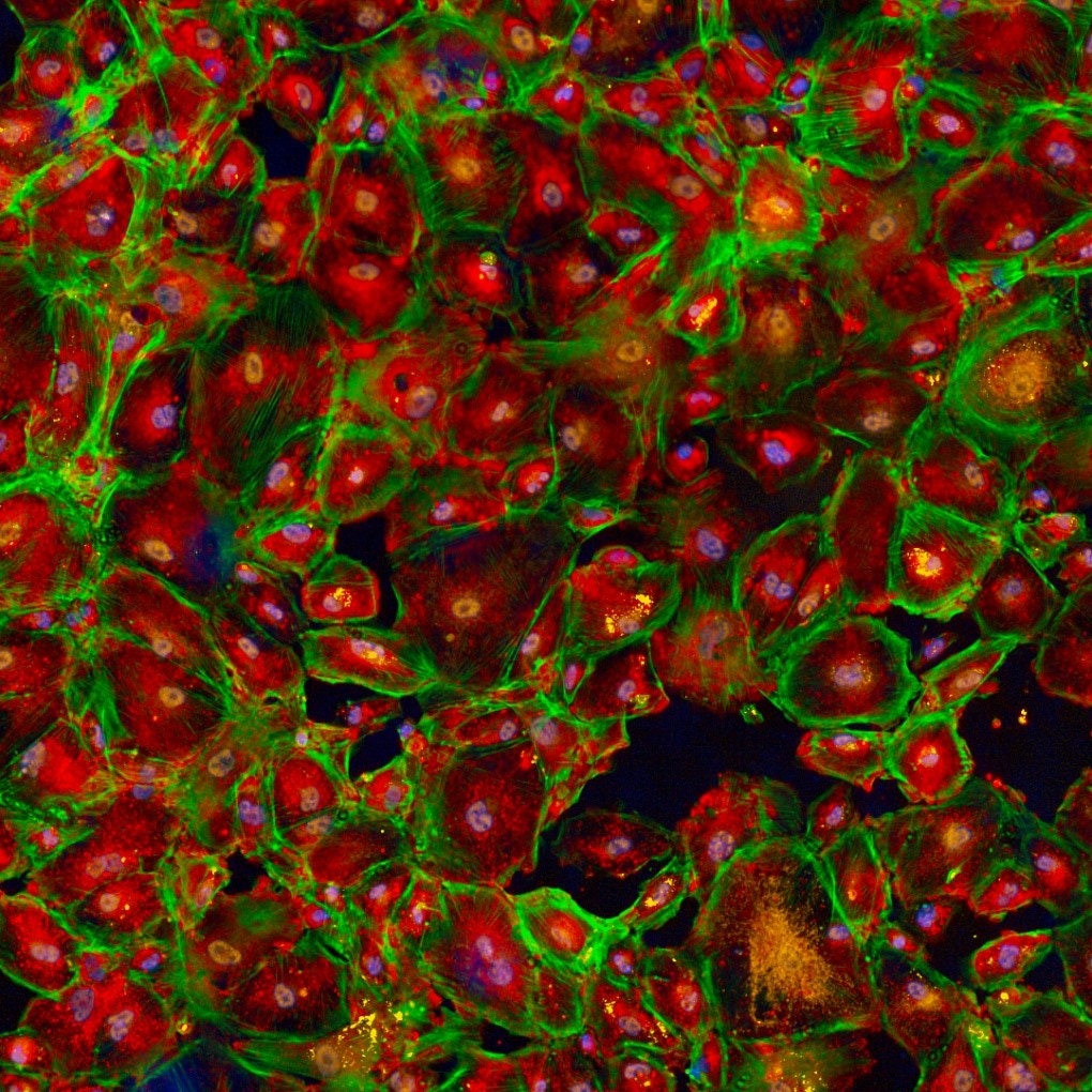
HepG2 cells imaged on the Opera Phenix high-content screening system.
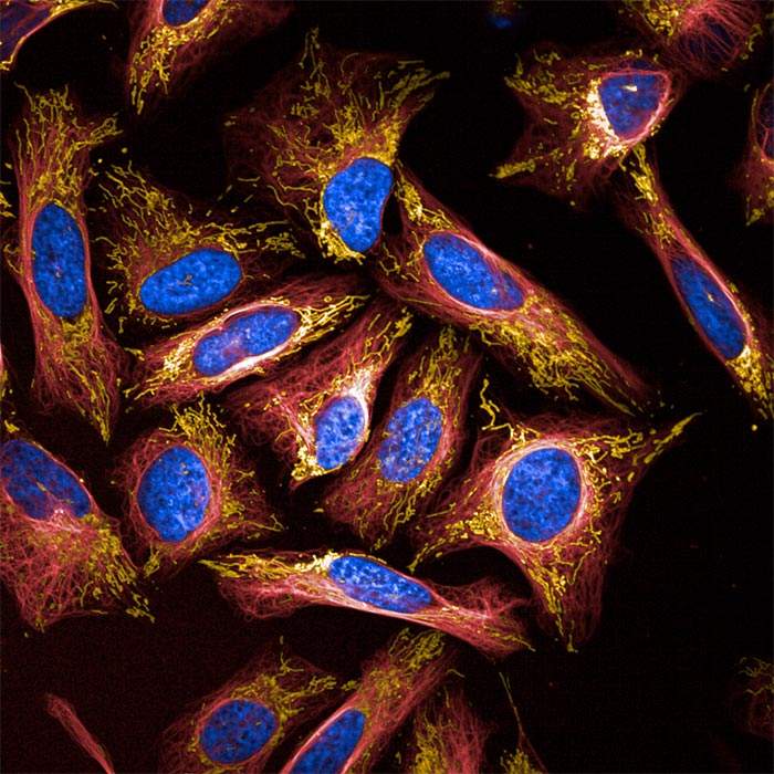
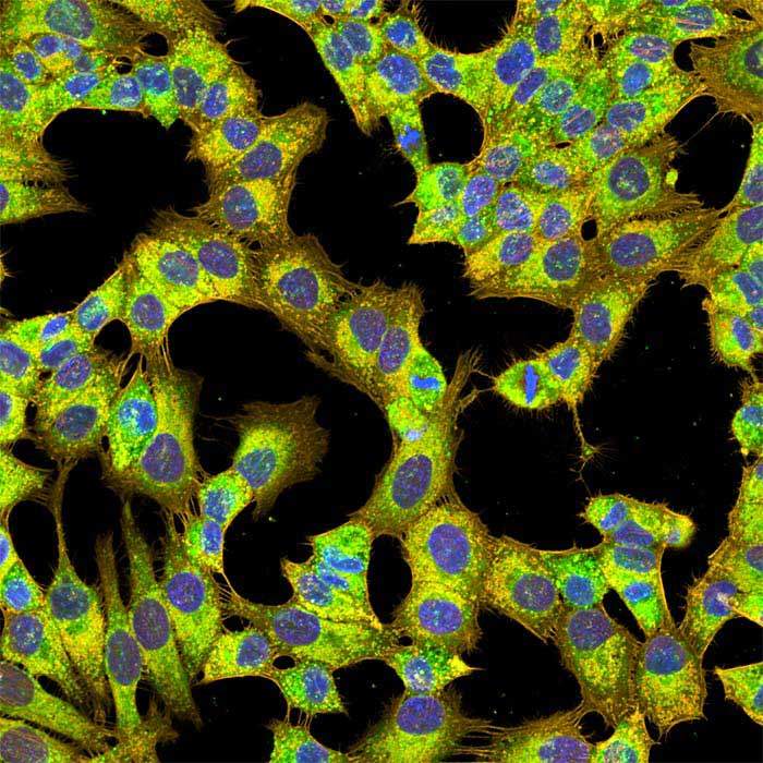
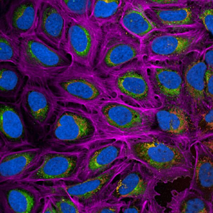
HeLa cells imaged on the Opera Phenix Plus high-content imaging system.
Induced pluripotent stem cells imaged on the Opera Phenix high-content screening system.
Immortalized human podocytes imaged on the Opera Phenix Plus high-content screening system.
5-plex phenotypic assay imaged on the Opera Phenix Plus high-content screening system.
A549 cells on a Semarion SemaCyte microcarrier imaged on the Opera Phenix Plus high-content screening system.
Neurons imaged on the Operetta CLS high-content analysis system
×
Progress faster with our verified solutions
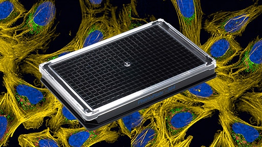
Microplates for high-content imaging
Utilize PhenoPlate™ 384-well microplates designed for optimal performance in high-content imaging applications. Employ CellCarrier Spheroid ULA plates for 3D cell models.

PhenoVue cellular imaging reagents
Ready-to-use kits and reagents with straightforward protocols. Extensively tested to provide optimal formulations and long-term stability.
Automation and workstations
Improve throughput, productivity, and reduce variability and reagent costs. Benefit from automated workflows using the explorer™ G3 workstations for cell painting, 3D cell culture, or phenotypic screening.
Efficient data management
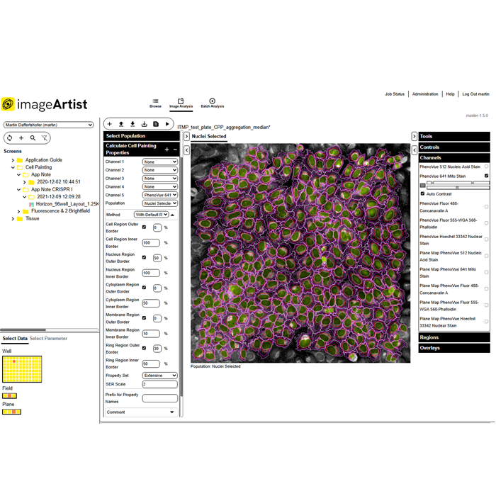
Export results automatically into the Image Artist™ platform that uses high performance computing and an industry standard object store to provide a scalable, multi-user solution for image analysis and management.

Product information
Overview
Opera Phenix Plus high-content screening system for your most demanding high-content applications.
- Modular design adapts to your changing application needs.
- Enhanced speed using a dual - or four-camera configuration with simultaneous imaging.
- Synchrony Optics™ combines a microlens-enhanced Nipkow spinning disk with a pinhole distance optimized for thick and 3D samples.
- Dual-view excitation of neighboring spectral channels minimizes crosstalk.
- Custom-designed high-NA water immersion objectives capture more photons and provide high-image resolution even in thick samples.
- Fast imaging frame rate of up to 100 fps and optional pipettor module captures fast cellular responses.
Specifications
| Dimensions | 134.0 cm (W) x 65.0 cm (D) x 47.0 cm (H) |
|---|
| Automation Compatible |
Yes
|
|---|---|
| Brand |
Opera Phenix Plus
|
| Imaging Modality |
Brightfield
Confocal
Digital phase contrast
Fluorescence
|
| Unit Size |
1 unit
|
Video gallery
Opera Phenix Plus High-Content Screening System
Citations
Resources
Are you looking for resources, click on the resource type to explore further.
Brochure
3D cell culture workflow solutions
More than ever, researchers are turning to 3D cell cultures, microtissues and organoids to bridge the gap between 2D cell cultures...
Technical Note
3D volumetric analysis of luminal spaces inside cysts or organoids
High-content assays using 3D objects such as cysts or organoids can be challenging from the perspectives of both image acquisition...
Technical Note
3D volumetric and zonal analysis of solid spheroids
Multicellular 3D “oids” (tumoroids, spheroids, organoids) have the potential to better predict the effects of drug candidates...
Whitepaper
A brand-new modality on the horizon: how targeted protein degradation can address the unmet need in drug discovery and development
Targeted protein degradation (TPD) is an emerging drug discovery modality that offers the potential to probe biological pathways...
Technical Note
A scalable and reproducible workflow for high-content analysis of cytotoxic effects in RASTRUM 3D cell cultures
Explore our Technical Note unveiling a robust workflow for advanced cytotoxicity analysis in 3D cell cultures. This method...
Case Study
A scalable method to monitor protein levels and localizations in cells
In this case study, we present a novel method developed by researchers at the CeMM Research Center for studying protein levels and...
Loading...


How can we help you?
We are here to answer your questions.

