


Powering scientific intelligence.
Revvity unlocks the full potential of AI with trusted biological intelligence.
Revvity brings together AI-powered informatics, automation, reagents, imaging, analytical technologies, and scientific expertise to power connected Lab-in-the-Loop workflows that transform biological data into actionable scientific insight.
See how AI-enabled science can support your work.
Connecting AI predictive modeling with wet lab experimentation.
Lab-in-the-Loop approaches help close this gap by creating continuous feedback loops between AI-driven prediction and wet lab experimentation.
Revvity supports these next-generation research environments with solutions that help laboratories:
- Capture and manage high-quality R&D data
- Connect in-silico insights with wet lab results
- Scale experimental workflows
- Improve reproducibility across studies
- Accelerate iteration from prediction to validation
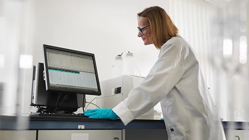

Built for the AI era of discovery.
Revvity enables researchers to turn AI-driven predictions into confident scientific decisions.
Our integrated reagents, platforms, automation, imaging, and informatics solutions connect the full Lab-in-the-Loop workflow, from model creation and experimentation to biological validation and continuous learning.
AI in the news






Enabling the flow between AI insight and scientific validation.
Make
Lay the foundation for AI-enabled discovery by creating biologically relevant models for experimentation.
Revvity provides the tools researchers need to engineer, modify, and prepare biological systems for downstream testing and validation.
Sample prep, automation and plate handling:
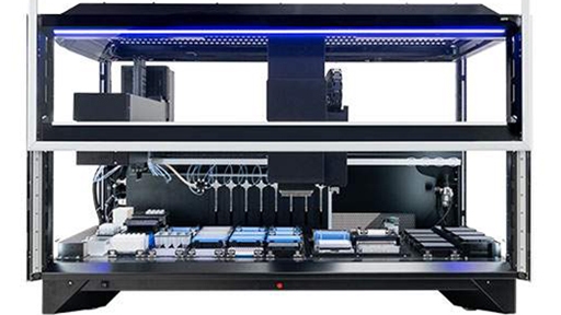
Fontus Workstations
With easy-to-use software, optimized deck access, verified protocols, and reformatting capabilities to simplify your workflows and improve turnaround times
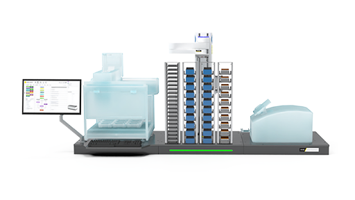
Explorer G3 SmartBench automation solutions
Affordable, scalable laboratory automation with the explorer™ G3 SmartBench workstations, that integrate 150+ instruments with collaborative robotics for seamless workflows.
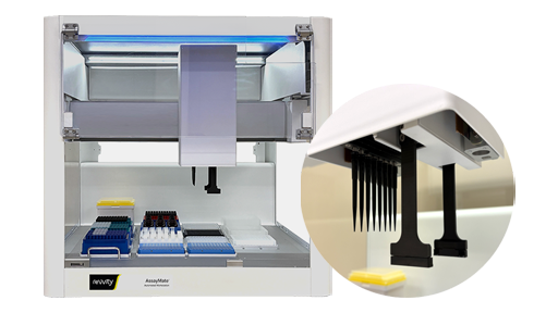
AssayMate Workstations
Automation performance, now for labs of any size: affordable, benchtop-sized, automated liquid handler designed to optimize a wide range of lab applications.
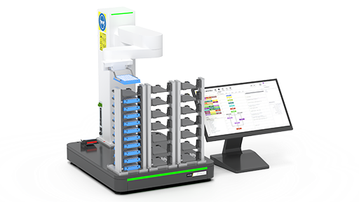
plate::handler Connect
Overcome manual workflow bottlenecks with the plate::handler™ Connect, a collaborative, four-axis SCARA robotic arm for a compact and cost-effective entry point into automated processing.
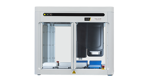
Zephyr Workstations
The Zephyr™ G3 workstation’s small footprint makes it ideal for workbench operation, while the convenient deck design provides ready access
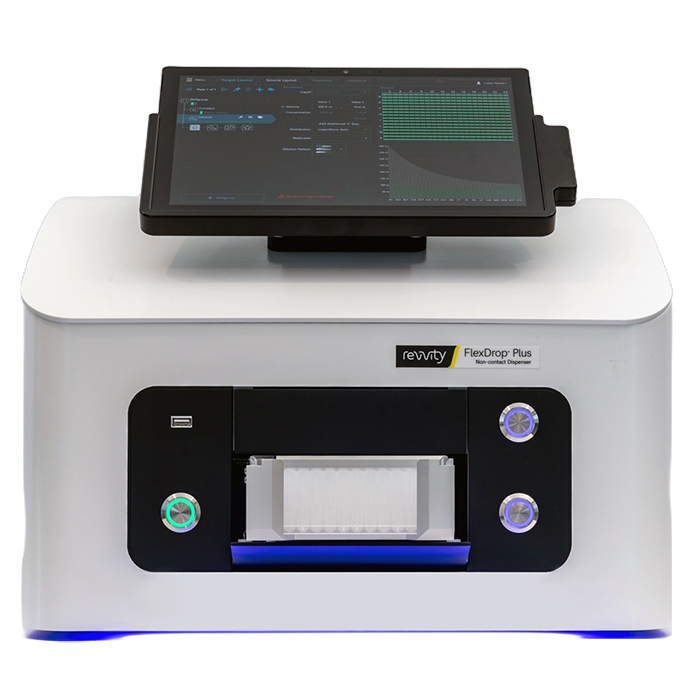
FlexDrop Plus Non-Contact Dispenser
The FlexDrop Plus platform redefines the user experience around nanoliter, low volume liquid handling automation, providing any laboratory with the means to consistently dispense as low as 8 nL with the push of a button in an easy-to-use device that runs from an integrated touch screen PC.
The FlexDrop Plus platform uses positive pressure in eight independent controlled channels. Each channel can dispense up to 100 droplets per second. It generates droplets between 8 nanoliters (nL) and 50 nL in size to achieve the target dispense volume. The system includes droplet verification, which verifies the exact number of droplets have been dispensed and provides a verification report post-run. The proprietary source well plate features 100 µm hole at the bottom which facilitates the droplets under positive pressure.
The touch-screen software control is seamless and simple to use. No previous programming or automation experience is needed. Users can set-up and start dispensing in minutes.
Includes:
- FlexDrop Plus instrument
- (4) Power cords (EU, UK, US, AU) Fuse change may be required.
- Air tubing - 6mm tubing with Quick Connect to instrument and fitting to connect to house air or compressor
- (4) Black adapter plates (1, 2, 4 and 8 mm) - for target plate heights
- FlexDrop source plates with 80 uL wells in startup kit
- Waste PCR tubes
Immunoassays:
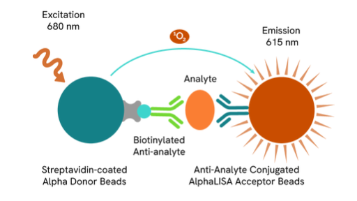
Alpha Assays
Virtually any target can be quickly and easily measured using Alpha technology, an improved alternative to quantitative ELISA assays and ELISA reagents.
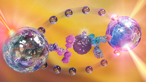
AlphaLISA™ SureFire® Ultra™
AlphaLISA® SureFire® Ultra™ is a homogeneous, no-wash assay platform designed for the sensitive detection of endogenous and tagged phosphorylated or non-phosphorylated proteins in cell lysates.
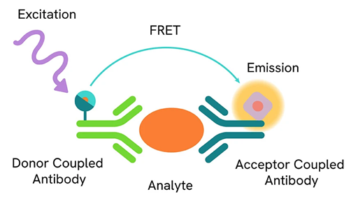
HTRF
HTRF technology performs as cellular or biochemical assays in competitive and non-competitive formats.
Biological model development:
TotalSeq™ reagents
Seamlessly add protein expression to single-cell multiomics workflows with oligonucleotide-conjugated antibodies, hashtags, and cocktails.
LEGENDplex™
Multiplex protein quantification for 14 analytes in a single sample. Select focused pre-defined panels or customize each target.
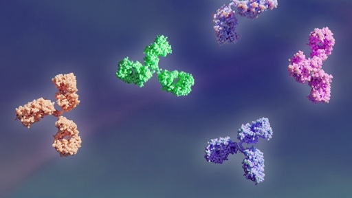
BioLegend Antibodies
Choose from thousands of primary and secondary antibodies. We offer directly conjugated or purified antibodies to match your needs.
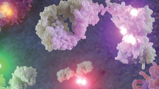
BioLegend Flow Cytometry
Phenotype immune cells by building bigger and better flow cytometry panels with novel fluorophores and highly specific antibodies.
Cell culture reagents:
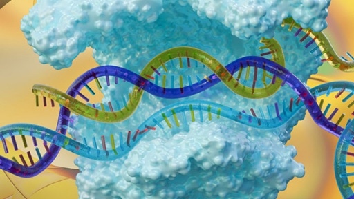
Dharmacon™ CRISPR reagents and libraries
Browse CRISPR based reagents including: sgRNA and Cas9 solutions for CRISPR knock-out, CRISPR knock-in, CRISPR activation, CRISPR interference, and CRISPR derived base editing.
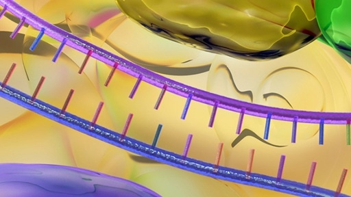
Dharmacon™ RNAi reagents and libraries
Choose from a broad portfolio of synthetic and lentiviral RNAi reagents including: siRNA, shRNA, and microRNA for highly specific and efficient gene silencing.

Pin-point™ base editing reagents
Base editing reagents including custom sgRNAs, CBE or ABE mRNA, validated controls, and non-targeting controls for precise, DSB-free single-nucleotide edits.
Cell monitoring:
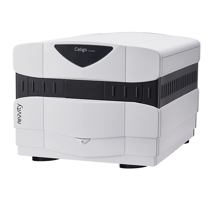
Celigo Image Cytometer
Celigo is a plate-based benchtop brightfield and fluorescent imaging system designed for whole-well live-cell analysis and cell sample characterization. The Celigo system images and analyzes cells in various types of vessels including 6 - 1536 well plates, T25, T75 flasks, 10 cm dishes, and glass slides without disturbing their natural state.
Individual cell level analysis is easily generated, providing cell level insights unlike ELISA or protein-based assays, and at a faster rate than flow cytometry. A broad range of complex cell-based assays have been optimized for the Celigo cytometer.
Convenient workflow designed for biologists
-
Multiple object-driven algorithms and a variety of assay-based applications
- Accessible adaptation with easy-to-follow software
- Save customizable experiments as well as default settings
- Built-in gating parameters allow easy data analysis and visualization
- Real-time graphic feedback allows multiple parameters to be measured simultaneously for intuitive analysis
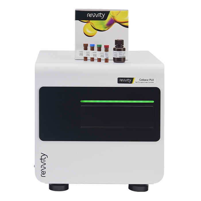
Cellaca PLX High-throughput Image Cytometer
The Cellaca PLX image cytometer provides versatile cell analysis ranging from simple cell counts, concentration, and viability readouts, to more complex multiplexed assays, all using only 15-50 µL of sample. The sensitivity of the system allows for the analysis of up to 24 samples using dual fluorescence and brightfield imaging in under 5 minutes, and single sample multiplexing with four channels in 1 minute.
Multiplex panels for immunophenotyping, apoptosis, and fluorescent protein expression shorten the time to downstream processing with Cellaca PLX low fluorescence slides, dedicated reagents, built-in assays, and configurable data reports. The Cellaca PLX features user friendly software, automation compatibility, and is 21 CFR Part 11 ready. Expert support is provided to integrate the system into current workflows to streamline cell line development, cell isolation, gene delivery, and various research applications.
Key features and benefits of the Cellaca PLX:
- Elevated throughput - run up to 24 samples in under 5 minutes using dual fluorescence and brightfield imaging to count both isolated nuclei and cells
- Multiplexing capabilities - shorten time to downstream processing (one minute per sample with four channels) using our custom reagents kits for surface marker, apoptosis, and fluorescent protein analysis
- Small sample volume - as little as 15 µL of cell sample needed
- Intuitive workflows - including a favorites feature for saving customized experiments and commonly used assays with automatic data analysis and reporting
- Automation ready - robotic integration ability with via API
- 21 CFR Part 11 ready - add-on that includes audit trail, user access control, and digital signature

Cytotoxicity and Cell Proliferation Assays
Cytotoxicity and cell proliferation assays are commonly used in the drug discovery process to assess a compound’s ability to cause or block a biologic activity without having toxic effects on cells. Adenosine TriPhosphate (ATP) – monitoring assays allow for the quantitative evaluation of proliferation and cytotoxicity.
Protein and nucleic acid QC:
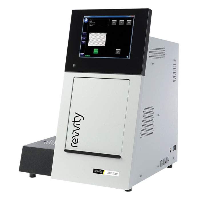
LabChip GXII Touch HT Protein Characterization System
LabChip GXII Touch instrument simplifies sample analysis
User friendly operation:
- Load sample plate and chip
- Select samples (up to 384 in a run)
- Select assay type
- Touch 'Run' to start
- Automatically export data directly to your network or LIMS system
Observe runs in real time:
- Sample analysis in as few as 42 seconds
- View electropherogram in real time during data collection
- Overlay collected data to compare sample profiles
- Select from various run time analytical feature annotations
See data in real time or export for later analysis:
- Choose "display" in E-gram, virtual gel, or data table format (Figure 1)
- Pull multiple archived plates into data review or analytical comparisons
- Apply data mining filter functions on key attributes
- Highlight expected peaks
- Track relevant user access and data history parameters with FDA 21 CFR Part 11 compatible software
Easy set-up for protein and nucleic acid characterization
The LabChip GXII Touch protein characterization system operator controls are designed to allow users to easily set up and execute a run in as few as three easy steps. Run templates can also be imported with the operator control features. Run templates can include well selections, sample names, peak tables, which facilitate operator ease of use, and data can be automatically exported to network or LIMS directories for subsequent analysis. Every instrument comes with a full software package for data review, allowing analysis from current or archived data sets.
Designed to facilitate integration of quality by design initiatives into workflow
The LabChip GXII Touch protein characterization system offers rapid quantification and quality control throughout biotherapeutics and genomic workflows. For example, automating the characterization process allows multiple critical quality attributes to be obtained significantly faster. Researchers can now screen for optimal protein characteristics earlier in the process and integrate Quality by Design initiatives into their biotherapeutics development workflow.
21CFR Part 11 compatible software is available with the LabChip GXII Touch protein characterization system
LabChip GXII Touch and Reviewer Software contain built-in technical controls and features specifically designed to support FDA 21 CFR Part 11 regulations. These features include a shared user account database, access controls, device check, enforced sequencing of run steps, audit trails, record copying, record retention, system documentation, and electronic signature controls.
To assist in your compliance planning, LabChip GxP Security Software is available which allows the LabChip GXII Touch protein characterization system to run in an environment consistent with CFR21 Part 11 compliance and validation (Figure 2). This includes access security, data security and verification, and a complete set of audit logging functionality.
LabChip CE: How does it work?
LabChip CE is performed on a small, microfluidic chip. Prior to analysis, reagents are loaded into the individual wells of the chip. These wells are connected to tiny microchannels about the size of a human hair etched within the quartz microfluidic chip. When the chip is loaded into the LabChip GXII Touch protein characterization system, the chip's wells interface with platinum electrodes that provide voltage and current control. The instrument moves the microtiter plate directly under the chip's capillary 'sipper', and approximately 150 nL of sample is aspirated onto the chip. Sample staining and destaining are performed automatically on the instrument platform. Individual sample analytes are separated electrophoretically and the bands detected via laser induced fluorescence in the capillaries of the chip. Sizing and concentration for each band are determined using ladder and internal markers. Rinsing the sipper between samples minimizes cross contamination.
Computational design and ideation:

Signals One™ software
AI‑enhanced, unified, cloud‑native R&D solution that helps you reduce complexity and collaborate seamlessly across drug discovery, materials science, and food science.
Test
Transform biological models into meaningful scientific data with Revvity's integrated portfolio of reagents, assays, and imaging solutions.
Rapidly measure cellular responses, characterize biomarkers, and generate reproducible experimental results.
High-content imaging instrument, software and reagents:
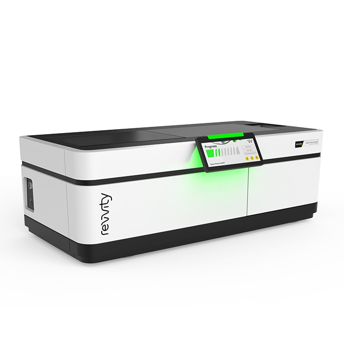
Opera Phenix OptIQ High-Content Screening System
Opera Phenix OptIQ high-content screening system for your most demanding high-content applications.
- Enhanced speed using a dual - or four-camera configuration with simultaneous imaging.
- Synchrony Optics™ combines a microlens-enhanced Nipkow spinning disk with a pinhole distance optimized for thick and 3D samples.
- Dual-view excitation of neighboring spectral channels minimizes crosstalk.
- Custom-designed high-NA water immersion objectives capture more photons and provide high-image resolution even in thick samples.
- Fast imaging frame rate of up to 100 fps and optional pipettor module captures fast cellular responses.
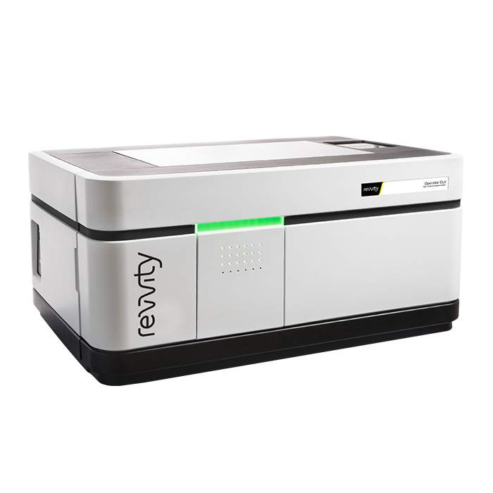
Operetta CLS High-Content Analysis System
The Operetta CLS system combines speed and sensitivity with the powerful and intuitive data analysis you’ve come to trust from the Operetta platform. The all-new Operetta CLS delivers everything you need from the high-content analysis. What’s more, the Operetta CLS system is part of our comprehensive HCS workflow – everything from HCS systems and microplates to automation and informatics for every application. All from one knowledgeable, trusted vendor. Put that together with our Harmony high-content imaging and analysis software - the easy-to-learn, easy-to-use software that empowers biologists to do their own analysis - and you have everything you need to run your every day (and complex) analyses right away.
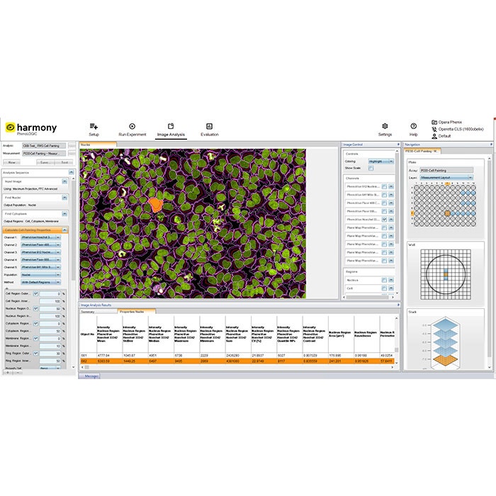
Harmony High-Content Imaging and Analysis Software
Designed for biologists, Harmony’s workflow-based interface makes the whole process of high-content analysis straightforward, even for new users with little microscopy or programming knowledge.
One high-content analysis software for all your applications
- Phenotypic Screening
- Get robust phenotypic fingerprints based on intensity, morphology, texture and fluorescence distribution parameters
- Easily train the software to recognize relevant phenotypes
- Quantify even subtle phenotypic differences
- Live Cell Imaging
- Follow phenotypic changes over time
- Accurately quantify label-free live cell images
- Track individual cells over time or analyze wound-healing assays
- Analyze cell signaling dynamics using e.g. FRET based assays
- Perform fast response assays such as calcium flux or cardiomyocyte beat rate with imaging frame rate of up to 100fps.
- Set-up "dispense and read" assays on the Opera Phenix Plus system with liquid handling module
- Enhance your live-cell assay capabilities through improved time-series flexibility
- 3D Cell Models
- Speed up 3D image acquisition by targeted imaging of microtissues
- Better understand your cell models by exploring them in the new 3D viewer and the XYZ viewer
- Measure morphologies and volumes in 3D, count nuclei within spheroids
- Enhance Maximum Intensity Projection Analysis using PlaneMap
- One software for image acquisition, 3D visualization and analysis
- Rare Cell Phenotypes
- Quickly capture your rare cell phenotypes with PreciScan™
- Pre-screen at low resolution and acquire objects of interest at high resolution
- Shorten your image acquisition times
- Reduce your data volume
- Routine Assays
- Utilize more than 30 ready-made-solutions for standard assays
- Ensure repeatability by running established protocols
- Optional modules
- Phenologic.AI offers an efficient and reliable method for identifying cells and cellular nuclei within fluorescent and brightfield images by harnessing the power of pre-trained deep neural networks (DNNs).
- Connect Harmony software to your favorite image analysis tools. Access greater flexibility with the Harmony Application Connector. Integrate third-party applications, including Cellpose, to access advanced AI-driven analysis and user trained AI-models maintaining a streamlined workflow within the Harmony environment.1,2
One high-content analysis software for all users
- New Users
- Get quantification for your cell images
- Easy to learn – even for users with little microscopy or programming knowledge
- Get started with 30 ready-made solutions
- Logical workflow for easy control of all aspects of image acquisition and analysis
- Simple building blocks to assemble image analysis sequences (step-by-step)
- Expert Users
- Quantify difficult phenotypes with advanced texture and morphology readouts
- Utilize advanced building blocks to enable e.g. ratiometric FRET imaging
- Create “stitched” global images from multiple fields of view
- Examine samples at multiple scales – regions found in the global image can be used for analysis in the original images
- For Lab Managers
- Keep up with increasing research needs – analyze larger sample sizes
- Quickly train new users to set up the instrument and analyze their images
- Find images, metadata and results quickly via the integrated sortable database
- View and analyze data from any computer with installation of Harmony software
- Get application support by image analysis experts
1Third-party applications are provided and licensed separately by their respective ownersunder separate third-party terms. Customers are responsible for obtaining, licensing, deploying, and using any third-party software with the Harmony Application Connector in compliance with all applicable license terms. Revvity does not control, support or warrant third-party applications.
2Cellpose is a third-party open-source software tool and is not affiliated with, sponsored by or endorsed by Revvity. Cellpose and related names are the property of their respective owners.

Phenologic.AI
The Phenologic.AI module represents a significant advancement in the field of cellular imaging. By harnessing the power of pretrained deep neural networks (DNNs), it offers an efficient and reliable method for identifying cells and cellular nuclei within fluorescent and brightfield images.
Phenologic.AI introduces a Find Nuclei AI building block to Harmony and Image Artist software which simplifies the detection of cellular nuclei in brightfield images - traditionally a challenging task without the use of nuclear staining. This innovation saves time by reducing the need for parameter fine-tuning and also allows the analysis of cell images to be conducted swiftly and with precision. The ability to reliably locate cellular nuclei without staining is achieved through the advanced Phenologic.AI algorithms.
Trained on a variety of diverse cell lines, Phenologic.AI is a deep neural network model offering robust identification of cellular structures, such as nuclei, and providing reliable phase contrast imaging to delineate the cytoplasmic regions of individual cells.
Phenologic.AI benefits from the use of Graphics Processing Units (GPUs), which are the computing system of choice for image analysis and significantly enhance the processing capabilities of AI-driven software modules allowing for rapid and precise analysis that is critical in biological research settings. Phenologic.AI also supports Central Processing Unit (CPU) execution.
Phenologic.AI streamlines cell imaging workflows and opens up new possibilities for understanding cellular behaviors and disease mechanisms.
Phenologic.AI is available as a software module for Harmony High Content Imaging and Analysis Software and Image Artist Image Data Management and Analysis System.
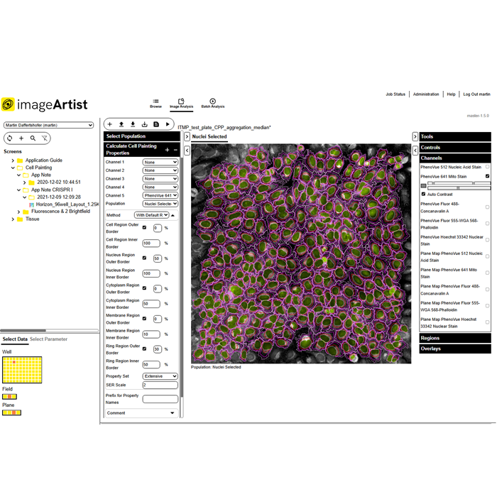
Image Artist
Next generation image analysis and management
Image Artist has powerful capabilities for processing, analyzing, storing, and sharing all your high-content screening and cell imaging data – so you can significantly cut your time to results.
- Fast image data processing and image analysis even for complex assays powered by high performance computing
- Easy-to-use assay building blocks with integrated Artificial Intelligence (AI) that make advanced image analysis
straightforward – now with improved segmentation and analysis capabilities for 3D cell applications - A central location to store all your image data with associated instrument metadata for an enduring picture of
experiments - Compatible with all major high-content screening and cell imaging systems
- Multi-user solution that can support your entire lab without compromising performance
- Scalable data storage to expand with your labs needs over time
- Seamless integration with Revvity Opera Phenix OptIQ™ and Operetta CLS™ high-content screening systems operated
by Harmony™ software, as well as Signals One
for profiling image data, hit selection, and more - NEW! The latest version is compatible with data from a broader range of systems including the Celigo™ image
cytometer - NEW! Wider range of on-premise and cloud options, with AWS S3 support. Now featuring flexible tier-based storage
options, and Intelligent-Tiering for automatic cost-savings. - NEW! Phenologic.AI is an optional module that provides an efficient and reliable method for identifying cells
and cellular nuclei within fluorescent and brightfield images by harnessing the power of pre-trained deep neural
networks (DNNs).
It is also part of our complete workflow solution for high-content screening that includes PhenoVue™ cellular imaging reagents, PhenoPlate™ imaging microplates, HCS instruments, image cytometers, software, and automation.
For all your high-content screening and cellular imaging data
High-content screening and cellular imaging experiments generate huge amounts of image data. With the latest applications such as phenotypic screening, cell painting, and 3D imaging which use more complex,
physiologically-relevant disease models, the volume of data continues to grow.
To maximize all this valuable data, you need a powerful solution for image processing, management and storage, and for sharing data with colleagues.
Image Artist allows you to bring together cell imaging data from a wide range of sources on a single software platform so that you can store, share, analyze, and re-analyze it seamlessly.
Not only does it make managing image data easier for research scientists but also for your IT team. With cloud deployment options (such as AWS S3), your IT team have just one platform and installation to manage and store research image data which can easily be scaled. They can also have peace of mind with enhanced security for cloud deployments. What's more, with AWS S3, you can choose from a range of storage classes based on your cost and data access requirements, and optional Intelligent-Tiering allows automatic transfer of data to more cost-effective tiers, based on access frequency.
Image analysis designed for biologists
Whether you are performing phenotypic screening, cell painting, 3D imaging, live cell imaging, or more routine assays, Image Artist is designed to make it easy for biologists to perform sophisticated image analysis without any coding experience.
The software platform’s image analysis building blocks encapsulate many years of knowledge and expertise in cellular imaging and analysis - so that you can focus on the biology. By simply adding together building blocks such as “Find Nuclei” and “Calculate Cell Painting Properties”, users can quickly create image analysis protocols in just a few simple steps.
With 3D cell models bridging the gap between 2D cell cultures and in vivo animal models, Image
Artist has been further optimized for 3D cell applications, with improved segmentation and analysis capabilities – so you can generate more accurate results than ever.
Built-in Artificial Intelligence (AI) and machine learning technologies allow users to train the software to develop image analysis algorithms. While other systems may require an image analysis expert to create an algorithm, Image Artist uses proprietary machine-learning technology to make it easy for you to do it on your own. Using a learn-by-example approach, in just a few clicks segmented images can be classified quickly and easily.
This is all powered by high performance computing, so you can get your answers faster.
- Get started without extensive training
- Measure complex and subtle phenotypic responses
- Compare multiple samples, plates, or batches to quality check your results
- Kinetic measurement capabilities to track cells, reveal changes in cell properties, and provide information on
cell movement - Use with Signals One to perform screening data analysis and validation, QC analyses,
calculate reliable normalization, multivariate hit stratification, dose response curves, and drug response
profiling
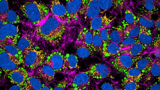
Cellular Imaging Reagents
Revvity's PhenoVue™ suite of cellular imaging reagents – including cell painting kits, organelle and cell compartment stains
Plate-based assay validation:
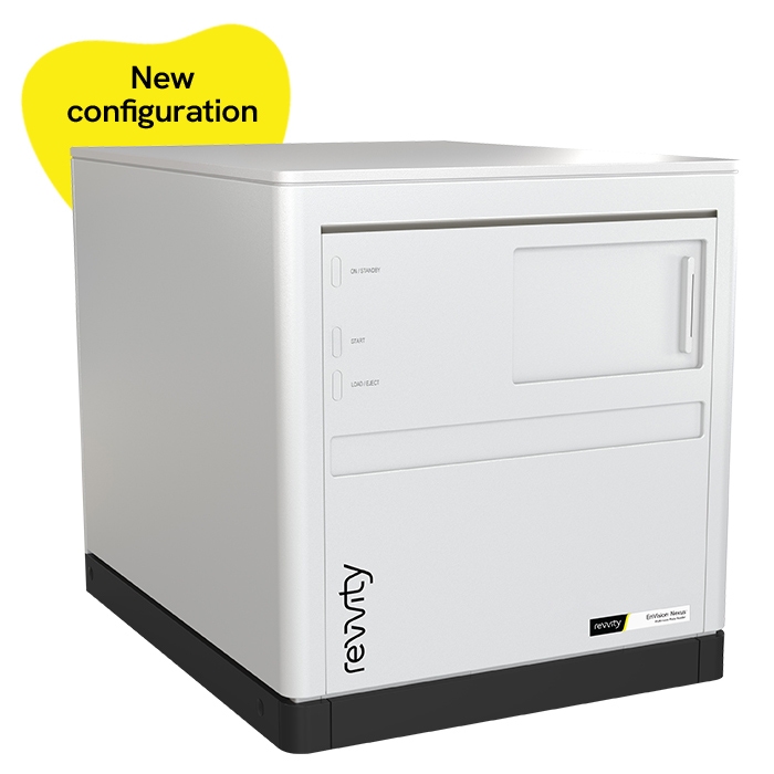
EnVision Nexus Multimode Microplate Reader Family
The EnVision Nexus family supports high-throughput screening applications, providing consistent performance and reliable data across multiple detection technologies. With enhanced speed, high throughput capabilities, and sensitive detection across technologies, the EnVision Nexus family offers flexible configurations to help match your specific workflow needs.
EnVision Nexus One provides advanced detection performance in a streamlined single-detector configuration.
EnVision Nexus features dual detectors for simultaneous dual wavelength reading, supporting high-throughput screening environments with demanding workflow requirements.
Key features:
- Enhanced sensitivity across detection technologies
- Magnetic filter attachment for easy configuration
- Flexible detection options to help match your application needs
Main detection:
- Absorbance
- Fluorescence intensity
- Luminescence
- Time-resolved fluorescence
- Fluorescence polarization
Advanced detection:
- HTS Alpha
- TRF laser
- Ultrasensitive luminescence
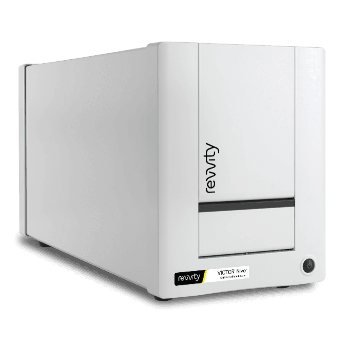
VICTOR Nivo Multimode Microplate Reader
Data analysis and compliance
The VICTOR Nivo software features intuitive data analysis with optional compliance tools. Perform rapid on-instrument calculations such as standard curves, plate statistics, and curve fitting directly after your assay. For advanced analysis, MyAssays™ Desktop Pro enables deeper data evaluation beyond the lab. In regulated environments, the Enhanced Security software supports 21 CFR Part 11 compliance through secure logins, defined user roles, electronic signatures, and full audit trails to ensure data integrity and traceability.
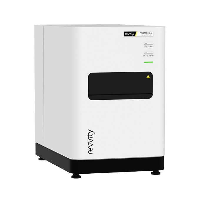
VICTOR Kira Multimode Microplate Reader
The VICTOR Kira is useful for a wide range of applications, including by helping to enhance workflow efficiency with its advanced monochromator-based technology. It features an intuitive software interface with built-in data analysis for a pleasant user experience. Plus, our expert service and application support team is here to support you.
The VICTOR Kira multimode plate reader is equipped with three detection modes. Additional options are available such as temperature control and shaking, it’s useful for cell-based and kinetic measurements.
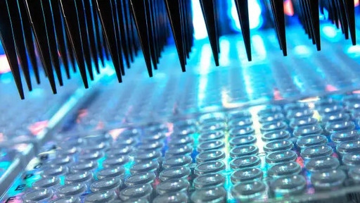
Microplates
Choosing the right microplate is a critical, often overlooked, part of an assay. The right microplate helps provides valuable data, whereas the wrong microplate can lead to missed or inaccurate data leading to missed project timelines and ultimately higher costs.
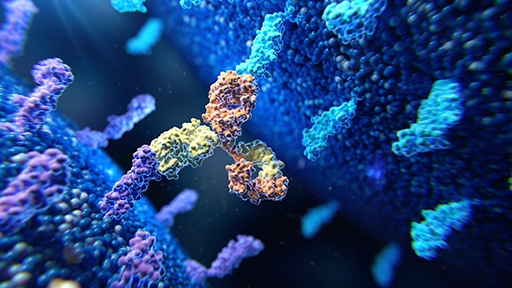
pHSense Reagents
Discover a powerful new plate reader tool specifically designed for cell surface target internalization studies for ADC and GPCR research.
In vivo imaging, software, and reagents:
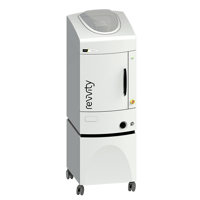
IVIS Spectrum 2 In Vivo Imaging System
Building upon the IVIS Spectrum in vivo optical system with proven 2D bioluminescence and 2D fluorescence imaging and 3D optical tomography capabilities combined in a single system, the IVIS Spectrum 2 is our next generation in optical imaging. This advanced imaging system incorporates a CCD camera with eXcelon® coating that enables detection of more signal at higher efficiency across a broader spectrum of wavelengths. With exclusivity to this innovative camera for in vivo imaging, the IVIS Spectrum 2 preclinical optical imaging system delivers the sensitivity you demand for non-invasive longitudinal imaging to better understand early disease-related biological changes, track disease progression, and help guide the drug development process.
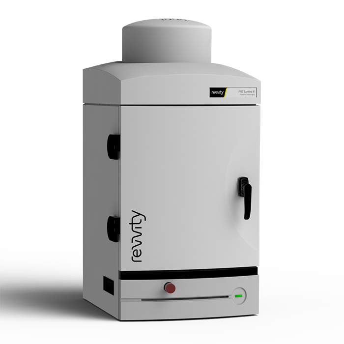
IVIS Lumina III In Vivo Imaging System
The IVIS Lumina III imaging system is capable of imaging both fluorescent and bioluminescent reporters. The system is equipped with up to 26 filter sets that can be used to image reporters that emit from green to near-infrared. Superior spectral unmixing can be achieved with the IVIS Lumina III system's optional high resolution short cut off filters.
Features and Benefits
- Market trusted technology offering the fullest suite of leading imaging technologies, reagents and support
- Exquisite sensitivity in bioluminescence
- Full fluorescence tunability through the NIR spectrum
- Compute Pure Spectrum spectral umixing for ultimate fluorescence sensitivity
- Expandable system tailored to your workflow
- Complimentary Living Image™ software licenses are provided with the IVIS systems and upon request.

In Vivo Imaging Reagents
Our comprehensive range of IVISbrite™ bioluminescent and IVISense™ fluorescent imaging reagents provides researchers with the necessary tools to better understand early disease-related biological changes, track disease progression, help guide the drug discovery process, and evaluate efficacy and safety of drug candidates.
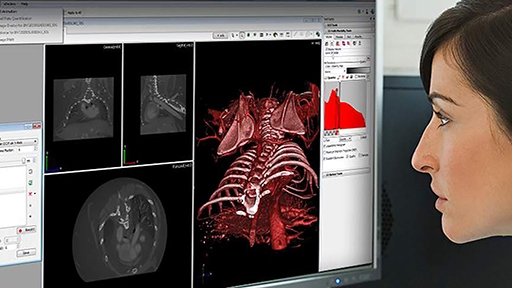
In Vivo Imaging Software
Analyze even the most complex in vivo imaging data with ease using Revvity's portfolio of imaging software. With intuitive interfaces and seamless workflow, our software tools streamline acquisition through analysis simplifying even the most advanced imaging protocol to help facilitate success of your research.
Immunoassays:
Alpha Assays
Virtually any target can be quickly and easily measured using Alpha technology, an improved alternative to quantitative ELISA assays and ELISA reagents.
AlphaLISA™ SureFire® Ultra™
AlphaLISA® SureFire® Ultra™ is a homogeneous, no-wash assay platform designed for the sensitive detection of endogenous and tagged phosphorylated or non-phosphorylated proteins in cell lysates.
HTRF
HTRF technology performs as cellular or biochemical assays in competitive and non-competitive formats.
Biological insights:
TotalSeq™ reagents
Seamlessly add protein expression to single-cell multiomics workflows with oligonucleotide-conjugated antibodies, hashtags, and cocktails.
LEGENDplex™
Multiplex protein quantification for 14 analytes in a single sample. Select focused pre-defined panels or customize each target.
BioLegend Antibodies
Choose from thousands of primary and secondary antibodies. We offer directly conjugated or purified antibodies to match your needs.
Preclinical services:
Preclinical Services
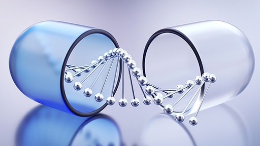
Outsource the biological validation of your AI-based drug screening with our preclinical services, delivering the quality biological data you need to support go/no-go decisions and accelerate your AI-driven drug discovery.
Computational design and ideation:
Signals One™ software
AI‑enhanced, unified, cloud‑native R&D solution that helps you reduce complexity and collaborate seamlessly across drug discovery, materials science, and food science.
Validate
Confirm scientific insights and validate AI-generated predictions with trusted experimental evidence.
Revvity's integrated imaging, detection, assay, and biological analysis solutions help researchers confirm targets, characterize biological responses, and generate reproducible results for confident decisions.
Sample prep, automation, and plate handling:
Fontus Workstations
With easy-to-use software, optimized deck access, verified protocols, and reformatting capabilities to simplify your workflows and improve turnaround times
Explorer G3 SmartBench automation solutions
Affordable, scalable laboratory automation with the explorer™ G3 SmartBench workstations, that integrate 150+ instruments with collaborative robotics for seamless workflows.
AssayMate Workstations
Automation performance, now for labs of any size: affordable, benchtop-sized, automated liquid handler designed to optimize a wide range of lab applications.
plate::handler Connect
Overcome manual workflow bottlenecks with the plate::handler™ Connect, a collaborative, four-axis SCARA robotic arm for a compact and cost-effective entry point into automated processing.
FlexDrop Plus Non-Contact Dispenser
The FlexDrop Plus platform redefines the user experience around nanoliter, low volume liquid handling automation, providing any laboratory with the means to consistently dispense as low as 8 nL with the push of a button in an easy-to-use device that runs from an integrated touch screen PC.
The FlexDrop Plus platform uses positive pressure in eight independent controlled channels. Each channel can dispense up to 100 droplets per second. It generates droplets between 8 nanoliters (nL) and 50 nL in size to achieve the target dispense volume. The system includes droplet verification, which verifies the exact number of droplets have been dispensed and provides a verification report post-run. The proprietary source well plate features 100 µm hole at the bottom which facilitates the droplets under positive pressure.
The touch-screen software control is seamless and simple to use. No previous programming or automation experience is needed. Users can set-up and start dispensing in minutes.
Includes:
- FlexDrop Plus instrument
- (4) Power cords (EU, UK, US, AU) Fuse change may be required.
- Air tubing - 6mm tubing with Quick Connect to instrument and fitting to connect to house air or compressor
- (4) Black adapter plates (1, 2, 4 and 8 mm) - for target plate heights
- FlexDrop source plates with 80 uL wells in startup kit
- Waste PCR tubes
Plate-based assay validation:
EnVision Nexus Multimode Microplate Reader Family
The EnVision Nexus family supports high-throughput screening applications, providing consistent performance and reliable data across multiple detection technologies. With enhanced speed, high throughput capabilities, and sensitive detection across technologies, the EnVision Nexus family offers flexible configurations to help match your specific workflow needs.
EnVision Nexus One provides advanced detection performance in a streamlined single-detector configuration.
EnVision Nexus features dual detectors for simultaneous dual wavelength reading, supporting high-throughput screening environments with demanding workflow requirements.
Key features:
- Enhanced sensitivity across detection technologies
- Magnetic filter attachment for easy configuration
- Flexible detection options to help match your application needs
Main detection:
- Absorbance
- Fluorescence intensity
- Luminescence
- Time-resolved fluorescence
- Fluorescence polarization
Advanced detection:
- HTS Alpha
- TRF laser
- Ultrasensitive luminescence
VICTOR Nivo Multimode Microplate Reader
Data analysis and compliance
The VICTOR Nivo software features intuitive data analysis with optional compliance tools. Perform rapid on-instrument calculations such as standard curves, plate statistics, and curve fitting directly after your assay. For advanced analysis, MyAssays™ Desktop Pro enables deeper data evaluation beyond the lab. In regulated environments, the Enhanced Security software supports 21 CFR Part 11 compliance through secure logins, defined user roles, electronic signatures, and full audit trails to ensure data integrity and traceability.
VICTOR Kira Multimode Microplate Reader
The VICTOR Kira is useful for a wide range of applications, including by helping to enhance workflow efficiency with its advanced monochromator-based technology. It features an intuitive software interface with built-in data analysis for a pleasant user experience. Plus, our expert service and application support team is here to support you.
The VICTOR Kira multimode plate reader is equipped with three detection modes. Additional options are available such as temperature control and shaking, it’s useful for cell-based and kinetic measurements.
Microplates
Choosing the right microplate is a critical, often overlooked, part of an assay. The right microplate helps provides valuable data, whereas the wrong microplate can lead to missed or inaccurate data leading to missed project timelines and ultimately higher costs.
In Vivo Imaging, software and reagents:
IVIS Spectrum 2 In Vivo Imaging System
Building upon the IVIS Spectrum in vivo optical system with proven 2D bioluminescence and 2D fluorescence imaging and 3D optical tomography capabilities combined in a single system, the IVIS Spectrum 2 is our next generation in optical imaging. This advanced imaging system incorporates a CCD camera with eXcelon® coating that enables detection of more signal at higher efficiency across a broader spectrum of wavelengths. With exclusivity to this innovative camera for in vivo imaging, the IVIS Spectrum 2 preclinical optical imaging system delivers the sensitivity you demand for non-invasive longitudinal imaging to better understand early disease-related biological changes, track disease progression, and help guide the drug development process.
IVIS Lumina III In Vivo Imaging System
The IVIS Lumina III imaging system is capable of imaging both fluorescent and bioluminescent reporters. The system is equipped with up to 26 filter sets that can be used to image reporters that emit from green to near-infrared. Superior spectral unmixing can be achieved with the IVIS Lumina III system's optional high resolution short cut off filters.
Features and Benefits
- Market trusted technology offering the fullest suite of leading imaging technologies, reagents and support
- Exquisite sensitivity in bioluminescence
- Full fluorescence tunability through the NIR spectrum
- Compute Pure Spectrum spectral umixing for ultimate fluorescence sensitivity
- Expandable system tailored to your workflow
- Complimentary Living Image™ software licenses are provided with the IVIS systems and upon request.
In Vivo Imaging Reagents
Our comprehensive range of IVISbrite™ bioluminescent and IVISense™ fluorescent imaging reagents provides researchers with the necessary tools to better understand early disease-related biological changes, track disease progression, help guide the drug discovery process, and evaluate efficacy and safety of drug candidates.
In Vivo Imaging Software
Analyze even the most complex in vivo imaging data with ease using Revvity's portfolio of imaging software. With intuitive interfaces and seamless workflow, our software tools streamline acquisition through analysis simplifying even the most advanced imaging protocol to help facilitate success of your research.
Immunoassays:
Alpha Assays
Virtually any target can be quickly and easily measured using Alpha technology, an improved alternative to quantitative ELISA assays and ELISA reagents.
AlphaLISA™ SureFire® Ultra™
AlphaLISA® SureFire® Ultra™ is a homogeneous, no-wash assay platform designed for the sensitive detection of endogenous and tagged phosphorylated or non-phosphorylated proteins in cell lysates.
HTRF
HTRF technology performs as cellular or biochemical assays in competitive and non-competitive formats.
Biological insights:
TotalSeq™ reagents
Seamlessly add protein expression to single-cell multiomics workflows with oligonucleotide-conjugated antibodies, hashtags, and cocktails.
LEGENDplex™
Multiplex protein quantification for 14 analytes in a single sample. Select focused pre-defined panels or customize each target.
BioLegend Antibodies
Choose from thousands of primary and secondary antibodies. We offer directly conjugated or purified antibodies to match your needs.
Preclinical services:
Preclinical Services
Outsource the biological validation of your AI-based drug screening with our preclinical services, delivering the quality biological data you need to support go/no-go decisions and accelerate your AI-driven drug discovery.
Computational design and ideation:
Signals One™ software
AI‑enhanced, unified, cloud‑native R&D solution that helps you reduce complexity and collaborate seamlessly across drug discovery, materials science, and food science.
Generate
Transform scientific knowledge into the next breakthrough.
Revvity's computational design and ideation solutions help generate hypotheses, design experiments to loop AI-driven insights with the next cycle of discovery.
Computational design and ideations:
Signals One™ software
AI‑enhanced, unified, cloud‑native R&D solution that helps you reduce complexity and collaborate seamlessly across drug discovery, materials science, and food science.
Signals ChemDraw™ software
The #1 Trusted Chemical Drawing Solution. A complete suite for creating, managing, sharing, and presenting your chemistry research and discoveries.
Predict
Transform scientific data into predictive intelligence.
Revvity's AI-enabled analytics and modeling solutions help identify patterns, prioritize opportunities, and design smarter experiments to loop computational insights with experimental discovery.
Data modeling and predictive analysis:

Signals Xynthetica™ software
AI-augmented molecular design service combining generative design, property prediction, and virtual screening.
vScreen
Virtual screening helps identify the most promising opportunities before moving into experimental workflows.
Revvity's informatics solutions loop computational screening with wet lab validation, for faster iteration and efficient discovery.
Virtual screening workflows:
Signals One™ software
AI‑enhanced, unified, cloud‑native R&D solution that helps you reduce complexity and collaborate seamlessly across drug discovery, materials science, and food science.
Explore our solutions


Featured resources
Pin-point™ base editing reagents are available for research use only and are not for diagnostic use or direct administration into humans or animals. The Pin-point™ base editing platform technology is available for clinical or diagnostic study and commercialization under a commercial license from Revvity.
For research use only. Not for use in diagnostic procedures.

How can we help you?
Contact us to see how AI-enabled science can support your work.
