


US
Revvity Sites Globally
Select your location.
*e-commerce not available for this region.

Operetta CLS High-Content Analysis System
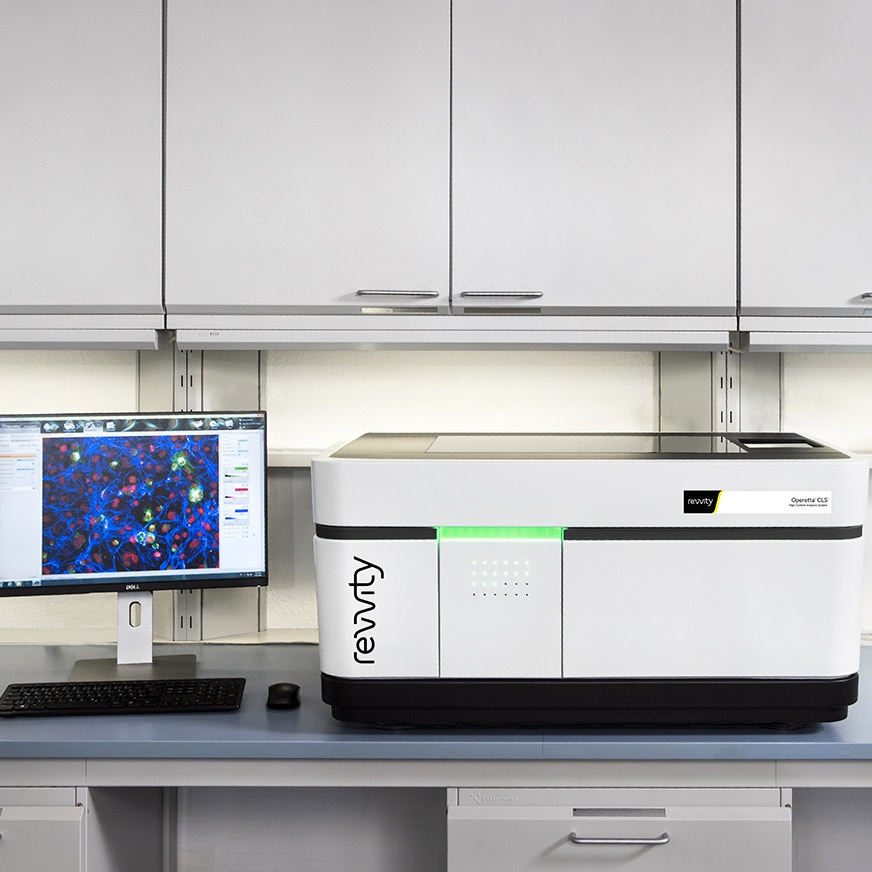
Uncover deep biological understanding in your everyday assays and innovative applications with the Operetta CLS™ high-content analysis system. Featuring a unique combination of technologies, the system delivers the speed, sensitivity and resolution you need to reveal fine sub-cellular details, and our simple yet powerful Harmony™ software, Operetta CLS lets you find even subtle phenotypic changes.
For research use only. Not for use in diagnostic procedures.
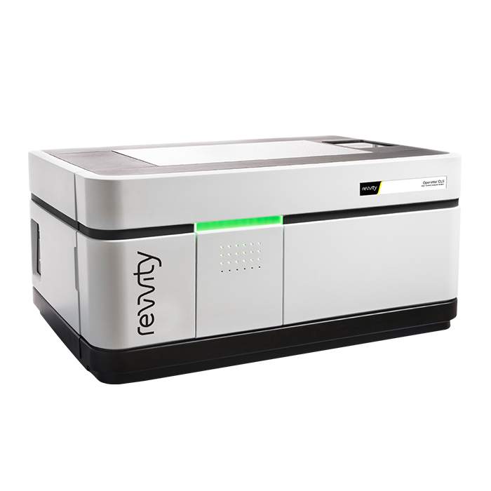


Operetta CLS High-Content Analysis System
Operetta CLS High-Content Analysis System
Part #:
HH16000020
Imaging Modality:
Brightfield, Confocal, Digital phase contrast, Fluorescence
Loading...
Your complete HCS workflow
The Operetta CLS combines speed and sensitivity with intuitive analysis. Integrated with our complete HCS workflow and Harmony software, it empowers biologists to run complex analyses independently - right out of the box.
Get superior results with our PhenoPlate™ microplates for high content screening.
Use together with our PhenoVue™ cellular imaging reagents including cell painting kits.
Improve throughput and productivity by automating your Operetta CLS system, or benefit from our automated cellular workflows and drug discovery workflows.
Export results automatically into Image Artist™ Image Analysis and Management platform, so you can access, re-analyze, store, and share all your cell image data from Operetta CLS and other HCS systems.
Combine with Signals One™ for powerful multivariate statistical methods and unsupervised machine learning techniques so you can identify parameters that best define distinct cellular fingerprints.
Video overview
Key features
Intelligent image acquisition
Target more accurately your object of interest for significantly reduced acquisition and analysis times, particularly valuable for 3D microtissue and rare event studies.
Machine learning
Easily create algorithms without being an image analysis expert using our PhenoLOGIC proprietary machine-learning technology.
Easily quantify cellular phenotypes in complex 3D models
Explore your cell models by visualizing them in a 3D- and an XYZ-viewer and quantify volumetric and other 3D related phenotypic readouts.
Powerfully simple analysis capabilities
Harmony high-content imaging and analysis software - is the easy-to-learn, easy-to-use software that can make you more productive - faster.
High efficiency excitation design
Direct coupling limits the light loss that is typical of light guides or fibers.
Fast frame rate imaging
Capture rapid cellular changes with an imaging frame rate up to 105 fps.
Automated water-immersion objectives
From the company with more than 20 years of experience - giving faster exposure times, increased resolution and reduced photodamage.
Confocal spinning-disk technology
Provides a gentle imaging process (especially for live-cell experiments) with efficient background rejection.
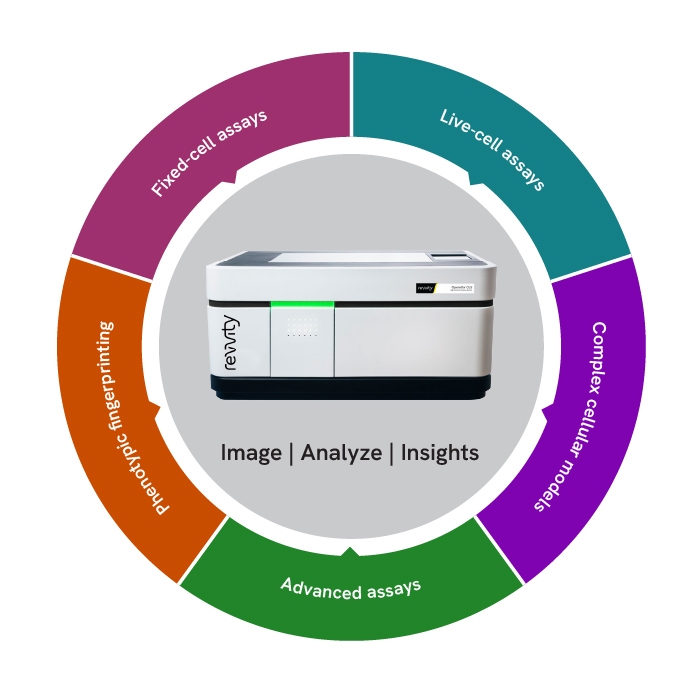
Operetta CLS applications
Fixed-cell assays
Up to eight high-power excitation sources and user-accessible emission filters enable maximum flexibility of fluorescent stains and labels, plus the system features widefield and spinning-disk fluorescent imaging.
Live-cell assays
The spinning disk confocal optics and synchronized LED illumination provides stable excitation and minimize phototoxicity and bleaching for meaningful live-cell assays. You can also choose brightfield or digital-phase contrast imaging modes.
Complex cellular models
The large-format sCMOS camera with water-immersion objectives provides sensitivity and high resolution, while advanced software helps you address the imaging and analysis challenges presented by complex cellular models.
Advanced assays
FRET is a powerful tool for investigating conformational changes and protein-protein interactions. The Operetta CLS system’s sensitive imaging and dedicated analysis tools for ratiometric imaging, facilitate robust results.
Phenotypic fingerprinting
The Operetta CLS combines high-resolution imaging with advanced software tools to help create robust phenotypic fingerprints of the subtle differences at the core of successful phenotypic assays.
Read how the Operetta CLS assists in university research
Operetta CLS configurations

Operetta CLS Quattro
Ideal for common applications that need sensitivity and resolution, with the capacity to grow if the need arises.

Operetta CLS FLEX
Offers flexibility in excitation and imaging modes for many challenging applications – and it can be upgraded to even higher performance.

Operetta CLS LIVE
Ideal for gentle yet highly sensitive live-cell imaging.
Configuration details
| Quattro | FLEX | LIVE | ||
|---|---|---|---|---|
| System Options | Camera | sCMOS | sCMOS | sCMOS |
| Number of LEDs | 4 | 8 | 8 | |
| Confocal Disk | º | • | • | |
| Environmental Control | º | º | • | |
| Water Immersion | º | º | • | |
| Transmitted Light | • | • | • | |
| Robotics/Automation Compatible | • | • | • | |
| Imaging Modes | Fluorescence Widefield Imaging | • | • | • |
| Confocal Fluorescence | º | • | • | |
| Brightfield and Digital Phase Contrast | • | • | • | |
| Ratiometric FRET | • | • | • | |
| 3D Imaging | º | • | • | |
| Live-Cell Imaging | º | º | • |
• Includedº Optional
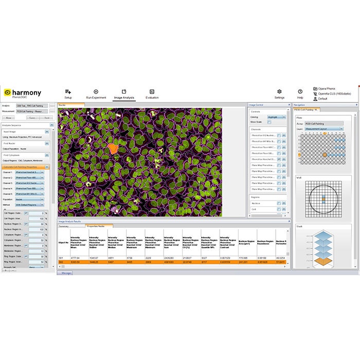
Image acquisition and analysis simplified with Harmony software
Intuitive workflow
Harmony software offers an intuitive user interface that guides you from image acquisition to analysis and evaluation.
Templates for quick set-up
Templates allow you to set up acquisition channels and parameters efficiently.
Ready-made solutions
Choose from pre-built solutions for common image analysis tasks, simplifying your workflow.
Customizable building blocks
Create, configure, and customize your own high-content analysis applications using image analysis building blocks.
Advanced features
Harmony includes advanced analysis capabilities, such as texture and STAR morphology analysis, providing detailed descriptions of cellular morphology and robust differentiation of phenotypes.
Data management
The software automatically stores analysis results and metadata, including assay layout, instrument settings, and user-defined keywords and annotations.
The Operetta CLS high-content analysis system highlights
3D cell models
Enrich your research
The Operetta CLS system can analyze 3D cell cultures to help generate more physiologically relevant data for better-informed decisions.
Brochure
Operetta CLS at a glance
Discover the features and benefits of the Operetta CLS high-content analysis system in detail.
Reagents
Ideal for the Operetta CLS
Revvity's PhenoVue suite of cellular imaging reagents complement our proven HCS instruments.
Meet our users
Hear from scientists
Read how researchers are using our HCS products to advance their research in a variety of fields.
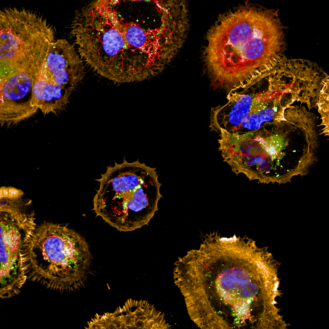
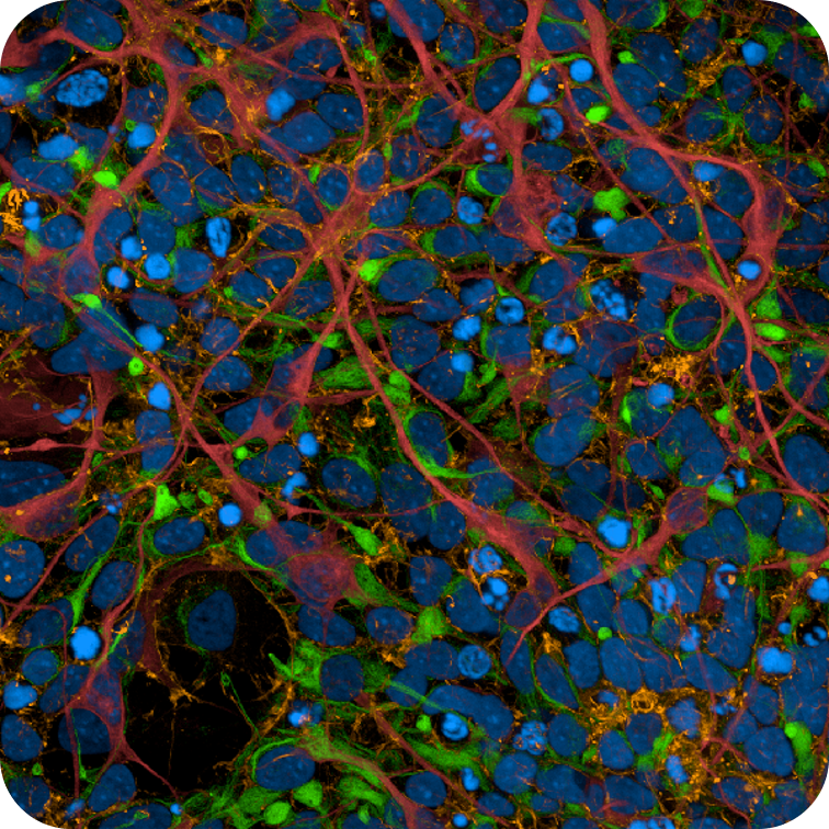
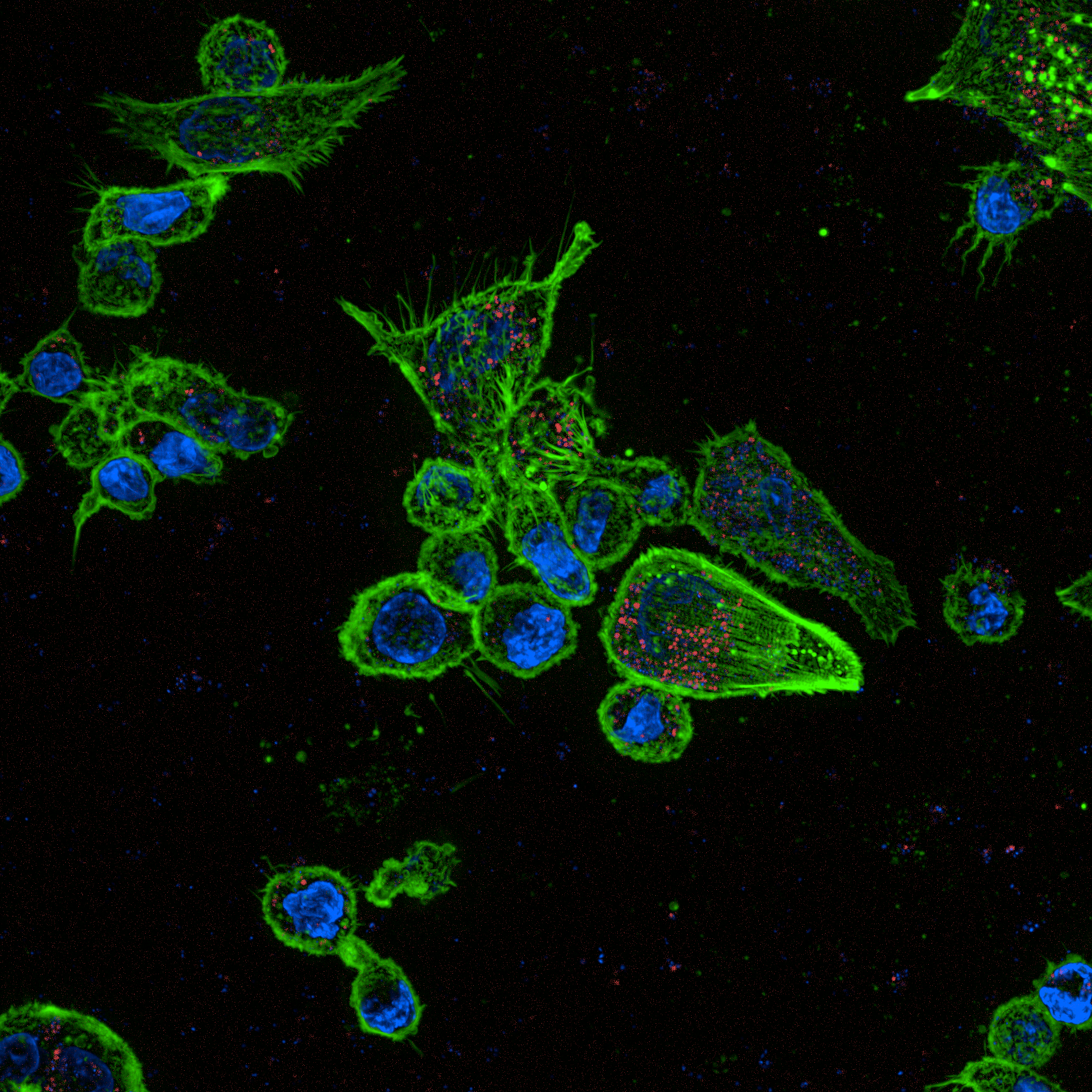
Image gallery
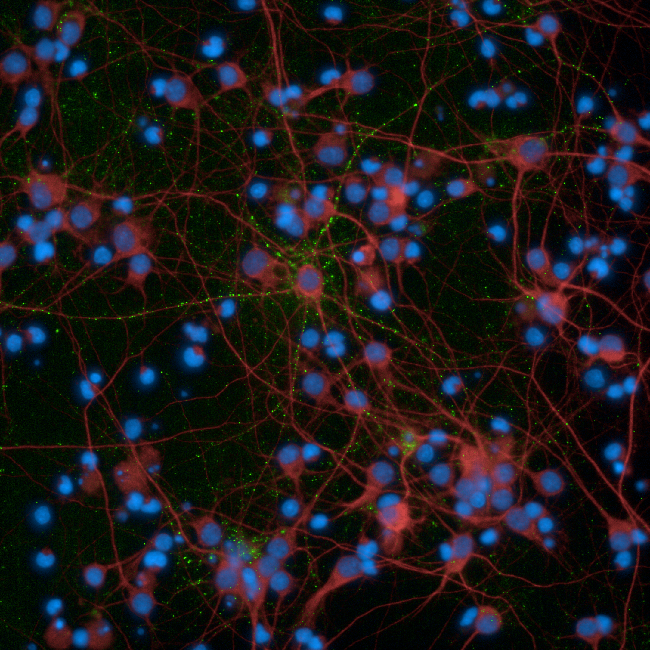
Primary rat neurons imaged on the Operetta CLS high-content analysis system
Human ovarian granulosa tumor cell line imaged on the Operetta CLS high-content analysis system
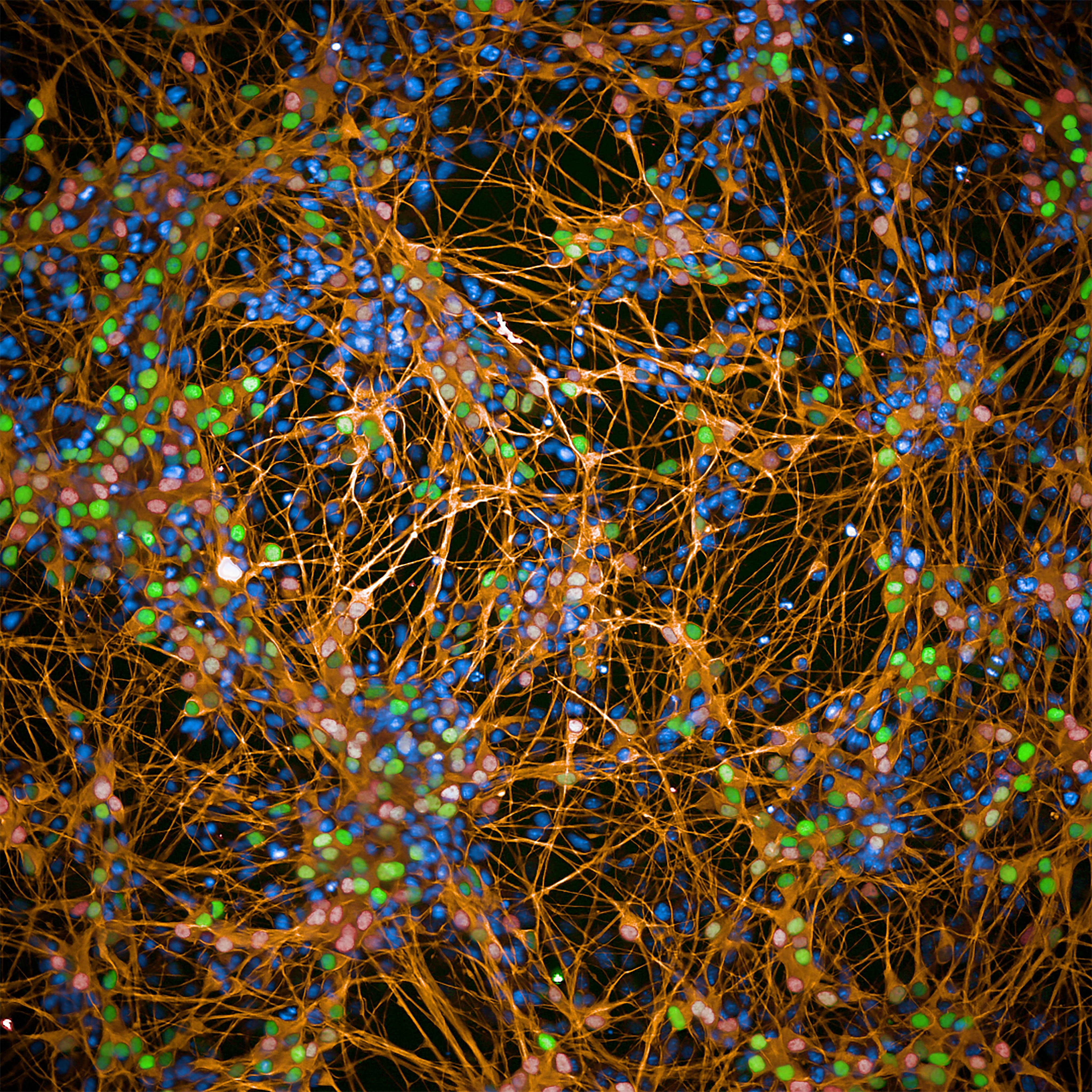
iPSC-derived human cortical neurons imaged on the Operetta CLS high-content analysis system
Macrophages imaged on the Operetta CLS high-content analysis system
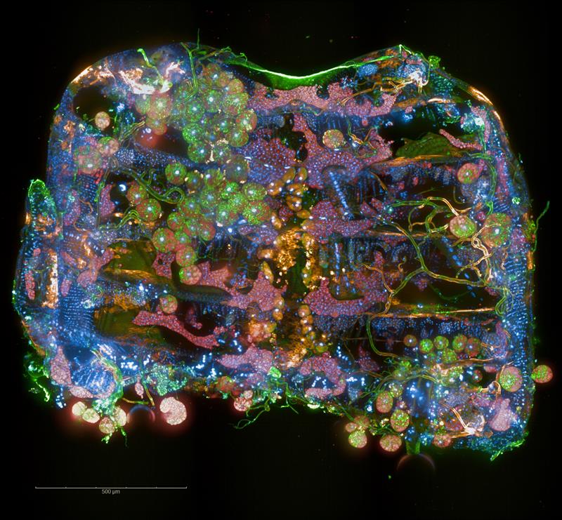
Drosophila abdomen imaged on the Operetta CLS high-content analysis system
iPSC-derived motor neurons imaged on the Operetta CLS high-content analysis system
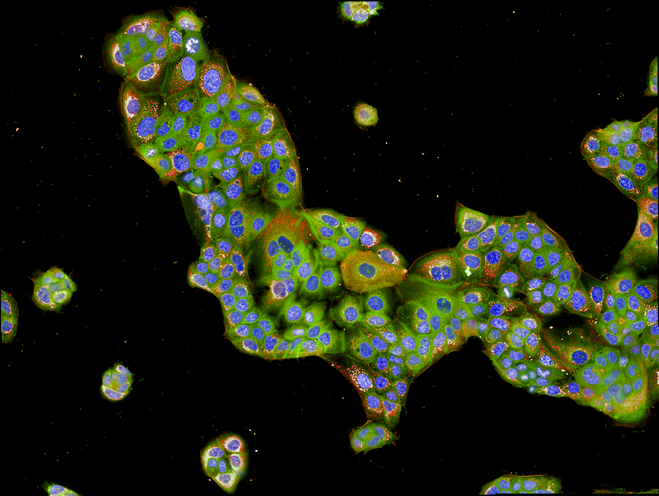
HPAF-II cells imaged on the Operetta CLS high-content analysis system.
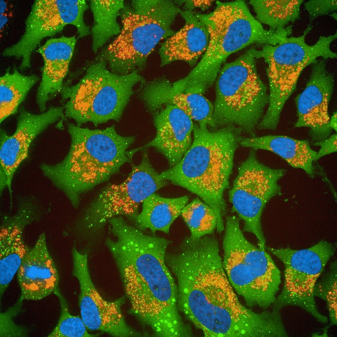
HeLa cells imaged on the Operetta CLS high-content analysis system.
×
Product information
Overview
The Operetta CLS system combines speed and sensitivity with the powerful and intuitive data analysis you’ve come to trust from the Operetta platform. The all-new Operetta CLS delivers everything you need from the high-content analysis. What’s more, the Operetta CLS system is part of our comprehensive HCS workflow – everything from HCS systems and microplates to automation and informatics for every application. All from one knowledgeable, trusted vendor. Put that together with our Harmony high-content imaging and analysis software - the easy-to-learn, easy-to-use software that empowers biologists to do their own analysis - and you have everything you need to run your every day (and complex) analyses right away.
Specifications
| Dimensions | 66.0 cm (W) x 98.0 cm (D) x 45.0 cm (H) |
|---|
| Automation Compatible |
Yes
|
|---|---|
| Brand |
Operetta CLS
|
| Imaging Modality |
Brightfield
Confocal
Digital phase contrast
Fluorescence
|
| Unit Size |
1 unit
|
Video gallery
Operetta CLS High-Content Analysis System
Citations
Resources
Are you looking for resources, click on the resource type to explore further.
Application Note
3D Analysis of Cell Invasion using Operetta
Here, we present a method for analyzing cell invasion into a 3D extracellular matrix using the Operetta® high-content analysis...
Brochure
3D cell culture workflow solutions
More than ever, researchers are turning to 3D cell cultures, microtissues and organoids to bridge the gap between 2D cell cultures...
Technical Note
3D volumetric analysis of luminal spaces inside cysts or organoids
High-content assays using 3D objects such as cysts or organoids can be challenging from the perspectives of both image acquisition...
Technical Note
3D volumetric and zonal analysis of solid spheroids
Multicellular 3D “oids” (tumoroids, spheroids, organoids) have the potential to better predict the effects of drug candidates...
Application Note
A multiparametric live-cell cytotoxicity analysis using the Operetta High-content Analysis System
The detection of compound cytotoxicity is an essential part of drug discovery. In this work we describe a rapid and flexible image...
Case Study
A workflow to characterize and benchmark human induced pluripotent stem cells
The UK-based Human Induced Pluripotent Stem Cell Initiative (HipSci) aims to offer the scientific community access to a vast panel...
Loading...


How can we help you?
We are here to answer your questions.

