


US
Revvity Sites Globally
Select your location.
*e-commerce not available for this region.

HTRF Human and Mouse Phospho-p38 MAPK (Thr180/Tyr182) Detection Kit, 96 Assay Points


HTRF Human and Mouse Phospho-p38 MAPK (Thr180/Tyr182) Detection Kit, 96 Assay Points


generic HTRF phospho primary image
The phospho-p38 (Thr180/Tyr182) kit enables the cell-based quantitative detection of phosphorylated p38 as a readout of the MAPK pathway.
| Feature | Specification |
|---|---|
| Application | Cell Signaling |
| Sample Volume | 16 µL |
The phospho-p38 (Thr180/Tyr182) kit enables the cell-based quantitative detection of phosphorylated p38 as a readout of the MAPK pathway.
Product variants
Unit Size: 96 assay points
Part #:
64P38PET
List price
USD 739.00
Your price:
Unit Size: 500 assay points
Part #:
64P38PEG
List price
USD 2,387.00
Your price:
Unit Size: 10,000 assay points
Part #:
64P38PEH
List price
USD 13,884.00
Your price:
For research use only. Not for use in diagnostic procedures. All products to be used in accordance with applicable laws and regulations including without limitation, consumption, and disposal requirements under European REACH regulations (EC 1907/2006).
HTRF Human and Mouse Phospho-p38 MAPK (Thr180/Tyr182) Detection Kit, 96 Assay Points
generic HTRF phospho primary image
Loading...
Product information
Overview
This HTRF cell based assay conveniently and accurately quantifies phosphorylated p-38-MAPK at Thr180/Tyr182. This kit has applications in inflammation, immune response, and oncology research. Using a streamlined protocol, amenable to low-volume formats, this kit can be used from basic research to High Throughput drug screening.
HTRF assays offer many advantages over other technologies:
- Homogeneous add-and-read format
- No wash steps
- Low background
- Straightforward miniaturization from 96- or 384-well microplates to high density assay formats such as 384-well low volume and 1536-well plates
- Stable signal, providing flexibility in time of readout or size of assays
How it works
Phospho-p38 MAPK (Thr180/Tyr182) assay principle
The Phospho-p38 MAPK (Thr180/Tyr182) assay measures p38 MAPK when phosphorylated at Thr180/Tyr182. Contrary to Western Blot, the assay is entirely plate-based and does not require gels, electrophoresis or transfer. The Phospho-p38 MAPK (Thr180/Tyr182) assay uses 2 labeled antibodies: one with a donor fluorophore, the other one with an acceptor. The first antibody is selected for its specific binding to the phosphorylated motif on the protein, the second for its ability to recognize the protein independent of its phosphorylation state. Protein phosphorylation enables an immune-complex formation involving both labeled antibodies and which brings the donor fluorophore into close proximity to the acceptor, thereby generating a FRET signal. Its intensity is directly proportional to the concentration of phosphorylated protein present in the sample, and provides a means of assessing the proteins phosphorylation state under a no-wash assay format.
Phospho-p38 MAPK (Thr180/Tyr182) 2-plate assay protocol
The 2 plate protocol involves culturing cells in a 96-well plate before lysis then transferring lysates to a 384-well low volume detection plate before adding phospho-p38 MAPK (Thr180/Tyr182) HTRF detection reagents. This protocol enables the cells' viability and confluence to be monitored.

Phospho-p38 MAPK (Thr180/Tyr182) 1-plate assay protocol
Detection of Phosphorylated p38 MAPK (Thr180/Tyr182) with HTRF reagents can be performed in a single plate used for culturing, stimulation and lysis. No washing steps are required. This HTS designed protocol enables miniaturization while maintaining robust HTRF quality.

Assay validation
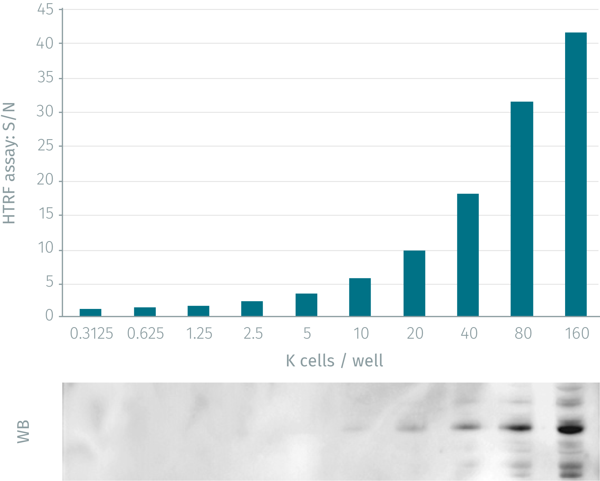
HTRF assay compared to WB using phospho-p38 MAPK cellular assay
HeLa cells were grown in a T175 flask 37°C, 5% Co2, 2 days. Stimulation was done with anisomycin 10µM for 15min. After elimination of cell culture medium, 3ml of supplemented lysis buffer were added and incubated for 30min. Soluble supernatants were collected after 10min centrifuging. Equal amounts of lysates were used for a side by side comparison of WB and HTRF. HTRF assay shows better sensitivity than Western Blot: 2500 cells for HTRF compared to10,000 cells for WB.
Anisomycin dose-response on NIH 3T3 cells
Murine NIH 3T3 cells (50,000 cells/well) were stimulated for 45 minutes at 37°C with various concentrations of anisomycin. After a 30 minutes lysis incubation time, phosphorylated p38 MAPK was measured using the two-plate assay protocol.

Anisomycin dose-response on Hela cells
Various concentrations of HeLa cells (25, 50 and 100K cells/well) were incubated for 45 minutes at 37°C with various concentrations of anisomycin. After a 30 minutes lysis incubation time, phosphorylated p38 MAPK was measured using the two-plate assay protocol.

Simplified pathway
Phospho-p38 MAPK (Thr180/Tyr182) simplified pathway
p38 MAPK is activated by a variety of cellular stresses, including osmotic shock, inflammatory cytokines, lipopolysaccharides (LPS), UltraViolet light, and growth factors. It is involved in cell differentiation and apoptosis. Activated p38 MAPK has been shown to phosphorylate and activate MAPKAP kinase 2 as well as phosphorylate the transcription factors ATF-2 , Max , and MEF2 . p38 MAPK plays a critical role in inflammation, immune response, apoptosis, cell differentiation, cell-cycle regulation and tumorigenesis.

Specifications
| Application |
Cell Signaling
|
|---|---|
| Automation Compatible |
Yes
|
| Brand |
HTRF
|
| Detection Modality |
HTRF
|
| Lysis Buffer Compatibility |
Lysis Buffer 1
|
| Molecular Modification |
Phosphorylation
|
| Product Group |
Kit
|
| Sample Volume |
16 µL
|
| Shipping Conditions |
Shipped in Dry Ice
|
| Target |
P38 MAPK
|
| Target Class |
Phosphoproteins
|
| Target Species |
Human
Mouse
|
| Technology |
TR-FRET
|
| Therapeutic Area |
Cardiovascular
Infectious Diseases
NASH/Fibrosis
Neuroscience
Oncology & Inflammation
|
| Unit Size |
96 assay points
|
Video gallery
HTRF Human and Mouse Phospho-p38 MAPK (Thr180/Tyr182) Detection Kit, 96 Assay Points
Resources
Are you looking for resources, click on the resource type to explore further.
Guide
HTRF solutions, guide to major applications
This guide provides you an overview of HTRF applications in several therapeutic areas.
Flyer
Reagent solutions for autoimmunity research.
Advance your autoimmune disease research and benefit from Revvity broad offering of reagent technologies
Loading...


How can we help you?
We are here to answer your questions.

