


US
Revvity Sites Globally
Select your location.
*e-commerce not available for this region.

HTRF Human Phospho-H2AX (Ser139) Detection Kit, 500 Assay Points


HTRF Human Phospho-H2AX (Ser139) Detection Kit, 500 Assay Points


generic HTRF phospho primary image
The HTRF Human Phospho-H2AX (Ser139) Detection Kit is an immunoassay designed for the detection and quantitation of Human H2AX when phosphorylated at Ser139 in cellular lysates, in a homogeneous (no-wash steps, no separation steps) format.
| Feature | Specification |
|---|---|
| Application | Cell Signaling |
| Sample Volume | 16 µL |
The HTRF Human Phospho-H2AX (Ser139) Detection Kit is an immunoassay designed for the detection and quantitation of Human H2AX when phosphorylated at Ser139 in cellular lysates, in a homogeneous (no-wash steps, no separation steps) format.
Product variants
Unit Size: 500 assay points
Part #:
64H2XPEG
List price
USD 2,340.00
Your online price:
Unit Size: 10,000 assay points
Part #:
64H2XPEH
List price
USD 13,611.00
Your online price:
For research use only. Not for use in diagnostic procedures. All products to be used in accordance with applicable laws and regulations including without limitation, consumption, and disposal requirements under European REACH regulations (EC 1907/2006).
HTRF Human Phospho-H2AX (Ser139) Detection Kit, 500 Assay Points
generic HTRF phospho primary image
Loading...
Product information
Overview
The phospho-H2AX (Ser139) kit offers quantitative detection of the histone variant H2AX, phosphorylated on serine 139. This specific phosphorylation may occur when double-stranded DNA breaks, either due to ionizing radiation or to endogenous physiological processes. This modification, called gamma-H2AX (phospho-Ser139), triggers cell cycle arrest and DNA damage response repair, which makes this assay particularly appropriate for studying genome stability, cell cycle, DNA repair, and more broadly as a biomarker for cancer and for aging process investigations.
How it works
Phospho-H2AX (Ser139) assay principle
The Phospho-H2AX (Ser139) assay measures H2AX when phosphorylated at Ser139. Contrary to Western Blot, the assay is entirely plate-based and does not require gels, electrophoresis or transfer. The Phospho-H2AX (Ser139) assay uses 2 labeled antibodies: one with a donor fluorophore, the other one with an acceptor. The first antibody is specific for the phosphorylated motif on the protein, the second one recognizes the protein independent of its phosphorylation state. Protein phosphorylation enables an immune-complex formation involving both labeled antibodies and which brings the donor fluorophore into close proximity to the acceptor, thereby generating a FRET signal. Its intensity is directly proportional to the concentration of phosphorylated protein present in the sample, and provides a means of assessing the proteins phosphorylation state under a no-wash assay format.
Phospho-H2AX (Ser139) 2-plate assay protocol
The 2 plate protocol involves culturing cells in a 96-well plate before lysis then transferring lysates to a 384-well low volume detection plate before adding phospho-H2AX (Ser139) HTRF detection reagents. This protocol enables the cells' viability and confluence to be monitored.

Phospho-H2AX (Ser139) 1-plate assay protocol
Detection of Phosphorylated H2AX (Ser139) with HTRF reagents can be performed in a single plate used for culturing, stimulation and lysis. No washing steps are required. This HTS designed protocol enables miniaturization while maintaining robust HTRF quality.

Assay validation
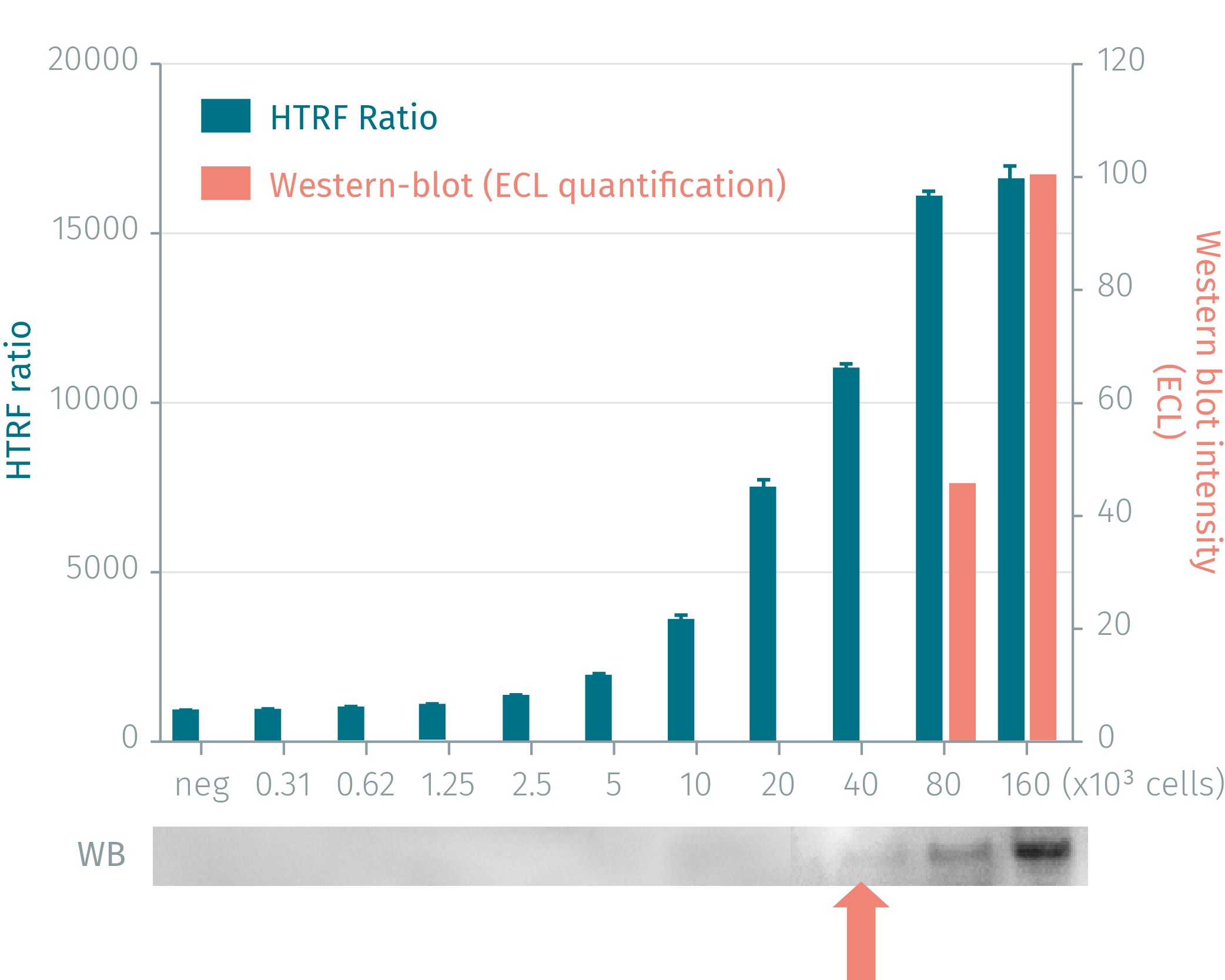
HTRF phospho-H2AX cellular assay compared to Western Blot
HEK-293 cells were grown in a T175 flask to 80% confluency. Cell culture were then lysed and pelleted by centrifugation. Serial dilutions of the cell lysate were performed in the supplemented lysis buffer and 16 µL of each dilution were dispensed and analyzed side-by-side by Western Blot and by HTRF. The HTRF cellular assays were found to be at least 16-fold more sensitive than the Western Blot.
Neocarzinostatin dose-response on Jurkat cells
25,000 human Jurkat cells were plated in 96 well plates. After incubation with increasing concentrations of neocarzinostatin (30min), cells were lysed with 10µl of lysis buffer 4X for 30min at RT under gentle shaking. 16 µL of lysate were transferred into a 384-well sv white microplate and 4 µL of the HTRF phospho-H2AX (Ser139) detection reagents were added. The HTRF signal was recorded after an overnight incubation.

Phospho-H2AX(Ser139) activation on Jurkat cells: detection kinetics
25,000 human Jurkat cells were plated in 96 well plates. After incubation with increasing concentrations of neocarzinostatin (30min), cells were lysed with 10µl of lysis buffer 4X for 30min at RT under gentle shaking. 16 µL of lysate were transferred into a 384-well small volume white microplate and 4 µL of the HTRF phospho-H2AX (Ser139) detection reagents were added. The HTRF signal was recorded after different incubation times.

Simplified pathway
Phospho-H2AX (Ser139) cellular assay kit simplified pathway
DNA Double-Strand Breaks (DSB) are the most deleterious type of DNA damage, consecutive to endogenous processes or exogenous factors (e.g. ionizing & UV radiations, specific anticancer drugs). H2AX is a variant of the histone H2A family, which gets phosphorylated at Ser139 upon the event of a DSB. This modification is called ?-H2AX and is spread within minutes to thousands of H2AX proteins in proximity to the damage site. It is crucial is crucial to activating the DNA damage response pathway, a complex molecular mechanism to detect and repair DNA damage.

Specifications
| Application |
Cell Signaling
|
|---|---|
| Brand |
HTRF
|
| Detection Modality |
HTRF
|
| Lysis Buffer Compatibility |
Lysis Buffer 1
Lysis Buffer 4
Lysis Buffer 5
|
| Molecular Modification |
Phosphorylation
|
| Product Group |
Kit
|
| Sample Volume |
16 µL
|
| Shipping Conditions |
Shipped in Dry Ice
|
| Target Class |
Phosphoproteins
|
| Target Species |
Human
|
| Technology |
TR-FRET
|
| Therapeutic Area |
Oncology & Inflammation
|
| Unit Size |
500 assay points
|
Video gallery
HTRF Human Phospho-H2AX (Ser139) Detection Kit, 500 Assay Points
Resources
Are you looking for resources, click on the resource type to explore further.
Brochure
HTRF assays and reagents product list
Discover the versatility and precision of Homogeneous Time-Resolved Fluorescence (HTRF) technology. Our HTRF portfolio offers a...
Guide
HTRF solutions, guide to major applications
This guide provides you an overview of HTRF applications in several therapeutic areas.
Flyer
Solutions to advance your CART-T cell research
Chimeric antigen receptor (CAR) T-cell therapy has transformed the field of immuno-oncology providing a novel approach to treating...
Loading...


How can we help you?
We are here to answer your questions.

