
HTRF Human Phospho-eNOS (Ser1177) Detection Kit, 500 Assay Points


This HTRF kit enables the cell-based quantitative detection of phosphorylated eNOS (endothelial nitric oxide synthase) at Serine 1177.
| Feature | Specification |
|---|---|
| Application | Cell Signaling |
| Sample Volume | 16 µL |
This HTRF kit enables the cell-based quantitative detection of phosphorylated eNOS (endothelial nitric oxide synthase) at Serine 1177.


Product information
Overview
This HTRF cell-based assay conveniently and accurately detects phosphorylated eNOS at Serine 1177.
eNOS is expressed primarily in endothelial cells, where its function is to control blood vessel dilation, blood pressure, and numerous other anti-atherosclerotic and vasoprotective effects.
Serine 1177 is the most important phosphorylation site of eNOS, positively modulating its activity. Phosphorylation at serine 1177 is under the control of several kinases, including AKT, PKA, AMPK, CaMKK2b, and CHKI
This HTRF cell-based assay conveniently and accurately detects phosphorylated eNOS at Serine 1177.
HTRF assays offer many advantages over other technologies:
- Homogeneous add-and-read format
- No wash steps
- Low background
- Straightforward miniaturization from 96- or 384-well microplates to high density assay formats such as 384-well low volume and 1536-well plates
- Stable signal, providing flexibility in time of readout or size of assays
How it works
Phospho-eNOS (Ser1177) assay principle
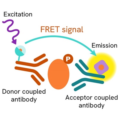
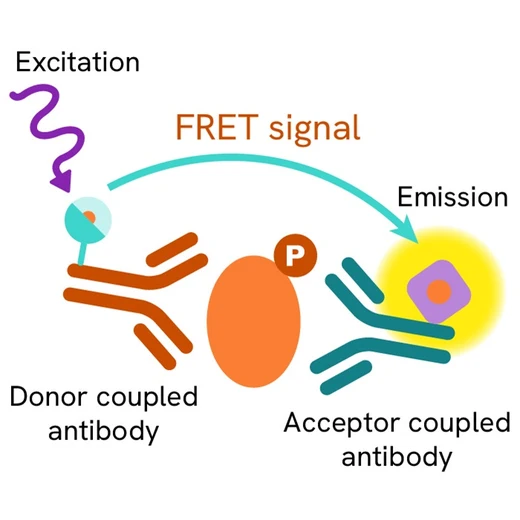
The Phospho-eNOS (S1177) assay measures eNOS when phosphorylated at serine 1177. Unlike Western Blot, the assay is entirely plate-based and does not require gels, electrophoresis, or transfer. The assay uses 2 antibodies, one labeled with a donor fluorophore and the other with an acceptor. The first antibody is selected for its specific binding to the phosphorylated motif on the protein, the second for its ability to recognize the protein independently of its phosphorylation state. Protein phosphorylation enables an immune-complex formation involving both labeled antibodies, and which brings the donor fluorophore into close proximity to the acceptor, thereby generating a FRET signal. Its intensity is directly proportional to the concentration of phosphorylated protein present in the sample, and provides a means of assessing the protein's phosphorylation state under a no-wash assay format.

Phospho-eNOS (Ser1177) two-plate assay protocol
The two-plate protocol involves culturing cells in a 96-well plate before lysis, then transferring lysates to a 384-well low volume detection plate before the addition of Phospho-eNOS (Ser1177) HTRF detection reagents. This protocol enables the cells' viability and confluence to be monitored.

Phospho-eNOS (Ser1177) one-plate assay protocol
Detection of Phosphorylated eNOS (Ser1177) with HTRF reagents can be performed in a single plate used for culturing, stimulation, and lysis. No washing steps are required. This HTS designed protocol enables miniaturization while maintaining robust HTRF quality.

Assay validation
Serum starvation and phosphorylation accumulation
Huvec cells were seeded in a 160mm Petri Dish at 80% of confluence in 37°C, 5% Co2 conditions, and an overnight serum starvation was done or not. Then a solution of pervanadate at 30µM for 30 minutes was added or not. Lysis buffer was added to each dish after culture medium removal, and incubated for 30 minutes under shaking. Then 16µl of lysate were transferred into a 384 well plate for Total and Phospho Ser1177 eNOS detection.
Starvation seems to induce a slight decrease in protein, but a real decrease in the phosphorylation level. Phosphorylation accumulation with pervanadate is efficient.

eNOS phosphorylation induced by fenofibrate stimulation through AMPK pathway
Huvec cells were seeded in a 100mm Petri Dish at 80% of confluence in 37°C, 5% Co2 conditions, and an overnight serum starvation was done. Then a fenofibrate solution at 30µM was added for 30 minutes or not. Lysis buffer was then dispensed after culture medium removal. 16µl of lysate were transferred into a 384 well plate for Total and Phospho Ser1177 eNOS detection.

WB versus HTRF assay for phospho-eNOS (Ser1177)
Huvec cells were grown in a 160mm Petri dish at 37°C, 5% Co2, for 2 days. Overnight serum starvation was carried out before an incubation of pervanadate at 30µM for 30 min. After removal of cell culture medium, 3 mL of supplemented lysis buffer were added and incubated for 30 min. Soluble supernatants were collected after 10 min centrifuging. Equal amounts of lysates were used for a side by side comparison of WB and HTRF. Phospho-eNOS (Ser1177)assay shows a better sensitivity than Western Blot: 37,500 cells/mL are sufficient to be detected by HTRF, while WB needs 75,000.

Specifications
| Application |
Cell Signaling
|
|---|---|
| Brand |
HTRF
|
| Detection Modality |
HTRF
|
| Lysis Buffer Compatibility |
Lysis Buffer 1
Lysis Buffer 2
Lysis Buffer 3
Lysis Buffer 4
Lysis Buffer 5
|
| Molecular Modification |
Phosphorylation
|
| Product Group |
Kit
|
| Sample Volume |
16 µL
|
| Shipping Conditions |
Shipped in Dry Ice
|
| Target Class |
Phosphoproteins
|
| Target Species |
Human
|
| Technology |
TR-FRET
|
| Therapeutic Area |
Cardiovascular
Metabolism/Diabetes
|
| Unit Size |
500 assay points
|
Video gallery
Resources
Are you looking for resources, click on the resource type to explore further.
Atherosclerosis pathogenesis, cellular actors, and pathways
Atherosclerosis is a common condition in which arteries harden and...
Discover the versatility and precision of Homogeneous Time-Resolved Fluorescence (HTRF) technology. Our HTRF portfolio offers a...
This guide provides you an overview of HTRF applications in several therapeutic areas.


Loading...
How can we help you?
We are here to answer your questions.


