VesselVue Microbubble Contrast Agent
Visualize microvasculature
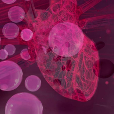
Revvity's VesselVue™ microbubble contrast agent is a lipid encapsulated micron-sized particle with a gas core that are injected intravenously into the animal. This agent is non-toxic to the animal, and with each microbubble being smaller than a red blood cell, circulate systemically and clear the body in minutes.
Contrast-enhanced ultrasound (CEUS) is a technique that uses microbubble-based contrast agents to improve the echogenicity of blood which in turn enhances and aids in the visualization of vessels and tissue vascularity.
VesselVue microbubble contrast agent can be used to study tissue perfusion and blood flow characteristics on ultrasound systems equipped with CEUS imaging modes. Using VesselVue in combination with our exclusive Acoustic Angiography mode on the Vega® ultrasound system, a unique form of CEUS imaging, researchers can obtain high resolution images of microvessel density and morphology, which are biomarkers useful for studying angiogenesis and vascular bed development.
For research use only. Not for use in therapeutic or diagnostic procedures.

VesselVue microbubble contrast agent is available in a P-formulation for your research
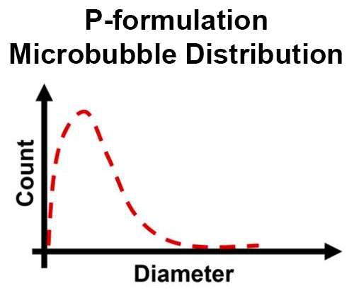
Wide microbubble size distribution with a smaller mean.
Structure of vesselVue microbubble contrast agent
Activation is required for VesselVue P-formulation.
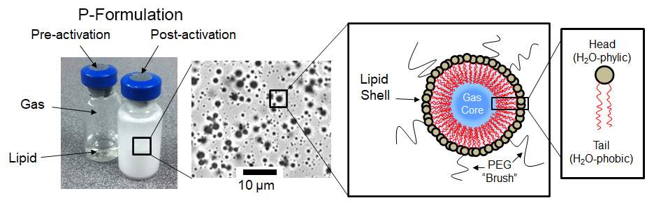
(Left) Activation of VesselVue P-formulation microbubble contrast agents pre and post mechanical agitation using a VesselVue Microbubble Mixer (sold separately). (Right) Structure of microbubbles.
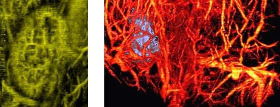
Kidney (left) and Tumor (right) vascularity using VesselVue microbubble contrast agent and acoustic angiography using the Vega ultrasound imaging system.
Visualize and quantify vascular network architecture and density of tumors or organs over time
- Evaluate tumor angiogenesis
- Explore response to therapy prior to tissue changes
- Collect high-resolution images of 3D microvessel trees in minutes and 2D capture in less than a second (using the Vega® ultrasound system)
- Polydisperse formulation for general CEUS imaging applications