


US
Revvity Sites Globally
Select your location.
*e-commerce not available for this region.

HTRF Human Total MKK4 Detection Kit, 500 Assay Points

HTRF Human Total MKK4 Detection Kit, 500 Assay Points


generic HTRF total primary image


This HTRF kit is designed to monitor the expression level of cellular MKK4, and can be used as a normalization assay for the phospho-MKK4 kit.
| Feature | Specification |
|---|---|
| Application | Cell Signaling |
| Sample Volume | 16 µL |
This HTRF kit is designed to monitor the expression level of cellular MKK4, and can be used as a normalization assay for the phospho-MKK4 kit.
Product variants
Unit Size: 500 assay points
Part #:
64NK4PEG
List price
USD 2,340.00
Your online price:
Unit Size: 10,000 assay points
Part #:
64NK4PEH
List price
USD 13,611.00
Your online price:
For research use only. Not for use in diagnostic procedures. All products to be used in accordance with applicable laws and regulations including without limitation, consumption, and disposal requirements under European REACH regulations (EC 1907/2006).
HTRF Human Total MKK4 Detection Kit, 500 Assay Points
generic HTRF total primary image
HTRF Human Total MKK4 Detection Kit, 500 Assay Points
Product information
Overview
HTRF Total MKK4 cellular assay monitors total MKK4, and can be used as a normalization assay with our phospho-MKK4 kit. This kit is compatible with buffers from phospho-MKK4 kit, so same lysate can be used analyses of both phosphorylated and total protein populations.
Phosphorylation of MKK4 on Serine 257 is induced by various MKKKinases such as TAK1, Tpl2, or ASK1, in response to cellular stresses and proinflammatory cytokines. Once phosphorylated, MKK4 prefentially triggers activation of JNK which regulates a range of biological processes implicated in tumorigenesis, neurodegenerative disorders, and fibrosis.
How it works
Total-MKK4 assay principle
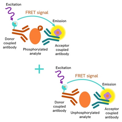
The Total MKK4 assay quantifies the expression level of MKK4 in a cell lysate. Unlike Western Blot, the assay is entirely plate-based and does not require gels, electrophoresis, or transfer. The Total-MKK4 assay uses two labeled antibodies, one coupled to a donor fluorophore and the other to an acceptor. Both antibodies are highly specific for a distinct epitope on the protein. In presence of MKK4 in a cell extract, the addition of these conjugates brings the donor fluorophore into close proximity with the acceptor and thereby generates a FRET signal. Its intensity is directly proportional to the concentration of the protein present in the sample, and provides a means of assessing the protein's expression under a no-wash assay format.
Total-MKK4 two-plate assay protocol
The two-plate protocol involves culturing cells in a 96-well plate before lysis, then transferring lysates to a 384-well low volume detection plate before the addition of Total-STING HTRF detection reagents. This protocol enables the cells' viability and confluence to be monitored.

Total-MKK4 one-plate assay protocol
Detection of Total MKK4 with HTRF reagents can be performed in a single plate used for culturing, stimulation, and lysis. No washing steps are required. This HTS designed protocol enables miniaturization while maintaining robust HTRF quality.

Assay validation
Anisomycin dose-response on Retinoic Acid (RA) differentiated SH-SHY5Y cells
Human SH-SY5Y cells were plated at 25,000 cells/well in a 96-well plate. After 24 h incubation at 37 °C, 5% CO2, the cells were incubated in differentiation medium containing 10µM of RA for 1 week in the dark. Note that due to the poor stability of RA, cell culture medium containing fresh RA was renewed daily. Once differentiated, SH-SY5Y were stimulated with increasing concentrations of anisomycin for 45 min. Then the medium was removed and 50 µL of supplemented lysis buffer 1X were added. After 30 min lysis at RT under gentle shaking, 16 µL of lysate were transferred into a 384-well low volume white microplate, and 4 µL of the HTRF phospho-MKK4 (Ser257) or total MKK4 detection reagents were added. The HTRF signal was recorded after an overnight incubation.
As described elsewhere, a dose dependent phosphorylation of MKK4 by Ser257 was induced by anisomycin, whereas total MKK4 slightly decreased under the same experimental conditions.

H2O2 stimulation on Retinoic Acid (RA) differentiated SH-SHY5Y cells
Human SH-SY5Y cells were plated at 400,000 cells/well in a 6-well plate. After 24 h incubation at 37 °C, 5% CO2, the cells were incubated in differentiation medium containing 10 µM of RA for 1 week in the dark. Note that due to the poor stability of RA, cell culture medium containing fresh RA was renewed daily. Once differentiated, SH-SY5Y were stimulated with 0.5 mM of H2O2 for 1h. Then the medium was removed and 500 µL supplemented lysis buffer 1X were added. After 30min lysis at RT under gentle shaking, 16 µL of lysate were transferred into a 384-well low volume white microplate and 4 µL of the HTRF phospho-MKK4 (Ser257) or total MKK4 detection reagents were added. The HTRF signal was recorded after an overnight incubation. H2O2 induced phosphorylation MKK4 on Ser257 residue, whereas the MKK4 expression level remained almost stable under the same experimental conditions.

Anisomycin dose-response on HepG2 cells correlated with Western Blot
Human HepG2 cells were plated at 100,000 cells/well in a 96-well plate. After an incubation of 24 h at 37 °C, 5% CO2, the cells were stimulated with increasing concentrations of anisomycin for 45 min. Then the medium was removed, and 50 µL of supplemented lysis buffer 1X were added. After 30 min lysis at RT under gentle shaking, 16 µL of lysate were transferred into a 384-well low volume white microplate and 4 µL of the HTRF phospho-MKK4 (Ser257) or total MKK4 detection reagents were added. The HTRF signal was recorded after an overnight incubation. The same amount of lysate was analyzed by Western Blot in a side by side experiment.
As shown on the graphs, both HTRF and Western Blot indicated an increase in MKK4 phosphorylation associated with a decrease of MKK4 expression.


HTRF total MKK4 cellular assay compared to Western Blot
The human HepG2 cell line was seeded in a T175 flask and incubated at 37 °C, 5% CO2. The cells were then stimulated with Sorbitol (1 M) for 30 min before lysis.
Serial dilutions of the cell lysate were performed in the supplemented lysis buffer, and 16 µL of each dilution were transferred into a low volume white microplate before the addition of 4 µL of HTRF total MKK4 detection reagents. Equal amounts of lysates were used for a side by side comparison between HTRF and Western Blot.
Using the HTRF total MKK4 assay, 1,250 cells/well were enough to detect a signal while 10,000 cells were needed using Western Blot which relies on an ECL detection. These results demonstrate that the HTRF total MKK4 assay is 8 times more sensitive than the Western Blot.

Simplified pathway
Simplified pathway for MKK4 assays
MKK4 (Mitogen-activated Kinase Kinase 4) is a member of MAP kinase kinase family that is activated by phosphorylation on Ser257 following activation of different MKKKs, for example TAK1 or ASK1, in response to stimuli such as GPCR activation, Growth factors, cellular stresses, or inflammatory cytokines. In turn, activated MKK4 phosphorylates JNK or p38 in order to activate c-jun, p53, or ATF2 and induce inflammation, cell survival/apoptosis, proliferation, or differentiation by regulating gene transcriptions.

Specifications
| Application |
Cell Signaling
|
|---|---|
| Brand |
HTRF
|
| Detection Modality |
HTRF
|
| Lysis Buffer Compatibility |
Lysis Buffer 1
Lysis Buffer 2
Lysis Buffer 3
Lysis Buffer 4
Lysis Buffer 5
|
| Molecular Modification |
Total
|
| Product Group |
Kit
|
| Sample Volume |
16 µL
|
| Shipping Conditions |
Shipped in Dry Ice
|
| Target |
MKK4
|
| Target Class |
Phosphoproteins
|
| Target Species |
Human
|
| Technology |
TR-FRET
|
| Therapeutic Area |
NASH/Fibrosis
Neuroscience
Oncology & Inflammation
|
| Unit Size |
500 assay points
|
Video gallery
HTRF Human Total MKK4 Detection Kit, 500 Assay Points
HTRF Human Total MKK4 Detection Kit, 500 Assay Points
Resources
Are you looking for resources, click on the resource type to explore further.
Guide
HTRF solutions, guide to major applications
This guide provides you an overview of HTRF applications in several therapeutic areas.
Flyer
Reagent solutions for autoimmunity research.
Advance your autoimmune disease research and benefit from Revvity broad offering of reagent technologies
Guide
Understanding obesity: exploring cellular pathways and mechanisms
Obesity is a complex condition characterized by excessive fat accumulation, posing significant health and socioeconomic challenges...


Loading...
How can we help you?
We are here to answer your questions.

