US
Revvity Sites Globally
Select your location.
*e-commerce not available for this region.


PhenoVue Fluor 555 - Concanavalin A
PhenoVue Fluor 555 - Concanavalin A

PhenoVue Fluor 555 - Concanavalin A
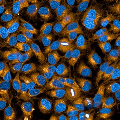
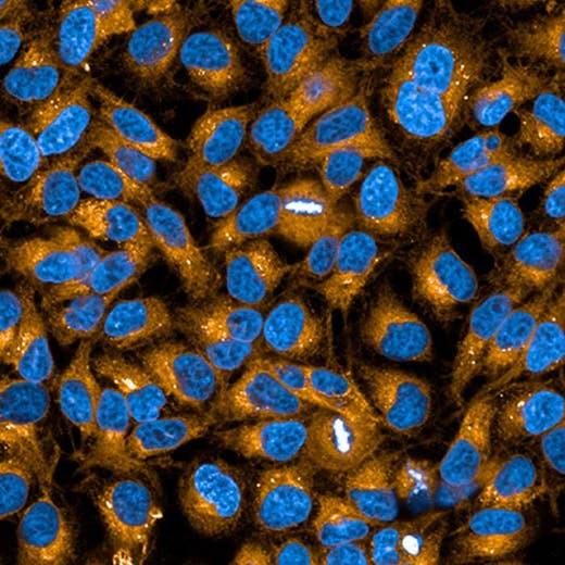
PhenoVue Fluor 555 - Concanavalin A is a fluorescent lectin which displays high affinity for glycoproteins and glycolipids present at the cellular membranes. It can be used for cellular membrane staining, particularly the endoplasmic reticulum. PhenoVue Fluor 555 - Concanavalin A exhibits bright green fluorescence and is validated for use in imaging microscopy and high-content screening applications. Part of Revvity's portfolio of cellular imaging reagents, PhenoVue Fluor 555 - Concanavalin A has a maximum excitation wavelength of 555 nm and a maximum emission wavelength of 570 nm.
View our extensive validation data in the Product Information Sheet within the Resources tab below.
| Feature | Specification |
|---|---|
| Color | Yellow |
| Filter | Cy3 |
| Fluorophore | PhenoVue™ Fluor 555 |
| Organelle and Cell Compartment |
Endoplasmic Reticulum Plasma membranes |
PhenoVue Fluor 555 - Concanavalin A is a fluorescent lectin which displays high affinity for glycoproteins and glycolipids present at the cellular membranes. It can be used for cellular membrane staining, particularly the endoplasmic reticulum. PhenoVue Fluor 555 - Concanavalin A exhibits bright green fluorescence and is validated for use in imaging microscopy and high-content screening applications. Part of Revvity's portfolio of cellular imaging reagents, PhenoVue Fluor 555 - Concanavalin A has a maximum excitation wavelength of 555 nm and a maximum emission wavelength of 570 nm.
View our extensive validation data in the Product Information Sheet within the Resources tab below.
Product variant
Quantity: 5 x 1 mg
Part #:
CP95551
List price
USD 393.00
Your online price:
For research use only. Not for use in diagnostic procedures.
PhenoVue Fluor 555 - Concanavalin A
PhenoVue Fluor 555 - Concanavalin A
Loading...
Product information
Overview
Concanavalin A is a plant homotetrameric lectin known to activate the immune system or induce apoptosis and autophagy. Concanavalin A displays high affinity for α-mannopyranosyl and α-glucopyranosyl residues of glycoproteins and glycolipids present at the cellular membranes. Fluorescent Concanavalin A derivatives are commonly used for staining the cellular membranes of mammalian cells, particularly the endoplasmic reticulum. PhenoVue Fluor 555 - Concanavalin A can be used to visualize cellular membranes in immunofluorescence, immunohistochemistry and flow cytometry, as well as high-content analysis and screening applications.
Specifications
| Color |
Yellow
|
|---|---|
| Form |
Lyophilized
|
| Maximum Emission Wavelength (Emmax) |
570 nm
|
| Maximum Excitation Wavelength (Exmax) |
555 nm
|
| Application |
High Content Imaging
Microscopy
|
|---|---|
| Brand |
PhenoVue™
|
| Detection Modality |
Fluorescence
|
| Filter |
Cy3
|
| Fluorophore |
PhenoVue™ Fluor 555
|
| Organelle and Cell Compartment |
Endoplasmic Reticulum
Plasma membranes
|
| Quantity |
5 x 1 mg
|
| Sample Type |
Live and fixed samples
|
| Shipping Conditions |
Shipped Ambient
|
| Storage Conditions |
2-8 °C, protected from light
|
| Type |
Individual reagent
|
Spectra viewer
Resources
Are you looking for resources, click on the resource type to explore further.
Loading...


How can we help you?
We are here to answer your questions.

