US
Revvity Sites Globally
Select your location.
*e-commerce not available for this region.


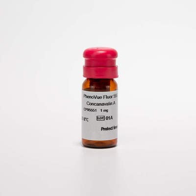
PhenoVue Fluor 555 - Concanavalin A
PhenoVue Fluor 555 - Concanavalin A

PhenoVue Fluor 555 - Concanavalin A
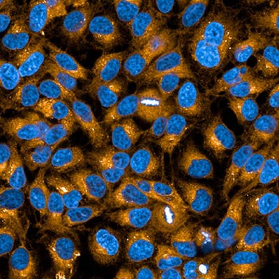
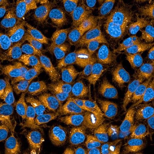
PhenoVue Fluor 555 - Concanavalin A is a fluorescent lectin which displays high affinity for glycoproteins and glycolipids present at the cellular membranes. It can be used for cellular membrane staining, particularly the endoplasmic reticulum. PhenoVue Fluor 555 - Concanavalin A exhibits bright green fluorescence and is validated for use in imaging microscopy and high-content screening applications. Part of Revvity's portfolio of cellular imaging reagents, PhenoVue Fluor 555 - Concanavalin A has a maximum excitation wavelength of 555 nm and a maximum emission wavelength of 570 nm.
View our extensive validation data in the Product Information Sheet within the Resources tab below.
| Feature | Specification |
|---|---|
| Color | Yellow |
| Filter | Cy3 |
| Fluorophore | PhenoVue™ Fluor 555 |
| Organelle and Cell Compartment |
Endoplasmic Reticulum Plasma membranes |
PhenoVue Fluor 555 - Concanavalin A is a fluorescent lectin which displays high affinity for glycoproteins and glycolipids present at the cellular membranes. It can be used for cellular membrane staining, particularly the endoplasmic reticulum. PhenoVue Fluor 555 - Concanavalin A exhibits bright green fluorescence and is validated for use in imaging microscopy and high-content screening applications. Part of Revvity's portfolio of cellular imaging reagents, PhenoVue Fluor 555 - Concanavalin A has a maximum excitation wavelength of 555 nm and a maximum emission wavelength of 570 nm.
View our extensive validation data in the Product Information Sheet within the Resources tab below.
Product variant
Quantity: 5 x 1 mg
Part #:
CP95551
List price
USD 393.00
Your online price:
For research use only. Not for use in diagnostic procedures.
PhenoVue Fluor 555 - Concanavalin A
PhenoVue Fluor 555 - Concanavalin A
Loading...
Product information
Overview
Concanavalin A is a plant homotetrameric lectin known to activate the immune system or induce apoptosis and autophagy. Concanavalin A displays high affinity for α-mannopyranosyl and α-glucopyranosyl residues of glycoproteins and glycolipids present at the cellular membranes. Fluorescent Concanavalin A derivatives are commonly used for staining the cellular membranes of mammalian cells, particularly the endoplasmic reticulum. PhenoVue Fluor 555 - Concanavalin A can be used to visualize cellular membranes in immunofluorescence, immunohistochemistry and flow cytometry, as well as high-content analysis and screening applications.
Specifications
| Color |
Yellow
|
|---|---|
| Form |
Lyophilized
|
| Maximum Emission Wavelength (Emmax) |
570 nm
|
| Maximum Excitation Wavelength (Exmax) |
555 nm
|
| Application |
High Content Imaging
Microscopy
|
|---|---|
| Brand |
PhenoVue™
|
| Detection Modality |
Fluorescence
|
| Filter |
Cy3
|
| Fluorophore |
PhenoVue™ Fluor 555
|
| Organelle and Cell Compartment |
Endoplasmic Reticulum
Plasma membranes
|
| Quantity |
5 x 1 mg
|
| Sample Type |
Live and fixed samples
|
| Shipping Conditions |
Shipped Ambient
|
| Storage Conditions |
2-8 °C, protected from light
|
| Type |
Individual reagent
|
Spectra viewer
Resources
Are you looking for resources, click on the resource type to explore further.
Flyer
PhenoVue Cellular Imaging Reagents Flyer
This flyer describes Revvity's PhenoVue cellular imaging reagents.
Product Info
PhenoVue Fluor - Concanavalin A Conjugates Product Information Sheet
Fluorescent Concanavalin A conjugates represent a method of choice for labelling the cellular membranes of mammalian cells...
Flyer
PhenoVue reagents & imaging microplates product list
PhenoVue reagents & imaging microplates product list
Loading...


How can we help you?
We are here to answer your questions.

