US
Revvity Sites Globally
Select your location.
*e-commerce not available for this region.


PhenoVue Hoechst 33342 Nuclear Stain 100mg
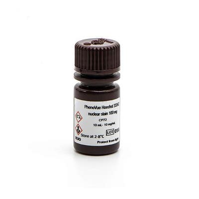
PhenoVue Hoechst 33342 Nuclear Stain 100mg


PhenoVue Hoechst 33342 Nuclear Stain 100mg
PhenoVue Hoechst 33342 nuclear stain is a cell-permeable organic molecule that binds to DNA.
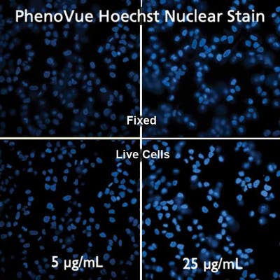
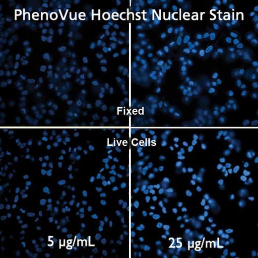
PhenoVue Hoechst 33342 nuclear stain exhibits bright blue fluorescence and is validated for use in imaging microscopy and high-content screening applications.
Part of Revvity's portfolio of cellular imaging reagents, the PhenoVue Hoechst 33342 nuclear stain has a maximum excitation wavelength of 357 nm and a maximum emission wavelength of 455 nm.
View our extensive validation data in the Product Information Sheet within the Resources tab below.
| Feature | Specification |
|---|---|
| Color | Blue |
| Filter | DAPI |
| Organelle and Cell Compartment | Nucleus |
PhenoVue Hoechst 33342 nuclear stain is a cell-permeable organic molecule that binds to DNA.
PhenoVue Hoechst 33342 nuclear stain exhibits bright blue fluorescence and is validated for use in imaging microscopy and high-content screening applications.
Part of Revvity's portfolio of cellular imaging reagents, the PhenoVue Hoechst 33342 nuclear stain has a maximum excitation wavelength of 357 nm and a maximum emission wavelength of 455 nm.
View our extensive validation data in the Product Information Sheet within the Resources tab below.
Product variants
Quantity: 1 x 10 mg
Part #:
CP71
List price
USD 61.00
Your online price:
Quantity: 1 x 100 mg
Part #:
CP72
List price
USD 118.00
Your online price:
For research use only. Not for use in diagnostic procedures.
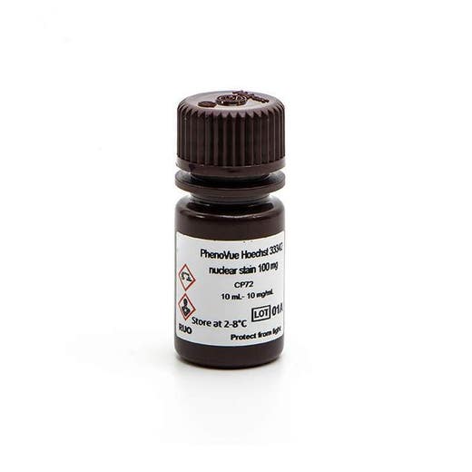
PhenoVue Hoechst 33342 Nuclear Stain 100mg
PhenoVue Hoechst 33342 Nuclear Stain 100mg
PhenoVue Hoechst 33342 Nuclear Stain 100mg
Loading...
Product information
Overview
PhenoVue Hoechst 33342 Nuclear stain (trihydrochloride form) is a cell-permeable organic molecule that binds preferentially to adenine-thymine (A-T) regions of the minor groove of DNA. When excited by ultraviolet light, Hoechst 33342 nuclear stain exhibits a strong fluorescence at 455 nm. It is commonly used to stain cells' nuclear compartment.
PhenoVue Hoechst 33342 Nuclear stain can be used to visualize cellular membranes in immunofluorescence, immunohistochemistry and flow cytometry, as well as high-content analysis and screening applications.
Typical working concentration: 2 µg/mL (3.24 µM)
Equivalent number of microplates:
- 1740 - 5200 x 96-well microplates
- 1450 - 5200 x 384-well microplates
- 2700 - 8140 x 1536-well microplates
See product information sheet for more information on fixed cell staining.
Specifications
| Color |
Blue
|
|---|---|
| Concentration |
10 mg/mL (16.23 mM)
|
| Form |
Solution in dd H2O
|
| Maximum Emission Wavelength (Emmax) |
455 nm
|
| Maximum Excitation Wavelength (Exmax) |
357 nm
|
| Application |
High Content Imaging
Microscopy
|
|---|---|
| Brand |
PhenoVue™
|
| Detection Modality |
Fluorescence
|
| Filter |
DAPI
|
| Organelle and Cell Compartment |
Nucleus
|
| Quantity |
1 x 100 mg
|
| Sample Type |
Live and fixed samples
|
| Shipping Conditions |
Shipped in Dry Ice
|
| Storage Conditions |
2-8 °C for short term (6 months) or -16 °C or below for long term (> 6 months), protected from light
|
| Type |
Individual reagent
|
Spectra viewer
Resources
Are you looking for resources, click on the resource type to explore further.


How can we help you?
We are here to answer your questions.

