
The challenge of interpreting negative liquid biopsy results
Plasma-based liquid biopsy offers a minimally invasive way to investigate tumor-associated genetic alterations by analyzing circulating DNA in blood. Blood plasma contains cell-free DNA (cfDNA) released from both healthy and tumor cells. The tumor-derived component, known as circulating tumor DNA (ctDNA), can carry genetic alterations such as single-nucleotide variants, insertions, deletions, and copy-number changes.
However, the amount of ctDNA present in plasma can vary considerably depending on tumor type, disease stage, tumor burden, and other biological factors. In some samples, tumor-derived DNA represents only a small fraction of the total circulating DNA.
Key highlights:
- A negative liquid biopsy result does not rule out mutations. Low tumor DNA levels may limit detection.
- Tumor fraction estimation adds context. sWGS and ichorCNA estimate tumor-derived DNA without requiring prior knowledge of mutations.
- 90% of mutation-negative samples had tumor fractions below 3%. This highlights the value of tumor fraction analysis as a complement to mutation testing.
This raises an important question when no mutation is detected: Is the mutation truly absent, or is there too little tumor-derived DNA in the sample to detect it?
The sensitivity of mutation detection depends on several factors, including variant allele frequency, DNA input, sequencing depth, and assay performance. A negative result therefore does not necessarily exclude the presence of tumor-associated mutations. Recent expert consensus recommendations emphasize the importance of recognizing these limitations when interpreting ctDNA findings.1
One way to provide additional context is to estimate tumor fraction, the proportion of cfDNA originating from tumor cells. Shallow whole-genome sequencing (sWGS), combined with the bioinformatics tool ichorCNA, can estimate tumor fraction from genome-wide copy-number patterns without requiring prior knowledge of specific tumor mutations. Originally described by Adalsteinsson and colleagues, this approach provides information complementary to mutation analysis.2
Investigating tumor fraction
Researchers at the Institut de Pathologie et de Génétique in Belgium evaluated whether tumor fraction estimation could help interpret mutation-negative liquid biopsy results. Their findings were presented at the EACR and ESMO conferences in 2026.3
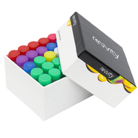
The study combined sWGS at approximately 0.4× genome coverage with ichorCNA analysis to estimate tumor fraction in plasma cfDNA. Libraries were prepared using the NEXTFLEX™ Cell Free DNA-Seq Library Prep Kit 2.0
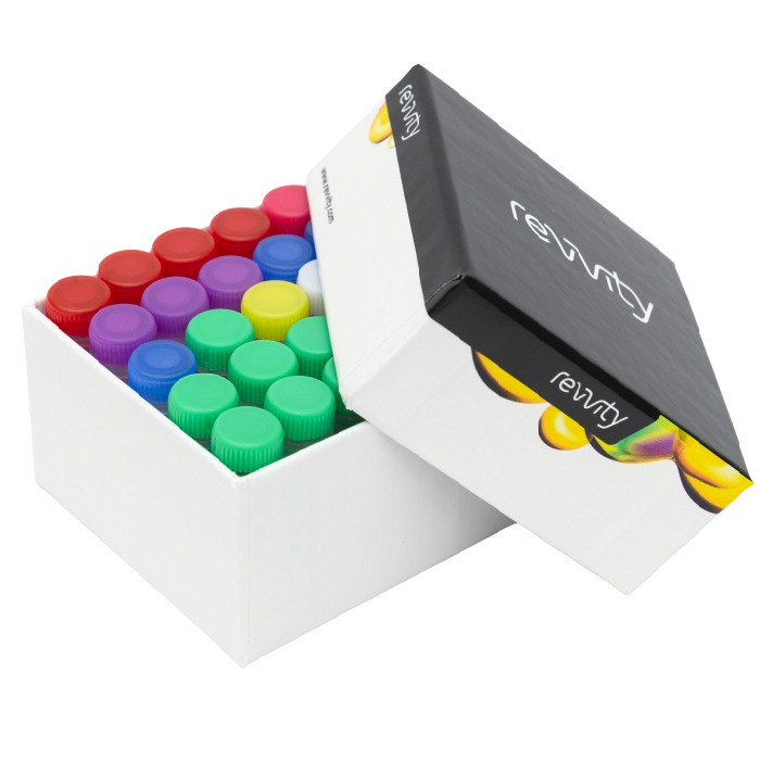 NEXTFLEX Cell Free DNA-Seq Library Prep Kit 2.0
from Revvity. The researchers assessed analytical performance using 37 negative reference samples and 20 clinical samples before applying the workflow to 55 routine samples.3
The study reported a limit of blank of 2.1% and a limit of detection of 3.0% for tumor fraction estimation. Median coefficients of variation were 13.9% for repeatability and 5.9% for inter-run reproducibility, reflecting the precision of the complete workflow under the study conditions.
Comparing tumor fraction estimates with mutation results from 55 routine samples revealed the following:
- 24 samples were mutation-positive: 19 had estimated tumor fractions above 3%.
- 31 samples were mutation-negative: 28 had estimated tumor fractions below 3%.
Thus, approximately 90% of mutation-negative samples had tumor fraction estimates below the workflow's detection limit. These results were considered non-informative rather than true negatives, although they do not establish that tumor DNA was absent or that mutations were missed.
Importantly, five mutation-positive samples also had tumor fraction estimates that did not exceed 3%. This shows why tumor fraction should complement, rather than replace, mutation testing. The two approaches measure different features: mutation assays detect specific sequence variants, whereas ichorCNA estimates tumor fraction from genome-wide copy-number patterns. The 3% detection limit therefore applies only to the tumor fraction workflow.
Tumor fraction estimates were generally higher than variant allele frequencies, although the relationship varied between samples. Tumor subclonality, copy-number alterations, and loss of heterozygosity may contribute to these differences.3
Together, these findings show how tumor fraction estimation can add context to mutation-negative results without being used to exclude samples from variant testing.
Conclusions: adding context to mutation-negative results
A negative liquid biopsy result does not necessarily mean that tumor-associated mutations are absent. When tumor-derived DNA is present at very low levels, mutations may go undetected, making it important to consider tumor fraction alongside mutation findings.
The research presented at EACR and ESMO demonstrates how sWGS and ichorCNA can provide valuable context for interpreting mutation-negative samples. Importantly, tumor fraction estimation should complement rather than replace mutation testing, as the two approaches have different analytical sensitivities and limitations.
References:
- de Jager VD, Giacomini P, Fairley JA, et al. Reporting of molecular test results from cell-free DNA analyses: expert consensus recommendations from the 2023 European Liquid Biopsy Society ctDNA Workshop. eBioMedicine. 2025;114:105636. https://doi.org/10.1016/j.ebiom.2025.105636
- Adalsteinsson VA, Ha G, Freeman SS, et al. Scalable whole-exome sequencing of cell-free DNA reveals high concordance with metastatic tumors. Nature Communications. 2017;8:1324. https://doi.org/10.1038/s41467-017-00965-y
- de Barsy M, Brohée S, De Rop C, Vannuffel P. Improving liquid biopsy informative value through robust tumor fraction estimation in non-invasive somatic mutation detection. Institut de Pathologie et de Génétique (IPG), Gosselies, Belgium. Research posters presented at EACR and ESMO, 2026.
Acknowledgment: We thank the Institut de Pathologie et de Génétique for granting permission to discuss their research findings.
For research use only. Not for use in diagnostic procedures.