US
Revvity Sites Globally
Select your location.
*e-commerce not available for this region.


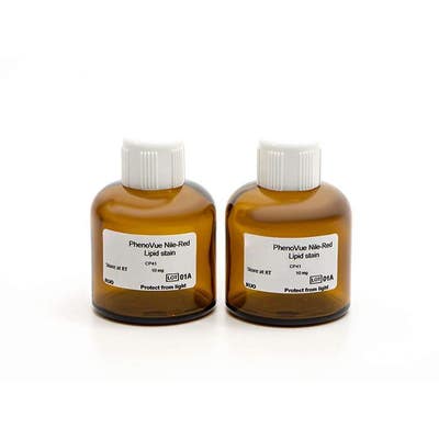
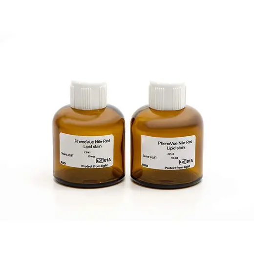
PhenoVue Nile Red Lipid Stain
 View All
PhenoVue Nile Red Lipid Stain

PhenoVue Nile Red Lipid Stain


| Feature | Specification |
|---|---|
| Color | Yellow/Orange |
| Filter | Cy3 |
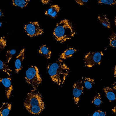
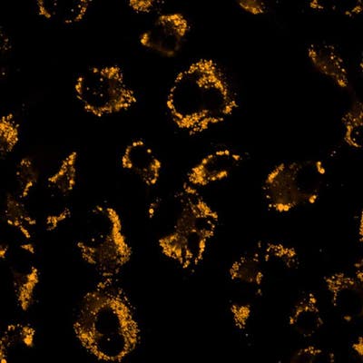
| Organelle and Cell Compartment | Lipid droplets |
Product variant
Quantity: 2 x 10 mg
Part #:
CP41
List price
USD 180.15
Your online price:
For research use only. Not for use in diagnostic procedures.
PhenoVue Nile Red Lipid Stain
PhenoVue Nile Red Lipid Stain
PhenoVue Nile Red Lipid Stain
Loading...
Product information
Overview
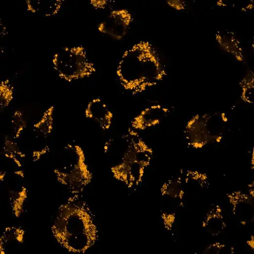
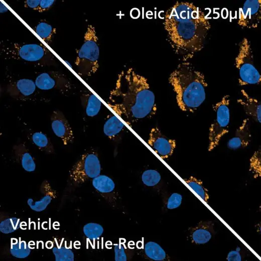
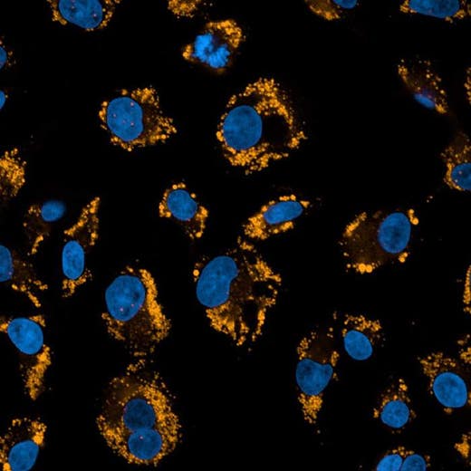
PhenoVue Nile Red Lipid stain is a lipophilic organic molecule that is almost nonfluorescent in water and polar solvents. In lipid-rich environments, Nile Red exhibits enhanced yellow fluorescence, as well as red fluorescence to a lesser extent.
Nile Red is commonly used for localization and quantification of intracellular lipid droplets which are involved in lipid synthesis, metabolism and transportation as well as protein storage and degradation or viral replication, all of which are related to pathophysiology, including dyslipidemia, obesity, lipodystrophy, diabetes, fatty liver diseases, or atherosclerosis.
Typical working concentration: 200 nM (0.008 ng/mL)
Equivalent number of microplates:
- 10,900 - 32,700 x 96-well plates
- 9,090 - 32,700 x 384-well plates
- 17,040 - 40,900 x 1536-well plates
See Product Information Sheet for more information on fixed cell staining.
Specifications
| Color |
Yellow/Orange
|
|---|---|
| Form |
Powder
|
| Maximum Emission Wavelength (Emmax) |
Triglycerides: 585 nm, phospholipids: 638 nm
|
| Maximum Excitation Wavelength (Exmax) |
Triglycerides: 512 nm, phospholipids: 552 nm
|
| Application |
High Content Imaging
Microscopy
|
|---|---|
| Brand |
PhenoVue™
|
| Detection Modality |
Fluorescence
|
| Filter |
Cy3
|
| Organelle and Cell Compartment |
Lipid droplets
|
| Quantity |
2 x 10 mg
|
| Sample Type |
Live and fixed samples
|
| Shipping Conditions |
Shipped Ambient
|
| Storage Conditions |
Room temperature or 2-8 °C, protected from light
|
| Type |
Individual reagent
|
Image gallery
PhenoVue Nile Red Lipid Stain
PhenoVue Nile Red Lipid Stain
Spectra viewer
Resources
Are you looking for resources, click on the resource type to explore further.


How can we help you?
We are here to answer your questions.

