Revvity Sites Globally
Select your location.
*e-commerce not available for this region.



PhenoVue DNA Damage and Apoptosis Staining Kit - 1 x 384 (gH2AX, cleaved PARP)
PhenoVue DNA Damage and Apoptosis Staining Kit - 1 x 384 (gH2AX, cleaved PARP)
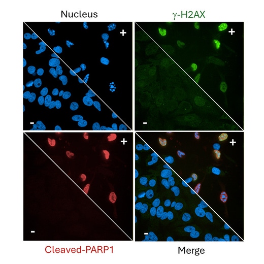
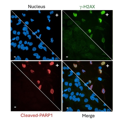
Staurosporine induces both γ-H2AX and cleaved PARP1 in HeLa cells. Untreated cells (−) display minimal staining, whereas treated cells (+) show strong induction of both markers.
The PhenoVue™ DNA Damage and Apoptosis Staining Kit provides a convenient solution for imaging DNA damage and apoptosis-associated markers in fixed cells. The kit combines PhenoVue™ Hoechst 33342 Nuclear Stain with antibodies targeting γ-H2AX and cleaved PARP1, enabling visualization of nuclear morphology together with DNA damage and apoptotic marker staining. With fluorescent labeling and compatibility with high-content imaging, the kit supports the characterization of compounds or conditions that induce DNA damage, activate DDR pathways, and/or trigger apoptosis-associated PARP1 cleavage.
The PhenoVue™ DNA Damage and Apoptosis Staining Kit provides a convenient solution for imaging DNA damage and apoptosis-associated markers in fixed cells. The kit combines PhenoVue™ Hoechst 33342 Nuclear Stain with antibodies targeting γ-H2AX and cleaved PARP1, enabling visualization of nuclear morphology together with DNA damage and apoptotic marker staining. With fluorescent labeling and compatibility with high-content imaging, the kit supports the characterization of compounds or conditions that induce DNA damage, activate DDR pathways, and/or trigger apoptosis-associated PARP1 cleavage.
Product variant
Quantity: 1 Kit
Part #:
PDDAP11
List price
USD 600.00
Your price:
For research use only. Not for use in diagnostic procedures.
PhenoVue DNA Damage and Apoptosis Staining Kit - 1 x 384 (gH2AX, cleaved PARP)
Staurosporine induces both γ-H2AX and cleaved PARP1 in HeLa cells. Untreated cells (−) display minimal staining, whereas treated cells (+) show strong induction of both markers.
Loading...
Product information
Overview
DNA is continuously exposed to insults that can result in strand breaks, replication stress, or chemical modifications. If left unrepaired, these lesions can compromise genome integrity and may lead to genomic instability, senescence, or cell death. Cells counteract these threats by activating the DNA Damage Response (DDR), a signaling network that detects lesions, promotes repair, and contributes to cell fate decisions.
Phosphorylation of histone H2AX at serine 139 (γ-H2AX) is a widely used marker of DNA double-strand breaks, while cleaved PARP1 serves as a marker of apoptotic pathway activation. Combined analysis of both markers provides complementary information on DNA damage signaling and apoptosis within fixed-cell imaging workflows.
The PhenoVue DNA Damage and Apoptosis Staining Kit combines PhenoVue Hoechst 33342 Nuclear Stain with antibodies targeting γ-H2AX and cleaved PARP1, compatible with high-content imaging systems for characterization of compounds or conditions that induce DNA damage or trigger apoptosis.
For enhanced flexibility, up to two additional biological markers can be included using the PhenoVue Fluor 555 and 400LS channels, allowing for deeper insights into DNA damage response (DDR) mechanisms.
Kit components and quantity:
- PhenoVue Hoechst 33342 Nuclear Stain, 1 vial of 70 µL (500X)
- PhenoVue anti-Phospho H2AX (S139) – mouse IgG1 Antibody (100X), 1 vial of 100 µL (100X)
- PhenoVue anti-cleaved PARP1 Antibody (Rabbit), 1 vial of 100 µL (100X)
- PhenoVue Fluor 488 - Goat anti-Mouse antibody Highly Cross-Adsorbed (100X), 1 vial of 100 µL (100X)
- PhenoVue Fluor 647 - Goat anti-Rabbit antibody Highly Cross-Adsorbed (100X) , 1 vial of 100 µL (100X)
- PhenoVue Paraformaldehyde, 4% Solution, 1 vial of 25 mL (1X)
- PhenoVue Permeabilization, 0.5% Triton X-100 Solution, 1 vial of 25 mL (1X)
- PhenoVue dye diluent A (5X), 1 vial of 8 mL (5X)
Specifications
| Application |
High Content Imaging
Microscopy
|
|---|---|
| Assay Points |
1 x 384-well microplate
|
| Brand |
PhenoVue™
|
| Product Group |
Kit
|
| Quantity |
1 Kit
|
| Sample Type |
Fixed cells only
|
| Shipping Conditions |
Shipped in Dry Ice
|
| Storage Conditions |
-16°C or below
|
Spectra viewer
Resources
Are you looking for resources, click on the resource type to explore further.

Product Note
PhenoVue DNA damage and apoptosis staining kit product information sheet
PhenoVueTM DNA Damage and Apoptosis Staining Kit (1×384-well) enables simultaneous detection of γ-H2AX and cleaved PARP1 in fixed...
Loading...


How can we help you?
We are here to answer your questions.