
HTRF Human Phospho-STAT5 (Tyr694) Detection Kit, 500 Assay Points


| Feature | Specification |
|---|---|
| Application | Cell Signaling |
| Sample Volume | 16 µL |


Product information
Overview
The Phospho-STAT5 (Tyr694) kit is designed to robustly and efficiently monitor STAT5 phosphorylation on Tyr694, as a readout of JAK/STAT pathway activation. With an optimized and streamlined mix-and-read, no-wash protocol, the phospho-STAT5 contains all reagents you need for your research to run faster and more efficiently.
How it works
Phospho-STAT5 (Tyr694) assay principle
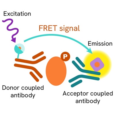
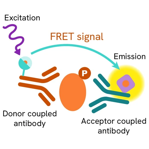
The Phospho-STAT5 (Tyr694) assay measures STAT5 when phosphorylated at Tyr694. Contrary to Western Blot, the assay is entirely plate-based and does not require gels, electrophoresis or transfer. The Phospho-STAT5 (Tyr694) assay uses 2 labeled antibodies: one with a donor fluorophore, the other one with an acceptor. The first antibody is selected for its specific binding to the phosphorylated motif on the protein, the second for its ability to recognize the protein independent of its phosphorylation state. Protein phosphorylation enables an immune-complex formation involving both labeled antibodies and which brings the donor fluorophore into close proximity to the acceptor, thereby generating a FRET signal. Its intensity is directly proportional to the concentration of phosphorylated protein present in the sample, and provides a means of assessing the proteins phosphorylation state under a no-wash assay format.

Phospho-STAT5 (Tyr694) 2-plate assay protocol
The 2 plate protocol involves culturing cells in a 96-well plate before lysis then transferring lysates to a 384-well low volume detection plate before adding phospho-STAT5 (Tyr694) HTRF detection reagents. This protocol enables the cells' viability and confluence to be monitored.

Phospho-STAT5 (Tyr694) 1-plate assay protocol
Detection of Phosphorylated STAT5 (Tyr694) with HTRF reagents can be performed in a single plate used for culturing, stimulation and lysis. No washing steps are required. This HTS designed protocol enables miniaturization while maintaining robust HTRF quality.

Assay validation
HTRF compared to western blot using phospho-STAT5 (Tyr694) assay kit
TF1 cells were grown in a T175 flask at 37°C until concentration reached 1.5M/ml of cell culture medium, then stimulated with IL3 for 10min. Cells were incubated for 30 minutess for lysis. Serial dilutions (1:2) were analyzed in parallel by HTRF and Western-blot. Note that 128KC corresponds roughly to 32µg of proteins. By using HTRF phospho-STAT5 (Tyr694) only 2000 cells are sufficient for minimal signal detection while 8000 cells are needed for a Western blot signal. The HTRF assay is at least 4-fold more sensitive than the Western blot and shows optimal correlation.


HTRF assay compared to ELISA using phospho-STAT5 (Tyr694) assay kit
TF1 cells were cultured and treated with increasing concentrations of a JAK inhibitor and stimulated with IL3 for 10min. Following lysis, serial dilutions of collected supernatant were analyzed in parallel by HTRF and ELISA. The results displayed in the graph show that for measuring STAT5 phosphorylation HTRF is more sensitive than ELISA, offers a bigger assay window and is more reproducible. Despite a good correlation between ELISA and HTRF, the overall performance of HTRF is more favorable.

Pharmacological response in TF1 cells using a two plate assay protocol
TF1 cells were plated in 25µl of cell culture medium treated for 30min with 5µl of increasing concentrations of the JAK inhibitor Lestaurtinib. After removal of cell culture medium, 10µl of 4X supplemented lysis buffer were added and incubated for 30 minutes prior to the addition of the HTRF detection reagents. 16µl of lysates were transferred in a 384sv white plate for analysis of STAT5 phosphorylation. The results clearly demonstrate, that the high sensitivity of the assay allows a 4-fold lower cell quantity to be used without a significant decrease of assay window.

Pharmacological response in HeLa cells using a two plate assay protocol
100KC HeLa cells were plated in 25µl of cell culture medium treated for 2h with 5µl of increasing concentrations of the JAK inhibitor Lestaurtinib and then stimulated with 20 nM IFN for 10min. After removal of cell culture medium, 10µl of 4X supplemented lysis buffer were added and incubated for 30 minutes prior to the addition of the HTRF detection reagents. 16µl of lysates were transferred in a 384sv white plate for analysis of STAT5 phosphorylation.

Simplified pathway
STAT5 cell signaling
The activation of Stat5 proteins,is widely mediated by cytokines, and other growth factors, allowing rapid delivery of signals from the membrane to the nucleus. These STATs regulate the expression of target genes in a cytokine-specific fashion. In addition to their activation by cytokines, the activities of Stat5a and Stat5b are negatively controlled by CIS/SOCS/SSI proteins. JAK/STAT pathway activation requires ligand binding to the receptor, which in consequence activates the Janus kinases JAKs, a family of intracellular tyrosine kinases. Via phosphorylation of downstream signal transducers and activators of transcription (STAT), STAT5 forms homo or heterodimers in order to become active and translocate into the nucleus, where it binds to STAT5 response elements, inducing the transcription of specific sets of genes.

Specifications
| Application |
Cell Signaling
|
|---|---|
| Brand |
HTRF
|
| Detection Modality |
HTRF
|
| Molecular Modification |
Phosphorylation
|
| Product Group |
Kit
|
| Sample Volume |
16 µL
|
| Shipping Conditions |
Shipped in Dry Ice
|
| Target Class |
Phosphoproteins
|
| Target Species |
Human
|
| Technology |
TR-FRET
|
| Therapeutic Area |
Infectious Diseases
Neuroscience
Oncology & Inflammation
|
| Unit Size |
500 assay points
|
Video gallery
Resources
Are you looking for resources, click on the resource type to explore further.
Atherosclerosis pathogenesis, cellular actors, and pathways
Atherosclerosis is a common condition in which arteries harden and...
Discover the versatility and precision of Homogeneous Time-Resolved Fluorescence (HTRF) technology. Our HTRF portfolio offers a...
This guide provides you an overview of HTRF applications in several therapeutic areas.
Advance your autoimmune disease research and benefit from Revvity broad offering of reagent technologies


Loading...
How can we help you?
We are here to answer your questions.


