
HTRF Prostaglandin E2 Detection Kit, 500 Assay Points


The HTRF Prostaglandin E2 Detection Kit is an immunoassay designed for the detection and quantitation of Prostaglandin E2 in cell supernatants, in a homogeneous (no-wash steps, no separation steps) format.
| Feature | Specification |
|---|---|
| Application | Protein Quantification |
| Dynamic Range | 8 - 1,000 pg/mL |
| Limit of Detection | 10 pg/mL |
| Sample Volume | 10 µL |
The HTRF Prostaglandin E2 Detection Kit is an immunoassay designed for the detection and quantitation of Prostaglandin E2 in cell supernatants, in a homogeneous (no-wash steps, no separation steps) format.


Product information
Overview
Prostaglandin E2 (PGE2) is produced in multiple cell types from PGH2, a primary product of the arachidonic acid metabolism, via the prostaglandin synthase. PGE2 displays several biological functions, such as vasodilatation and smooth muscle relaxation, and is involved in pro- and anti-inflammation pathways. PGE2 production is a commonly used method for the detection of COX-1 and COX-2 modulation. The PGE2 kit is a highly sensitive method for quantifying PGE2, either in cell supernatant or directly in whole cells.
HTRF assays offer many advantages over other technologies:
- Homogeneous add-and-read format
- No wash steps
- Low background
- Straightforward miniaturization from 96- or 384-well microplates to high density assay formats such as 384-well low volume and 1536-well plates
- Stable signal, providing flexibility in time of readout or size of assays
How it works
Assay principle
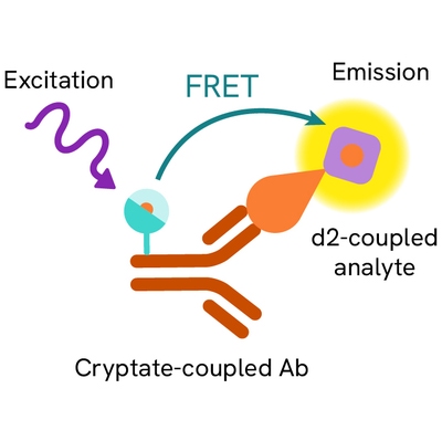
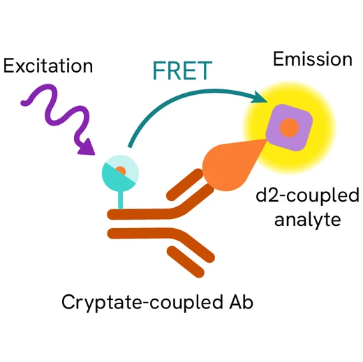
The PGE2 assay is based on the competition principle, where native PG2 produced by cells and d2-labelled PGE2 compete for binding to a monoclonal anti-PGE2 antibody labelled with Europium Cryptate. The assay can be run with several types of samples, such as cell supernatants or purified enzymes.

Assay Protocol
The PGE2 assay features a streamlined protocol with only 1 incubation step after sample and PGE2 detection reagents dispensing. This protocol requires a single 5-hour incubation period at RT.

Assay details
Key features
The PGE2 assay has a high level of flexibility. It can be performed using cell supernatants or directly on stimulated cells. The incubation time and temperature following addition of the detection reagents have little effect on the assay results, providing further assay flexibility.
| Detection limit | 10 pg/mL |
|---|---|
| Dynamic range | 8 to 1000 pg/mL |
| EC50 | 250 pg/mL |
| S/B | 31 |
| Z' | 0.9 for 20 µl assay volumes |

Assay validation
PGE2 modulation upon Indomethacin inhibitor treatment
One benefit of the HTRF PGE2 assay is its ability to quantify PGE2 in the presence of cells without sacrificing assay performance. The results comparing direct cell-based versus supernatant transfer did not differ significantly. Monocytes were stimulated to produce PGE2 in the presence or absence of indomethacin, a known inhibitor of PGE2 production. The IC50 of indomethacin determined using HTRF technology (1.0 ± 0.4 nM) was in agreement with previously published data.

Specifications
| Application |
Protein Quantification
|
|---|---|
| Brand |
HTRF
|
| Detection Modality |
HTRF
|
| Dynamic Range |
8 - 1,000 pg/mL
|
| Limit of Detection |
10 pg/mL
|
| Product Group |
Kit
|
| Sample Volume |
10 µL
|
| Shipping Conditions |
Shipped Ambient
|
| Target Class |
Biomarkers
|
| Technology |
TR-FRET
|
| Therapeutic Area |
Cardiovascular
Neuroscience
Oncology & Inflammation
|
| Unit Size |
500 assay points
|
Video gallery
Citations
Resources
Are you looking for resources, click on the resource type to explore further.
Fibrotic disorders are complex diseases believed to stem from uncontrolled and excessive wound healing processes. The exact causes...
Discover the versatility and precision of Homogeneous Time-Resolved Fluorescence (HTRF) technology. Our HTRF portfolio offers a...
This guide provides you an overview of HTRF applications in several therapeutic areas.


Loading...
How can we help you?
We are here to answer your questions.


