


US
Revvity Sites Globally
Select your location.
*e-commerce not available for this region.

HTRF Human and Mouse Phospho-p70 S6K (Thr389) Detection Kit, 10,000 Assay Points


HTRF Human and Mouse Phospho-p70 S6K (Thr389) Detection Kit, 10,000 Assay Points


generic HTRF phospho primary image


The phospho-P70S6K (Thr389) kit enables the cell-based quantitative detection of phosphorylated P70S6K at Thr389 as a readout of mTOR signaling.
| Feature | Specification |
|---|---|
| Application | Cell Signaling |
| Sample Volume | 16 µL |
The phospho-P70S6K (Thr389) kit enables the cell-based quantitative detection of phosphorylated P70S6K at Thr389 as a readout of mTOR signaling.
Product variants
Unit Size: 500 assay points
Part #:
64S6KPEG
List price
USD 2,340.00
Your online price:
Unit Size: 10,000 assay points
Part #:
64S6KPEH
List price
USD 13,611.00
Your online price:
For research use only. Not for use in diagnostic procedures. All products to be used in accordance with applicable laws and regulations including without limitation, consumption, and disposal requirements under European REACH regulations (EC 1907/2006).
HTRF Human and Mouse Phospho-p70 S6K (Thr389) Detection Kit, 10,000 Assay Points
generic HTRF phospho primary image
HTRF Human and Mouse Phospho-p70 S6K (Thr389) Detection Kit, 10,000 Assay Points
Product information
Overview
This HTRF cell based assay conveniently and accurately quantifies phosphorylated p70S6K at Thr389. Compatible with most cell types, this kit is the perfect tool for screening and studying compounds biologically impacting cell proliferation, survival, invasion, motility, and insulin receptor signaling. The kit has applications in oncology, metabolism (diabetes, obesity, aging), cardiovascular diseases, and neurodegenerative diseases.
HTRF assays offer many advantages over other technologies:
- Homogeneous add-and-read format
- No wash steps
- Low background
- Straightforward miniaturization from 96- or 384-well microplates to high density assay formats such as 384-well low volume and 1536-well plates
- Stable signal, providing flexibility in time of readout or size of assays
How it works
Phospho-p70S6K (Thr389) assay principle
The Phospho-p70S6K (Thr389) assay measures p70S6K when phosphorylated at Thr389. Contrary to Western Blot, the assay is entirely plate-based and does not require gels, electrophoresis or transfer. The Phospho-p70S6K (Thr389) assay uses 2 labeled antibodies: one with a donor fluorophore, the other one with an acceptor. The first antibody is selected for its specific binding to the phosphorylated motif on the protein, the second for its ability to recognize the protein independent of its phosphorylation state. Protein phosphorylation enables an immune-complex formation involving both labeled antibodies and which brings the donor fluorophore into close proximity to the acceptor, thereby generating a FRET signal. Its intensity is directly proportional to the concentration of phosphorylated protein present in the sample, and provides a means of assessing the proteins phosphorylation state under a no-wash assay format.
Phospho-p70S6K (Thr389) 2-plate assay protocol
The 2 plate protocol involves culturing cells in a 96-well plate before lysis then transferring lysates to a 384-well low volume detection plate before adding phospho-p70S6K (Thr389) HTRF detection reagents. This protocol enables the cells' viability and confluence to be monitored.

Phospho-p70S6K (Thr389) 1-plate assay protocol
Detection of Phosphorylated p70S6K (Thr389) with HTRF reagents can be performed in a single plate used for culturing, stimulation and lysis. No washing steps are required. This HTS designed protocol enables miniaturization while maintaining robust HTRF quality.

Assay validation
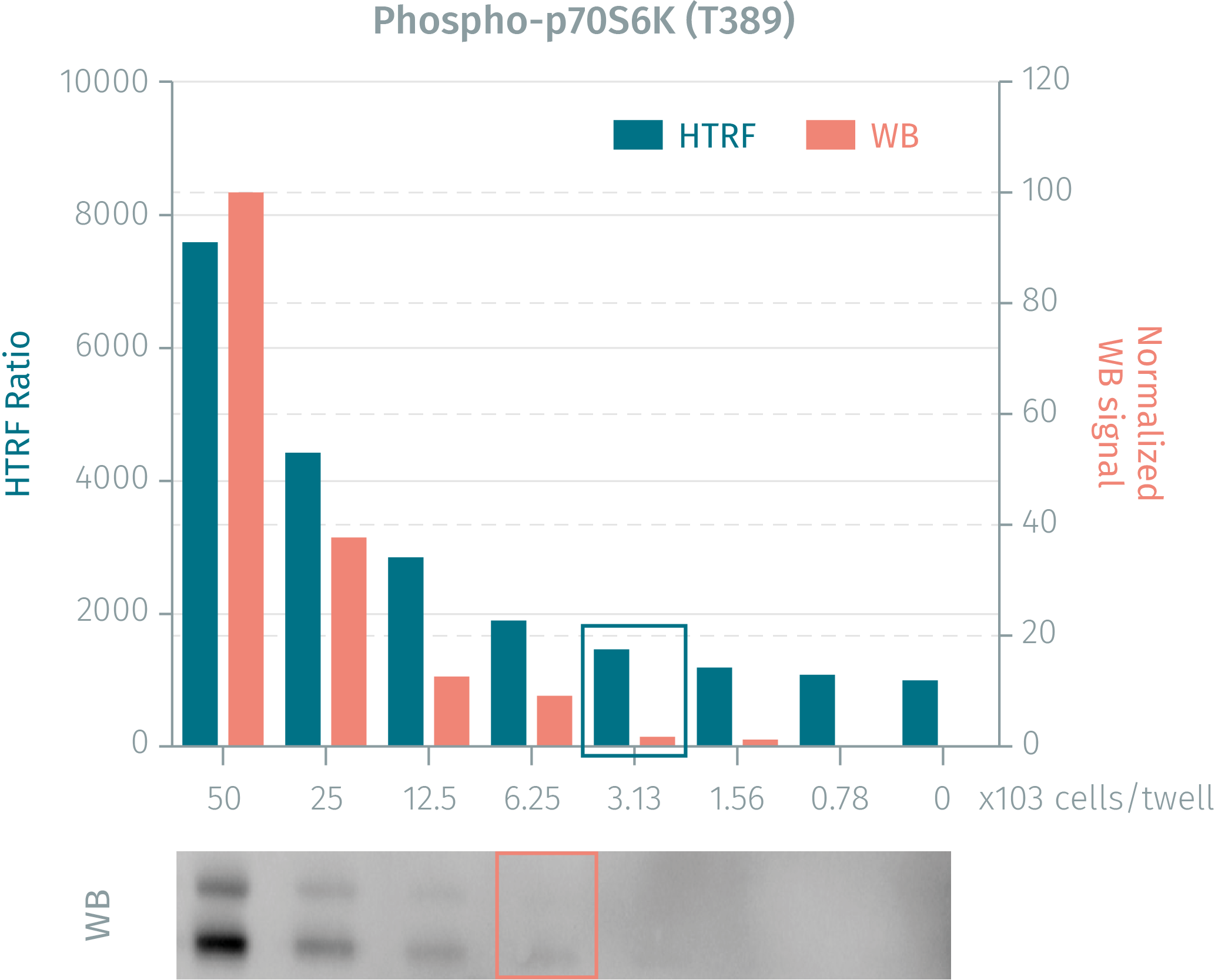
HTRF assay compared to WB using phospho-p70S6K cellular assay
Human HEK293 cells were grown in a T175 flask at 37°C, 5% CO2, for 1 day. At day2, after removal of cell culture medium, 3ml of supplemented lysis buffer were added and incubated for 30 minutes. Soluble supernatants were collected after a 10 minute centrifugation. Equal amounts of lysates were used for a side by side comparison of Western Blot and HTRF.3000 cells can be detected by using HTRF phospho-P70S6K (Thr389) whereas 6000 cells are needed for the Western Blot. The phospho-p70S6K HTRF assay is two-fold more sensitive than the WB.
Pharmacological response: agonist and antagonist action modes
Human HEK293 cells (100,000 cells/well) were incubated with two concentrations of the inhibitors indicated, Wortmanin and LY294002, followed by stimulation with 1.0 µM of Insulin for 30 minutes at 37°C. After 30 minutes of lysis incubation, phosphorylated P70S6K was measured using the two-plate assay protocol

Rapamycin inhibition on stimulated NIH3T3 cells
Murine NIH3T3 cells (100,000 / 50,000 / 25,000 cells/well) were incubated for 3 hours with varying concentrations of Rapamycin inhibitor, followed by stimulation with 1.0 µM of Insulin for 30 minutes at 37°C. After 30 minutes of lysis incubation, inhibition of P70S6K phosphorylation was measured using the HTRF phospho-P70S6K (Thr389) assay with two-plate protocol.

Simplified pathway
Phospho-p70S6K (Thr389) simplified pathway
P70S6K is a pro-survival factor which belongs to the family of serine/threonine protein kinases. P70S6K acts downstream of the mammalian target of Rapamycin (mTOR), and is crucial for the regulation of cell growth, proliferation, survival and migration by its signaling to several important downstream effectors, e.g. S6RP, elF4B and eEF2K, which induces protein synthesis. P70S6K has multiple functions: it is also involved in insulin receptor signaling by regulating the insulin substrate (IRS1), and has a survival effect by negatively regulating apoptosis via its control of the pro-apoptotic protein, Bad.

Specifications
| Application |
Cell Signaling
|
|---|---|
| Brand |
HTRF
|
| Detection Modality |
HTRF
|
| Lysis Buffer Compatibility |
Lysis Buffer 1
Lysis Buffer 3
|
| Molecular Modification |
Phosphorylation
|
| Product Group |
Kit
|
| Sample Volume |
16 µL
|
| Shipping Conditions |
Shipped in Dry Ice
|
| Target Class |
Phosphoproteins
|
| Target Species |
Human
Mouse
|
| Technology |
TR-FRET
|
| Therapeutic Area |
Metabolism/Diabetes
Neuroscience
|
| Unit Size |
10,000 assay points
|
Video gallery
HTRF Human and Mouse Phospho-p70 S6K (Thr389) Detection Kit, 10,000 Assay Points
HTRF Human and Mouse Phospho-p70 S6K (Thr389) Detection Kit, 10,000 Assay Points
Resources
Are you looking for resources, click on the resource type to explore further.
Guide
HTRF solutions, guide to major applications
This guide provides you an overview of HTRF applications in several therapeutic areas.


Loading...
How can we help you?
We are here to answer your questions.

