


US
Revvity Sites Globally
Select your location.
*e-commerce not available for this region.

HTRF Human and Mouse Phospho-NF-κB (Ser536) Detection Kit, 96 Assay Points


HTRF Human and Mouse Phospho-NF-κB (Ser536) Detection Kit, 96 Assay Points


generic HTRF phospho primary image
The phospho-NFkB (Ser536) kit enables the cell-based quantitative detection of phosphorylated NFkB at Ser536 as a readout of the NFkB pathway.
| Feature | Specification |
|---|---|
| Application | Cell Signaling |
| Sample Volume | 16 µL |
The phospho-NFkB (Ser536) kit enables the cell-based quantitative detection of phosphorylated NFkB at Ser536 as a readout of the NFkB pathway.
Product variants
Unit Size: 96 assay points
Part #:
64NFBPET
List price
USD 739.00
Your price:
Unit Size: 500 assay points
Part #:
64NFBPEG
List price
USD 2,387.00
Your price:
Unit Size: 10,000 assay points
Part #:
64NFBPEH
List price
USD 13,884.00
Your price:
For research use only. Not for use in diagnostic procedures. All products to be used in accordance with applicable laws and regulations including without limitation, consumption, and disposal requirements under European REACH regulations (EC 1907/2006).
HTRF Human and Mouse Phospho-NF-κB (Ser536) Detection Kit, 96 Assay Points
generic HTRF phospho primary image
Loading...
Product information
Overview
The Phospho-NFkB (Ser536) cellular assay kit is optimal for measuring phosphorylated NFkB (Nuclear Factor Kappa B) at Ser536 as a readout of NFkB pathway activation. This NF-κB activation assay kit is valuable for pre-clinical studies of autoimmunity, virology, and TLRs in vaccine adjuvants. NFKB is frequently observed in many cancers, and is a key player in the inflammatory response.
HTRF assays offer many advantages over other technologies:
- Homogeneous add-and-read format
- No wash steps
- Low background
- Straightforward miniaturization from 96- or 384-well microplates to high density assay formats such as 384-well low volume and 1536-well plates
- Stable signal, providing flexibility in time of readout or size of assays
How it works
Phospho-NFkB (Ser536) assay principle
The Phospho-NFkB (Ser536) assay measures NFkB when phosphorylated at Ser536. Contrary to Western Blot, the assay is entirely plate-based and does not require gels, electrophoresis or transfer. The Phospho-NFkB (Ser536) assay uses 2 labeled antibodies: one with a donor fluorophore, the other one with an acceptor. The first antibody is selected for its specific binding to the phosphorylated motif on the protein, the second for its ability to recognize the protein independent of its phosphorylation state. Protein phosphorylation enables an immune-complex formation involving both labeled antibodies and which brings the donor fluorophore into close proximity to the acceptor, thereby generating a FRET signal. Its intensity is directly proportional to the concentration of phosphorylated protein present in the sample, and provides a means of assessing the proteins phosphorylation state under a no-wash assay format.
Phospho-NFkB (Ser536) 2-plate assay protocol
The 2 plate protocol involves culturing cells in a 96-well plate before lysis then transferring lysates to a 384-well low volume detection plate before adding Phospho-NFkB (Ser536) HTRF detection reagents. This protocol enables the cells' viability and confluence to be monitored.

Phospho-NFkB (Ser536) 1-plate assay protocol
Detection of Phosphorylated NFkB (Ser536) with HTRF reagents can be performed in a single plate used for culturing, stimulation and lysis. No washing steps are required. This HTS designed protocol enables miniaturization while maintaining robust HTRF quality.

Assay validation
Detection of phospho-NFkB in various human/mouse cells
Human and murine cells in serum-deprived cell culture medium were plated at 40,000 cells per well in a 96-well plate and incubated for 24h at 37°C, 5% CO2. The phosphorylation state was induced by a 10 min stimulation time with 10 nM TNF alpha or 2 nM IL1 beta. After stimulation, medium was removed and cells were lysed with 50 µL of lysis buffer for 30 min at RT under gentle shaking. 16 µL of lysate were transferred into 384-well sv white microplate, and 4 µL of the HTRF phospho-NFkB (Ser536) detection reagents were added. The HTRF signal was recorded after an overnight incubation.

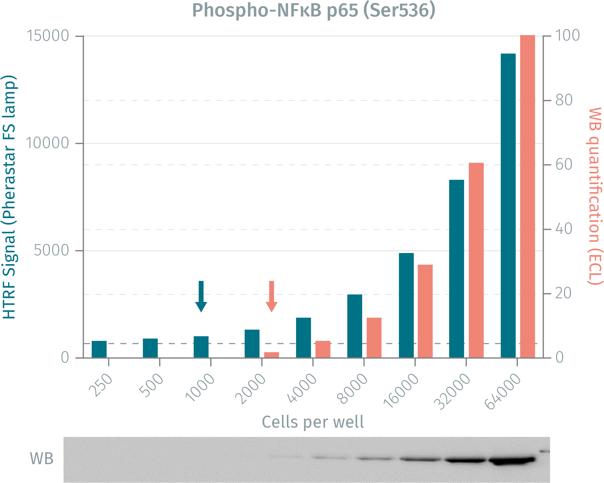
HTRF assay vs WB using phospho-NFkB assay
Human HeLa cells were cultured for 48 followed by TNFalpha stimualation. Following lysis, soluble fractions were collected after centrifugation. Serial dilutions of the cell lysate were analyzed side-by-side by Western Blot and by HTRF. Results show that HTRF Phospho-NFkB cellular assay is more sensitive than the Western Blot, as 1 000 cells are sufficient for minimal signal detection when using the HTRF phospho-NFkB assays while 2 000 cells are needed for a Western Blot signal.
TNFa dose-response on HeLa cells using phospho-NFkB assay
HeLa cells were plated and cultured for 24h before being exposed to increasing concentrations of TNFalpha. Following cell lysis, 16 µL of lysate were transferred into a 384-well sv white microplate and 4 µL of the phospho-NFkB (Ser536) detection reagents were added. The HTRF signal was recorded after an overnight incubation. Stimulation with increasing concentration of TNFa induced phosphorylation of NFkB.

Pharmacological response on Phospho and total NFkB of BAY 11-7085
40,000 cells of the U937 cell line were stimulated by increasing concentrations of BAY 11-7085 for a 3 hours, and co-stimulated for 10 min with 10 nM TNFalpha. Cells were lysed and transferred into a 384-well sv white microplate for detection of both HTRF phospho-NFkB.

Simplified pathway
Regulation of the NFkB pathway
NFkB is in a super-family with 5 members and consists of two subunits of either homo- or heterodimers that are involved in the regulation immune respose. Two main NFkB pathways exist. The classical pathway involves p65 & p50 and is stimulated by cytokines or TLR activation. The alternative pathway is mainly activated in lymphocyte generation. Inactive NFkB dimers are sequestered in the cytoplasm. Upon stimulation, the IB proteins are phosphorylated, ubiquitinylated and degraded, which activates the NF-B complex, causing it to translocate into the nucleus. Activated NFkB helps mediate gene expression, inflammatory response, cell survival and cellular proliferation. Deregulation of NFkB pathways have been found in several auto-immune disorders but also in some types of cancer.

Specifications
| Application |
Cell Signaling
|
|---|---|
| Automation Compatible |
Yes
|
| Brand |
HTRF
|
| Cellular or Signaling Pathway |
Inflammasome/Pattern Recognition Receptors (PRRs)
|
| Detection Modality |
HTRF
|
| Lysis Buffer Compatibility |
Lysis Buffer 3
Lysis Buffer 4
Lysis Buffer 5
|
| Molecular Modification |
Phosphorylation
|
| Product Group |
Kit
|
| Sample Volume |
16 µL
|
| Shipping Conditions |
Shipped in Dry Ice
|
| Target |
NF-κB
|
| Target Class |
Phosphoproteins
|
| Target Species |
Human
Mouse
|
| Technology |
TR-FRET
|
| Therapeutic Area |
NASH/Fibrosis
Neuroscience
Oncology & Inflammation
|
| Unit Size |
96 assay points
|
Video gallery
HTRF Human and Mouse Phospho-NF-κB (Ser536) Detection Kit, 96 Assay Points
Resources
Are you looking for resources, click on the resource type to explore further.
Guide
Benefit from a collection of important NAFLD pathways
Get a useful overview of today’s NAFLD knowledge with this booklet.
NASH disease is complex and follows many development pathways...
Guide
HTRF solutions, guide to major applications
This guide provides you an overview of HTRF applications in several therapeutic areas.
Guide
Protein degradation awareness in a single guide
An in-depth review of molecular and cellular pathways
The maintenance of proteostasis, the biological mechanisms that control the...
Flyer
Reagent solutions for autoimmunity research.
Advance your autoimmune disease research and benefit from Revvity broad offering of reagent technologies
Loading...


How can we help you?
We are here to answer your questions.

