


US
Revvity Sites Globally
Select your location.
*e-commerce not available for this region.

HTRF Human Phospho-IκKβ (Ser177/181) Detection Kit, 500 Assay Points


HTRF Human Phospho-IκKβ (Ser177/181) Detection Kit, 500 Assay Points


generic HTRF phospho primary image
The phospho-IKK-beta (Ser177/181) assay enables the cell-based detection of Ser177/181 phosphorylation of activated IKK-beta directly in whole cells.
| Feature | Specification |
|---|---|
| Application | Cell Signaling |
| Sample Volume | 16 µL |
The phospho-IKK-beta (Ser177/181) assay enables the cell-based detection of Ser177/181 phosphorylation of activated IKK-beta directly in whole cells.
Product variants
Unit Size: 500 assay points
Part #:
64KKBPEG
List price
USD 2,387.00
Your online price:
Unit Size: 10,000 assay points
Part #:
64KKBPEH
List price
USD 13,884.00
Your online price:
For research use only. Not for use in diagnostic procedures. All products to be used in accordance with applicable laws and regulations including without limitation, consumption, and disposal requirements under European REACH regulations (EC 1907/2006).
HTRF Human Phospho-IκKβ (Ser177/181) Detection Kit, 500 Assay Points
generic HTRF phospho primary image
Loading...
Product information
Overview
The Phospho-IKK-beta (Ser177/181) assay enables the detection of Ser177/181 phosphorylation of activated IKK-beta directly in whole cells. Using a streamlined protocol, amenable to low-volume formats, this kit can be used from basic research to High Throughput drug screening.
HTRF assays offer many advantages over other technologies:
- Homogeneous add-and-read format
- No wash steps
- Low background
- Straightforward miniaturization from 96- or 384-well microplates to high density assay formats such as 384-well low volume and 1536-well plates
- Stable signal, providing flexibility in time of readout or size of assays
How it works
Phospho-IKKß (Ser177/181) assay principle
The phospho-IKK-beta (Ser177/181) assay measures IKK beta when phosphorylated at Ser177/181. Contrary to Western Blot, the assay is entirely plate-based and does not require gels, electrophoresis or transfer. The phospho-IKK-beta (Ser177/181) assay uses 2 labeled antibodies: one with a donor fluorophore, the other one with an acceptor. The first antibody is selected for its specific binding to the phosphorylated motif on the protein, the second for its ability to recognize the protein independent of its phosphorylation state. Protein phosphorylation enables an immune-complex formation involving both labeled antibodies and which brings the donor fluorophore into close proximity to the acceptor, thereby generating a FRET signal. Its intensity is directly proportional to the concentration of phosphorylated protein present in the sample, and provides a means of assessing the proteins phosphorylation state under a no-wash assay format.
Phospho-IKKß (Ser177/181) 2-plate assay protocol
The 2 plate protocol involves culturing cells in a 96-well plate before lysis then transferring lysates to a 384-well low volume detection plate before adding Phospho-IKK beta (Ser177/181) HTRF detection reagents. This protocol enables the cells' viability and confluence to be monitored.

Phospho-IKKß (Ser177/181) 1-plate assay protocol
Detection of Phosphorylated IKK-beta (Ser177/181) with HTRF reagents can be performed in a single plate used for culturing, stimulation and lysis. No washing steps are required. This HTS designed protocol enables miniaturization while maintaining robust HTRF quality.

Assay validation
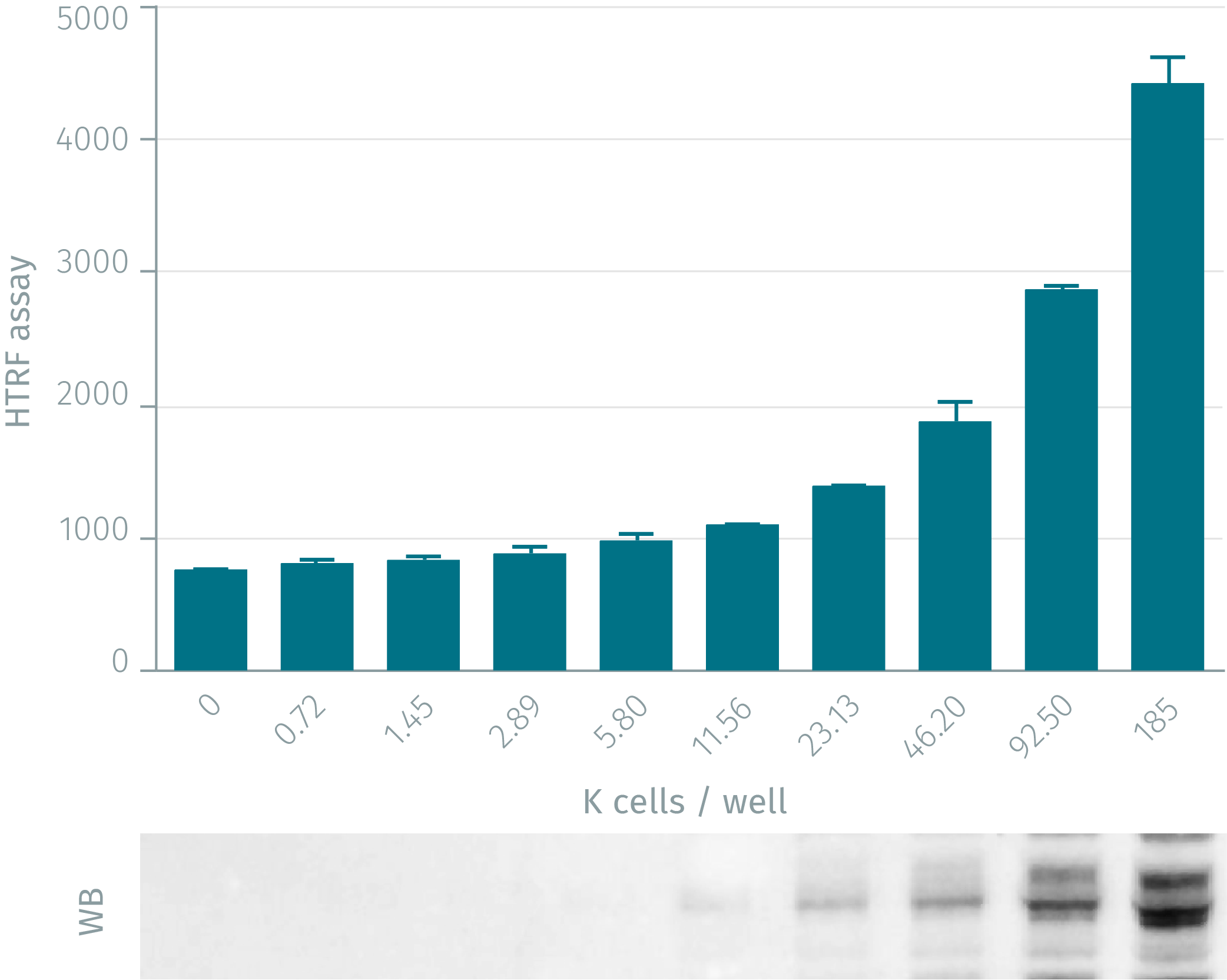
HTRF phospho-IKKß assay compared to Western Blot
HeLa cells were grown in a T175 flask 37°C, 5% Co2, 2 days. Stimulation was done with IL1b 0.5nM for 15min. After elimination of cell culture medium, 3 mL of supplemented lysis buffer was added and incubated for 45min. Soluble supernatants were collected after 10min centrifuging. Equal amounts of lysates were used for a side by side comparison of WB and HTRF. HTRF assay shows better sensitivity than Western Blot: 12,000 cells for HTRF compared to 46,000 cells for Western Blot.
TNFa dose-response on HeLa cells
Two concentrations of HeLa cells (100 and 200K cells/well) were incubated for 15 minutes at 37°C with various concentrations of TNF alpha. After a 30 minutes lysis incubation time, phosphorylated IKK-beta was measured using the two-plate assay protocol.

IL1ß dose-response on HeLa cells
Two concentrations of HeLa cells (100 and 200K cells/well) were incubated for 15 minutes at 37°C with various concentrations of IL1 beta. After a 30 minutes lysis incubation time, phosphorylated IKK-beta was measured using the two-plate assay protocol.

Detection of Phospho and total IKK-beta on HeLa cells
Different cell densities (200K and 100K) of HeLa cells were plated under 100µL in 96-well plate and incubated overnight. Media was aspirated and 50µL of different concentrations of IL-1beta was added during 15 minutes. After incubation, media was aspirated and cells were lysed with 50µL of lysis buffer 1X for 30 min at RT under gentle shaking. 16 µL of lysate were transferred into a 384-well sv white microplate and 4 µL of the HTRF phospho IKKbeta (Ser177/181) or total IKKbeta detection reagents were added. The HTRF signal was recorded after a 2 hour incubation at room temperature.

Simplified pathway
IKKß simplified pathway
Activation of the NF-?B is initiated by the signal-induced degradation of I?B proteins. This occurs primarily via activation of a kinase called the I?B kinase (IKK). IKK is composed of a heterotrimer of 3 subunits, IKK-alpha and IKK-beta (the two catalytic subunits) and IKK-gamma/NEMO (a regulatory component). Activated IKK-beta phosphorylates a protein called the inhibitor of NF-?B, I?B (I?Ba), which binds NF-?B to inhibit its function. Phosphorylated I?B is degraded via the ubiquitination pathway, freeing NF-?B and allowing its entry into the nucleus of the cell, where it activates various genes involved in inflammation and other immune responses. IKK-beta plays a significant role in brain cells following a stroke.

Specifications
| Application |
Cell Signaling
|
|---|---|
| Brand |
HTRF
|
| Cellular or Signaling Pathway |
Inflammasome/Pattern Recognition Receptors (PRRs)
|
| Detection Modality |
HTRF
|
| Lysis Buffer Compatibility |
Lysis Buffer 1
|
| Molecular Modification |
Phosphorylation
|
| Product Group |
Kit
|
| Sample Volume |
16 µL
|
| Shipping Conditions |
Shipped in Dry Ice
|
| Target Class |
Phosphoproteins
|
| Target Species |
Human
|
| Technology |
TR-FRET
|
| Therapeutic Area |
Infectious Diseases
Metabolism/Diabetes
NASH/Fibrosis
Neuroscience
Oncology & Inflammation
|
| Unit Size |
500 assay points
|
Video gallery
HTRF Human Phospho-IκKβ (Ser177/181) Detection Kit, 500 Assay Points
Resources
Are you looking for resources, click on the resource type to explore further.
Brochure
HTRF assays and reagents product list
Discover the versatility and precision of Homogeneous Time-Resolved Fluorescence (HTRF) technology. Our HTRF portfolio offers a...
Guide
HTRF solutions, guide to major applications
This guide provides you an overview of HTRF applications in several therapeutic areas.
Guide
Protein degradation awareness in a single guide
An in-depth review of molecular and cellular pathways
The maintenance of proteostasis, the biological mechanisms that control the...
Flyer
Reagent solutions for autoimmunity research.
Advance your autoimmune disease research and benefit from Revvity broad offering of reagent technologies
Application Note
TCR phospho signaling investigation with HTRF
Study your pathway of interest in PBMC and T cells
Combine models with a technique that maximizes each one’s relevance: this note...
Loading...


How can we help you?
We are here to answer your questions.

