


US
Revvity Sites Globally
Select your location.
*e-commerce not available for this region.

HTRF Human Phospho-HER3 (Tyr1289) Detection Kit, 10,000 Assay Points


HTRF Human Phospho-HER3 (Tyr1289) Detection Kit, 10,000 Assay Points


generic HTRF phospho primary image


The phospho-HER3 (Tyr1289) kit enables the cell-based detection of human epidermoid growth factor 3 (HR3, Erbb3) signaling activity when phosphorylated on Tyr1289
| Feature | Specification |
|---|---|
| Application | Cell Signaling |
| Sample Volume | 16 µL |
The phospho-HER3 (Tyr1289) kit enables the cell-based detection of human epidermoid growth factor 3 (HR3, Erbb3) signaling activity when phosphorylated on Tyr1289
Product variants
Unit Size: 500 assay points
Part #:
64HR3PEG
List price
USD 2,340.00
Your online price:
Unit Size: 10,000 assay points
Part #:
64HR3PEH
List price
USD 13,611.00
Your online price:
For research use only. Not for use in diagnostic procedures. All products to be used in accordance with applicable laws and regulations including without limitation, consumption, and disposal requirements under European REACH regulations (EC 1907/2006).
HTRF Human Phospho-HER3 (Tyr1289) Detection Kit, 10,000 Assay Points
generic HTRF phospho primary image
HTRF Human Phospho-HER3 (Tyr1289) Detection Kit, 10,000 Assay Points
Product information
Overview
The phospho-HER3 (Tyr1289) assay is designed for the robust quantification of HER3 (Erbb3) modulation when phosphorylated on Tyr1289, an indicator of many types of cancer pathologies. Overexpression of HER3, or human epidermoid receptor 3/ ErbB3, is considered as an indicator for a poor prognosis in human breast cancer. Heterodimerization of HER3 with HER2 is associated with an increase in the aggressiveness of a breast cancer. HER3 is also been found to be involved in prostate, gastric, colon, or other carcinomas. This makes HER3 a key target for anti-cancer therapies.
How it works
Phospho-HER3 (Tyr1289) assay principle
The Phospho-HER3 (Tyr1289) assay measures HER3 when phosphorylated at Tyr1289. Contrary to Western Blot, the assay is entirely plate-based and does not require gels, electrophoresis or transfer. The Phospho-HER3 (Tyr1289) assay uses 2 labeled antibodies: one with a donor fluorophore, the other one with an acceptor. The first antibody specifically binds to the phosphorylated motif on the protein, the second recognizes the protein independent of its phosphorylation state. Protein phosphorylation enables an immune-complex formation involving both labeled antibodies and which brings the donor fluorophore into close proximity to the acceptor, thereby generating a FRET signal. Its intensity is directly proportional to the concentration of phosphorylated protein present in the sample, and provides a means of assessing the proteins phosphorylation state under a no-wash assay format.
Phospho-HER3 (Tyr1289) 2-plate assay protocol
The 2 plate protocol involves culturing cells in a 96-well plate before lysis then transferring lysates to a 384-well low volume detection plate before adding Phospho-HER3 (Tyr1289) HTRF detection reagents. This protocol enables the cells' viability and confluence to be monitored.

Phospho-HER3 (Tyr1289) 1-plate assay protocol
Detection of Phosphorylated HER3 (Tyr1289) with HTRF reagents can be performed in a single plate used for culturing, stimulation and lysis. No washing steps are required. This HTS designed protocol enables miniaturization while maintaining robust HTRF quality.

Assay validation
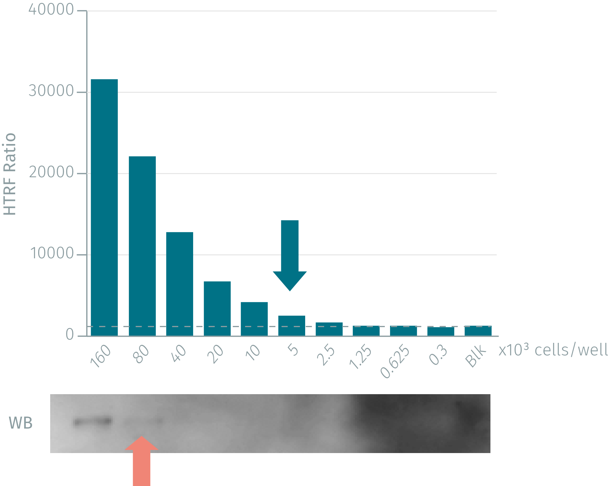
HTRF Phospho-HER3 assays compared to Western Blot
Breast cancer cells were grown in a T175 flask at 37 °C, 5% CO2 for 48 h. Cells were then stimulated with 100 nM HRG Beta1 for 10 min. After medium removal, the cells were lysed with 3 mL of supplemented lysis buffer for 30 min at room temperature. Soluble fractions were collected after a 10 min centrifugation. Serial dilutions of the cell lysate were performed in the supplemented lysis buffer and 16 µL of each dilution were dispensed and analyzed side-by-side by Western Blot and by HTRF. Using HTRF phospho-HER3 (Tyr1289) only 5,000 cells are sufficient for minimal signal detection, while 80,000 cells are needed for a Western Blot signal. The HTRF phospho-HER3 assay is at least 16-fold more sensitive than the Western Blot.
Phospho HER3 modulation in several breast adenocarcinoma cell lines
100,000 human recombinant HER2/HER3, MCF-7, SK-BR-3 and breast cancer cells were plated in 96 well plates and incubated for 24h at 37 °C-5% Co2. After incubation with increasing concentrations of HRG Beta3 (10min), medium was removed and cells were lysed with 50µl of Lysis buffer for 30min at RT under gentle shaking. 16 µL of lysate were transferred into a 384-well sv white microplate and 4 µL of the HTRF phospho-HER3 detection reagents were added. HTRF signal was recorded after an overnight incubation.

Inhibition of HER3 phosphorylation by tyrosine kinase inhibitors and therapeutic antibodies
100,000 cells were plated in 96-well plate and incubated for 24 h at 37 °C - 5% Co2. Then, after a pre-treatment with a dose-response of inhibitors, the cells were or were not stimulated with 100 nM HRG Beta1 at 37 °C, 5% CO2. After these steps, medium was removed and cells were lysed with 50 µL of Lysis buffer for 30 min at RT under gentle shaking. For detection, 16 µL of lysate were transferred into 384-well sv white microplates: 4 µL of the HTRF phospho-HER3 detection reagents were added to detect the phosphorylated HER3 and 4 µL of the HTRF total-HER3 assay to detect the total protein. The HTRF signal was recorded after an overnight incubation.

Intracellular inhibition by tyrosine kinase inhibitor Lapatinib
Human breast carcinoma and SKBR3 cells were pre-treated for 30 min with a dose-response of Lapatinib, a small tyrosine kinase inhibitor, then stimulated for 10 min with 100 nM HRG Beta1 for 10 min.


Inhibition of phospho-HER3 by monoclonal antibodies
Human breast carcinoma BXPC3 cells were pre-treated for 30 min with a dose-response of mAb-X or mAb-Y, then stimulated for 10 min with 100 nM HRG Beta1 for 10 min. Both mAbs were kindly provided by Dr Thierry Chardès (IRCM Montpellier, France).


Simplified pathway
HER3 Epidermal growth factor signaling
HER3 is a receptor tyrosine kinase and belongs to the ErbB family of epidermal growth factor receptors. HER3 is present on the cell surface and is the only EGFR family member for which no ligand has been found yet. HER3 receptor activation induces auto-phosphorylation of HER3 on Tyr1289. This provides docking sites for a variety of adaptor proteins, kinases & phosphatases that induce downstream activation of several signal transduction cascades. The HER3/ErbB receptor regulates various biological processes such as cell proliferation, differentiation, survival, adhesion, migration & angiogenesis. Hyperactivity of HER3 is associated with cancer which make HER3 a key target for anti-cancer therapies. Heterodimerization of HER3 with HER2 relates to an increase in cancer aggressiveness.

Specifications
| Application |
Cell Signaling
|
|---|---|
| Brand |
HTRF
|
| Detection Modality |
HTRF
|
| Lysis Buffer Compatibility |
Lysis Buffer 4
Lysis Buffer 5
|
| Molecular Modification |
Phosphorylation
|
| Product Group |
Kit
|
| Sample Volume |
16 µL
|
| Shipping Conditions |
Shipped in Dry Ice
|
| Target Class |
Phosphoproteins
|
| Target Species |
Human
|
| Technology |
TR-FRET
|
| Therapeutic Area |
Oncology & Inflammation
|
| Unit Size |
10,000 assay points
|
Video gallery
HTRF Human Phospho-HER3 (Tyr1289) Detection Kit, 10,000 Assay Points
HTRF Human Phospho-HER3 (Tyr1289) Detection Kit, 10,000 Assay Points
Resources
Are you looking for resources, click on the resource type to explore further.
Guide
HTRF solutions, guide to major applications
This guide provides you an overview of HTRF applications in several therapeutic areas.


Loading...
How can we help you?
We are here to answer your questions.

