


US
Revvity Sites Globally
Select your location.
*e-commerce not available for this region.

HTRF Human and Mouse Mutant Ataxin 2 Detection Kit, 500 Assay Points

HTRF Human and Mouse Mutant Ataxin 2 Detection Kit, 500 Assay Points
Generic-HTRF-mutant-detection-image
The HTRF Mutant Ataxin 2 kit is designed for the simple and rapid quantification of soluble mutant ataxin 2 proteins in cell/tissue lysates.
| Feature | Specification |
|---|---|
| Application | Protein Quantification |
| Sample Volume | 16 µL |
The HTRF Mutant Ataxin 2 kit is designed for the simple and rapid quantification of soluble mutant ataxin 2 proteins in cell/tissue lysates.
Product variants
Unit Size: 500 assay points
Part #:
64ATA2MPEG
List price
USD 1,503.00
Your online price:
Unit Size: 10,000 assay points
Part #:
64ATA2MPEH
List price
USD 15,973.00
Your online price:
For research use only. Not for use in diagnostic procedures. All products to be used in accordance with applicable laws and regulations including without limitation, consumption, and disposal requirements under European REACH regulations (EC 1907/2006).
HTRF Human and Mouse Mutant Ataxin 2 Detection Kit, 500 Assay Points
Generic-HTRF-mutant-detection-image
Loading...
Product information
Overview
Ataxin 2 is an RNA-binding protein that can be found in different species, organs (predominantly the brain), and cellular locations. In humans, the presence of an extended polyglutamine tract in ataxin 2 is linked to neurodegeneration and contributes to the progression of conditions such as amyotrophic lateral sclerosis (ALS) and spinocerebellar ataxia type 2 (SCA2). Some studies have shown that the polyglutamine expansion correlates with Ataxin 2 protein aggregation.
HTRF assays offer many advantages over other technologies:
- Homogeneous add-and-read format
- No wash steps
- Low background
- Straightforward miniaturization from 96- or 384-well microplates to high density assay formats such as 384-well low volume and 1536-well plates
- Stable signal, providing flexibility in time of readout or size of assays
How it works
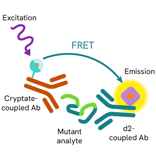
Principle of the HTRF human mutant Ataxin 2 assay
The HTRF Mutant Ataxin 2 assay is based on a TR-FRET sandwich immunoassay involving two specific antibodies, one labelled with Eu3+-cryptate (donor) and the other with d2 (acceptor). One antibody is directed against the total Ataxin 2 protein, and the second recognizes specifically the mutant polyQ domain. Both antibodies bind to soluble Mutant Ataxin 2, and the donor-acceptor proximity enables a fluorescent TR-FRET signal. The intensity of the signal is directly proportional to the concentration of soluble Mutant Ataxin 2 present in the sample (cell lysate or tissue lysate).
Protocol of the HTRF human mutant Ataxin 2 assay
The HTRF Mutant Ataxin 2 assay can be run in a 96- or 384-well low volume white detection plate (20 µL final). As described here, samples (cell/tissue lysates) or standards are dispensed directly into the assay plate for the detection of Mutant HTT by HTRF® reagents. The antibodies labelled with HTRF fluorophores may be pre-mixed and added in a single dispensing step. No washing steps are needed. The protocol can be further miniaturized or upscaled by simply resizing each addition volume proportionally.

Assay details
Human mutant Ataxin 2 specification table
| Sample size | 16 µL |
|---|---|
| Final assay volume | 20 µL |
| Time to result | Overnight at RT |
| Kit component | Frozen detection antibodies, frozen positive control, & buffers |
| Species | Human Not tested on mouse samples |
Analytical performance
Intra-assay precision table
Each of the 3 samples was measured 24 times, and the % CV was calculated for each sample. Samples were cell lysates from Mutant Ataxin 2 transfected cells.
| Sample | HTRF ratio | CV |
|---|---|---|
| 1 | 22,736 | 5.9% |
| 2 | 8,905 | 6.2% |
| 3 | 5,336 | 9.7% |
| Mean CV | 7.3% |
Inter-assay precision table
Each of the samples was measured in 3 independent experiments (3 days), and the % CV was calculated for each sample. Samples were cell lysates from Mutant Ataxin 2 transfected cells.
| Sample | HTRF Ratio | CV |
|---|---|---|
| 1 | 24,212 | 2% |
| 2 | 15,447 | 3% |
| 3 | 5,002 | 6% |
| Mean CV | 4% |
Assay validation
Validation of HTRF mutant Ataxin 2 detection kit on HAP1 KO cell line
Three Ataxin 2 plasmids were designed with 3 different repeats of glutamine. 30, 54 or 108 repeats were included in the protein sequence.
HAP1 Ataxin 2 KO cells were cultured upon 80% of confluency in a T175 flask. Transfections of cells took place for 6 hours with plasmids, then the medium was removed, and cells were incubated overnight in culture medium. Lysis buffer was added after the culture medium was removed. The flask was incubated for 30 minutes under gentle shaking.
Lysates were then collected and diluted to be tested with the Ataxin 2 and the Mutant Ataxin 2 detection kits. The Ataxin 2 concentration in the sample was estimated using the HTRF Human and Mouse Ataxin 2 Detection Kit.
As one antibody is specific to the polyQ region, the HTRF signal increased with the number of repeats even if the total concentration of protein was similar.

| Plasmid form |
30Q |
54Q |
108Q |
|---|---|---|---|
| [Ataxin2] (ng/mL) |
2.95 |
1.84 |
2.5 |
Normal and ALS patient human brain extract test using HTRF total and mutant Ataxin 2 detection kits
Human brain extracts from a normal patient and a patient with ALS were lysed and homogenized using a GentleMACS dissociator following the protocol described in the entitled application note "Best practices for analyzing brain samples with HTRF".
The soluble fractions were collected and the total amounts of protein were defined. The samples (0.125 mg/mL total protein) were then split to follow or not a disaggregation treatment using the Protein Disaggregation kit (part 64DAGGRPEG).
Samples were then tested with the HTRF Total Ataxin 2 and the Mutant Ataxin 2 Detection Kits.
The Ataxin 2 protein was more highly expressed in the ALS patient sample, with a high aggregation rate. The normal patient sample did not show any aggregation of the protein.
As expected, the mutant form of the protein was only detectable in the ALS patient sample. The disaggregation step resulted in a positive assay signal.


Specifications
| Application |
Protein Quantification
|
|---|---|
| Brand |
HTRF
|
| Detection Modality |
HTRF
|
| Product Group |
Kit
|
| Sample Volume |
16 µL
|
| Shipping Conditions |
Shipped in Dry Ice
|
| Target |
Ataxin 2
|
| Target Class |
Biomarkers
|
| Target Species |
Human
Mouse
|
| Technology |
TR-FRET
|
| Unit Size |
500 assay points
|
Resources
Are you looking for resources, click on the resource type to explore further.
Brochure
HTRF assays and reagents product list
Discover the versatility and precision of Homogeneous Time-Resolved Fluorescence (HTRF) technology. Our HTRF portfolio offers a...
Whitepaper
Shifting the treatment paradigm: the promise of gene therapy for neurodegenerative diseases
Neurodegenerative diseases, such as Alzheimer’s, Parkinson’s, and Huntington’s, are complex disorders that affect millions...
Loading...


How can we help you?
We are here to answer your questions.

