
HTRF Human and Mouse Phospho-HDAC5 (Ser259) Detection Kit, 1,000 Assay Points


| Feature | Specification |
|---|---|
| Application | Cell Signaling |
| Sample Volume | 16 µL |


Product information
Overview
Histone deacetylases (HDACs) regulate chromatin remodeling and subsequent gene transcription by controlling the status of histone acetylation. Histone deacetylation induces a condensed chromatin conformation, thus contributing to the repression of gene transcription, which is involved in diverse physiological processes.
HDAC5 belongs to class IIa HDACs, including HDAC4, HDAC7, and HDAC9. It plays a key role in the control of cellular proliferation, differentiation, migration, apoptosis, and metabolism.
How it works
Phospho-HDAC5 (Ser259) assay principle
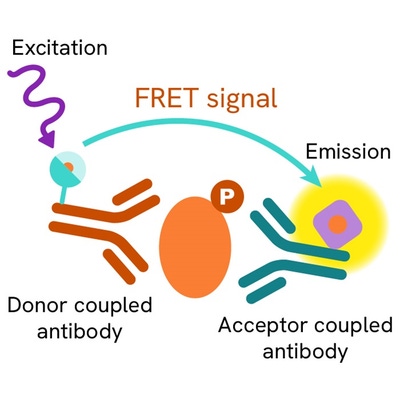
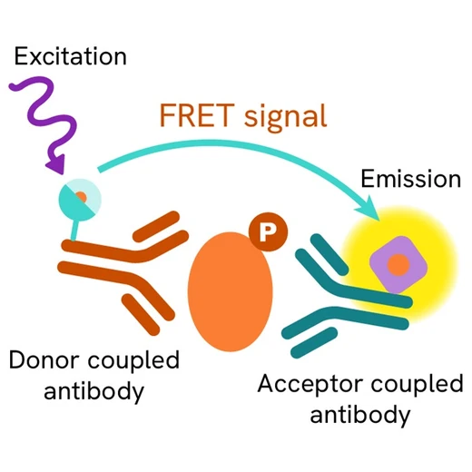
The HTRF Phospho-HDAC5 (Ser259) assay measures HDAC5 when phosphorylated at Ser259. Unlike Western Blot, the assay is entirely plate-based and does not require gels, electrophoresis, or transfer. The Phospho-HDAC5 (Ser259) assay uses 2 labeled antibodies: one with a donor fluorophore, the other with an acceptor. The first antibody was selected for its specific binding to the phosphorylated motif on the protein, the second for its ability to recognize the protein independent of its phosphorylation state. Protein phosphorylation enables an immune-complex formation involving the two labeled antibodies and bringing the donor fluorophore into close proximity to the acceptor, thereby generating a FRET signal. Its intensity is directly proportional to the concentration of phosphorylated protein present in the sample, and provides a means of assessing the protein’s phosphorylation state under a no-wash assay format.

Phospho-HDAC5 (Ser259) 2-plate assay protocol
The 2-plate protocol involves culturing cells in a 96-well plate before lysis, then transferring lysates into a 384-well low volume detection plate before adding Phospho-HDAC5 (Ser259) HTRF detection reagents. This protocol enables the cells' viability and confluence to be monitored.

Phospho-HDAC5 (Ser259) 1-plate assay protocol
Detection of Phosphorylated HDAC5 (Ser259) with HTRF reagents can be performed in a single plate used for culturing, stimulation, and lysis. No washing steps are required. This HTS designed protocol enables miniaturization while maintaining robust HTRF quality.

Assay validation
Inhibition of HDAC5 Ser259 phosphorylation on THP1 cells
THP1 Human cells (acute monocytic leukemia) were dispensed into a 96-well plate at 200,000 cells/well. They were treated with increasing concentrations of Staurosporine (kinase inhibitor) or Forskolin (Adenylate cyclase activator) for 2 hours at 37°C, 5% CO2, in complete cell culture medium. After treatment, 10 µl of supplemented Lysis Buffer#4 (4X) were dispensed into each well followed by 30 min at RT under gentle shaking (performed according to the suspension cell protocol). After cell lysis, 16 µL of lysates were transferred into a 384-well low volume white microplate and 4 µL of the HTRF Phospho-HDAC5 (Ser259) or Total HDAC5 detection antibodies were added. The HTRF signals were recorded after an overnight incubation. Note that the cell density was optimized beforehand to ensure HTRF detection within the dynamic range of the kit (data not shown).
As expected, our results show a dose-dependent inhibition in phosphorylated HDAC5 at Ser259 triggered by two different mechanisms: i) Staurosporine acts by CAMKII kinase inhibition ii) Forskolin activates the cAMP pathway, leading to PKD inhibition and consequently the decrease in phosphorylated HDAC5 ser259. The Total HDAC5 protein expression level remains almost constant using both compounds.


Specificity of HTRF Phospho-HDAC5 Ser259 assay using siRNA
THP1 cells were dispensed into a 96-well plate (300,000 cells/well), then transfected with 5µM of siRNAs specific for HDAC5 or for other Class IIa family members HDAC4, HDAC7, or HDAC9, as well as with a negative control siRNA. After 24h of incubation, the cells were lyzed with supplemented Lysis Buffer#4 (4X) and 16 µL of lysates were transferred into a 384-well low volume white microplate before the addition of 4 µL of the HTRF Phospho-HDAC5 Ser259 detection antibodies (performed according to the suspension cell protocol). The HTRF signals were recorded after an overnight incubation.
Cell transfection with specific HDAC5 siRNA led to a 77% signal decrease compared to the cells transfected with the negative siRNA, while no effect was observed using other HDAC4, HDAC7, or HDAC9 siRNAs.
Taken together, these data demonstrate that HTRF Phospho-HDAC5 Ser259 is specific and does not cross-react with other class IIa HDAC family members (HDAC4 shares more than 60% sequence homology with HDAC5).
Catalog siRNA references (Horizon Discovery): Accell Human HDAC4 siRNA #E-003497-00-0050; Accell Human HDAC5 siRNA #E-003498-00-0050; Accell Human HDAC7 siRNA #E-009330-00-0050; Accell Human HDAC9 siRNA #E-005241-00-0050

Validation on various Human and Mouse cell lines
The adherent cell lines HEK293, HEK293T (Human Embryonic Kidney), and NIH3T3 (Mouse fibroblast) were plated in 96-well culture plates and incubated for 24 hours at 37°C, 5% CO2. The cells were treated with Calyculin A (phosphatase inhibitor) at 100 nM for 1H. After medium removal, cells were lysed with 25 µL of supplemented lysis buffer #4 (1X) for 30 min at RT under gentle shaking.
The suspension Human cells THP1 (acute monocytic leukemia) were dispensed into a 96-well plate, and treated with Calyculin A (phosphatase inhibitor) at 100 nM for 1H at 37°C, 5% CO2. After treatment, cells were lysed with 10 µL of supplemented lysis buffer #4 (4X) for 30 min at RT under gentle shaking (performed according to the suspension cell protocol).
The phosphorylated HDAC5 ser259 expression level was assessed with the HTRF Phospho-HDAC5 Ser59 kit. Briefly, 16 µL of cell lysate were transferred into a low volume white microplate, followed by 4 µL of premixed HTRF detection reagents. The HTRF signals were recorded after an overnight incubation at RT. The dotted line corresponds to the non-specific HTRF signal. Note that the cell density was optimized beforehand to ensure HTRF detection within the dynamic range of the kit (data are shown for 200,000 cell/well for HEK293, HEK293T, and THP1 and 50,000 cells/well for NIH3T3, where the protein expression level was significantly higher).
The HTRF Phospho-HDAC5 Ser259 assay efficiently detects endogenous pS259-HDAC5 in various cellular models expressing different levels of the protein.

HTRF Phospho-HDAC5 Ser259 assay compared to Western Blot
THP1 cells were cultured in a T175 flask in complete culture medium at 37°C, 5% CO2. After 24H of incubation at 37°C, 5% CO2, the cells were lysed with 3 mL of supplemented lysis buffer #4 (1X) for 30 minutes at RT under gentle shaking.
Serial dilutions of the cell lysate were performed using supplemented lysis buffer#4, and 16 µL of each dilution were transferred into a low volume white microplate before the addition of 4 µL of HTRF Phospho-HDAC5 Ser259 detection reagents.
Equal amounts of lysates were used for a side-by-side comparison between HTRF and Western Blot.
In these conditions, the HTRF Phospho-HDAC5 Ser259 assay was 4-fold more sensitive than the Western Blot technique.

Simplified pathway
HDAC5 signaling Pathway
HDAC5 plays a key role in the control of genes involved in cellular functions such as proliferation, differentiation, migration and apoptosis. The nuclear accumulation of HDAC5 is governed by the balance between the activity of an N-terminal nuclear localization signal (NLS) and a C-terminal nuclear export signal. Thus, HDAC5 dynamically shuttles between nucleus and cytoplasm, depending on its phosphorylation status. . It was reported that cAMP signaling enhances the nuclear accumulation of HDAC5. It is phosphorylated by CAMK kinases leading to export to the cytoplasm and sequestration by 14-3-3 chaperon protein. While CaMKII does not bind directly to HDAC5, hetero-oligomerization of HDAC5 with HDAC4 enables CaMKII to phosphorylate HDAC5. 14-3-3 protein binding to either HDAC4 or HDAC5 is sufficient to induce nuclear export of the HDAC4-HDAC5 complex with consequent activation of transcription factors, such as MEF2, which are repressed by class IIa HDACs.

Specifications
| Application |
Cell Signaling
|
|---|---|
| Brand |
HTRF
|
| Detection Modality |
HTRF
|
| Lysis Buffer Compatibility |
Lysis Buffer 3
Lysis Buffer 4
|
| Molecular Modification |
Phosphorylation
|
| Product Group |
Kit
|
| Sample Volume |
16 µL
|
| Shipping Conditions |
Shipped in Dry Ice
|
| Target Class |
Phosphoproteins
|
| Target Species |
Human
Mouse
|
| Technology |
TR-FRET
|
| Unit Size |
500 assay points
|
Video gallery
Resources
Are you looking for resources, click on the resource type to explore further.
Discover the versatility and precision of Homogeneous Time-Resolved Fluorescence (HTRF) technology. Our HTRF portfolio offers a...
This guide provides you an overview of HTRF applications in several therapeutic areas.


Loading...
How can we help you?
We are here to answer your questions.


