


US
Revvity Sites Globally
Select your location.
*e-commerce not available for this region.

HTRF Human Phospho-LRRK2 (Ser1292) Detection Kit, 500 Assay Points


HTRF Human Phospho-LRRK2 (Ser1292) Detection Kit, 500 Assay Points

LRRK2 Phospho-Ser1292 Product-image
This HTRF kit allows for the cell-based quantitative detection of LRRK2 when phosphorylated at Ser1292.
| Feature | Specification |
|---|---|
| Application | Cell Signaling |
| Sample Volume | 16 µL |
This HTRF kit allows for the cell-based quantitative detection of LRRK2 when phosphorylated at Ser1292.
Product variants
Unit Size: 500 assay points
Part #:
64LRRKS1PEG
List price
USD 2,211.00
Your online price:
Unit Size: 10,000 assay points
Part #:
64LRRKS1PEH
List price
USD 12,865.00
Your online price:
For research use only. Not for use in diagnostic procedures. All products to be used in accordance with applicable laws and regulations including without limitation, consumption, and disposal requirements under European REACH regulations (EC 1907/2006).
HTRF Human Phospho-LRRK2 (Ser1292) Detection Kit, 500 Assay Points
LRRK2 Phospho-Ser1292 Product-image
Loading...
Product information
Overview
Leucine-Rich Repeat Kinase 2 (LRRK2) is a multi-domain protein that phosphorylates a subset of Rab GTPases. The LRRK2/Rab pathway operates at the interface of vesicular trafficking, lysosomal functioning, and autophagy, and thus plays a crucial role in immune response and neuroinflammation. Ser1292 autophosphorylation serves as an indicator of LRRK2 kinase activity. Pathogenic LRRK2 mutations, such as the common mutation G2019S, increase LRRK2 kinase activity, resulting in a toxic hyperactive protein that contributes to the Parkinson’s Disease (PD) phenotype. Consequently, LRRK2 inhibitors and degraders are considered promising therapeutic agents.
How it works
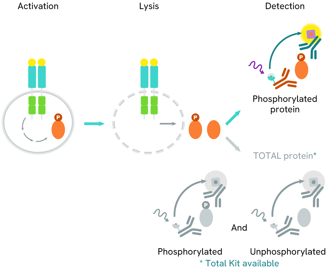
Phospho-LRRK2 (Ser1292) assay principle
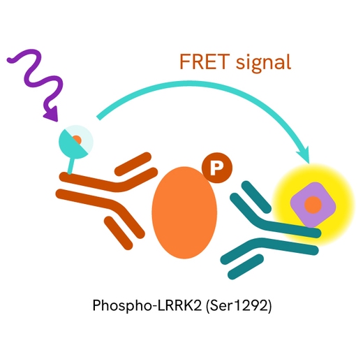
The Phospho-LRRK2 (Ser1292) assay measures LRRK2 when phosphorylated at Ser1292. Unlike Western Blot, the assay is entirely plate-based and does not require gels, electrophoresis, or transfer. The assay uses 2 antibodies, one labeled with a donor fluorophore and the other with an acceptor. The first antibody was selected for its specific binding to the phosphorylated motif on the protein, and the second for its ability to recognize the protein independently of its phosphorylation state. Protein phosphorylation leads to an immune-complex formation involving both labeled antibodies, and which brings the donor fluorophore into close proximity to the acceptor, thereby generating a FRET signal. Its intensity is directly proportional to the concentration of phosphorylated protein present in the sample and provides a means of assessing the protein's phosphorylation state under a no-wash assay format.
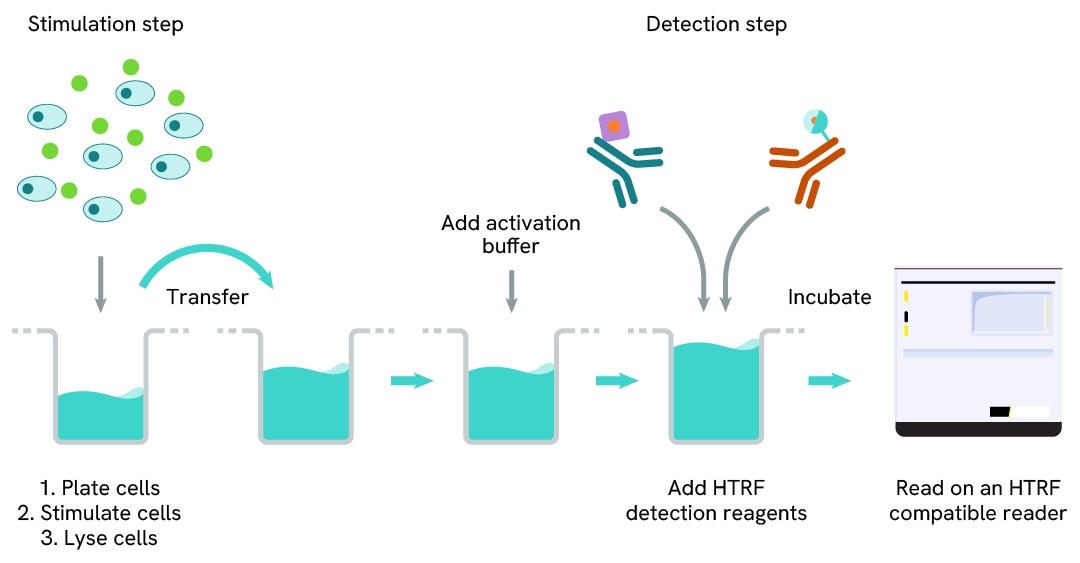
Phospho-LRRK2 (Ser1292) two-plate assay protocol
The two-plate protocol involves culturing cells in a 96-well plate before lysis, then transferring lysates into a 384-well low volume detection plate before the addition of Activation buffer followed by Phospho-LRRK2 (Ser1292) HTRF detection reagents. This protocol enables the cells' viability and confluence to be monitored.
Assay validation
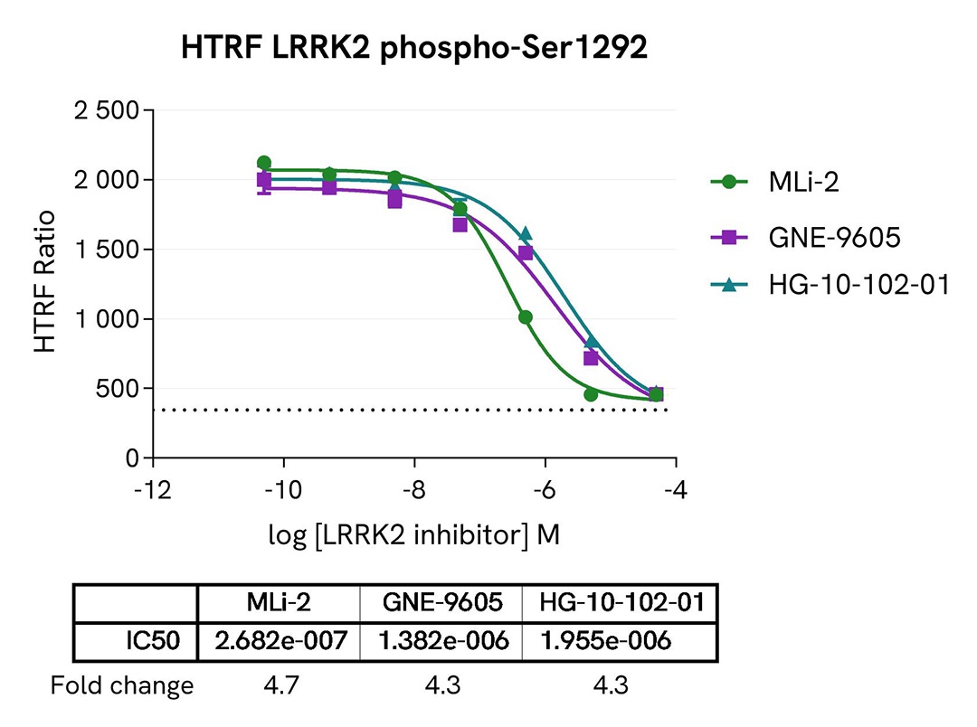
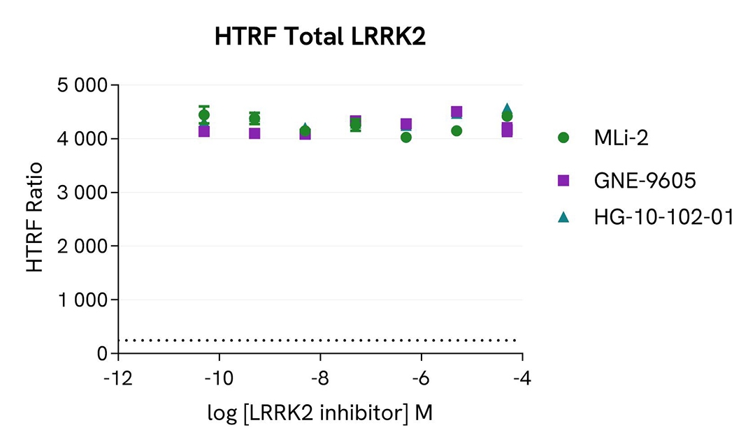
Inhibition of LRRK2 Ser1292 autophosphorylation by LRRK2 inhibitors
HEK293 cells were seeded in a 96-well culture plate (50,000 cells/well) and transfected for 24 hours with a plasmid encoding the human mutant LRRK2 G2019S protein. The cells were treated for 4 hours with increasing concentrations of the LRRK2 inhibitors MLi-2, GNE-9605, and HG-10-102-01, and then incubated for 30 minutes with 50 nM Calyculin-A. The cells were lysed with 50 µL of supplemented lysis buffer #4 for 30 minutes at room temperature under gentle shaking.
For the detection of phospho-LRRK2 (Ser1292), 14 µL of lysate were transferred into a 384-well low volume white microplate (ProxiPlate-384 Plus, Cat # 6008280/9) and supplemented with 2 µL Activation buffer before the addition of 4 µL of the HTRF Phospho-LRRK2 (Ser1292) detection reagents. For the detection of total LRRK2, 16 µL of lysate (previously diluted 50-fold in supplemented lysis buffer) were transferred into the same detection plate, and 4 µL of the HTRF Total LRRK2 detection reagents were added. The HTRF signal was recorded on an EnVision Nexus reader after the incubation time recommended for each assay (overnight for Phospho-LRRK2 and 2 hours for Total LRRK2). Cell viability was also assessed by transferring 5 µL of the same lysate into an HTRF 96-well low volume white plate (Cat # 66PL96005/025/100), followed by the addition of 25 µL of ATPlite 1step reagent (ATPlite 1step Luminescence Assay System, Cat # 6016736/1/9). The luminescence signal was measured on an EnVision Nexus reader after a 10-minute incubation in the dark.
As expected, the three inhibitors induced a dose-dependent decrease in LRRK2 autophosphorylation at Ser1292, while the expression level of LRRK2 remained unchanged. The cell viability monitored with the ATPlite 1step assay was not affected by the treatment with the inhibitors (data not shown here).
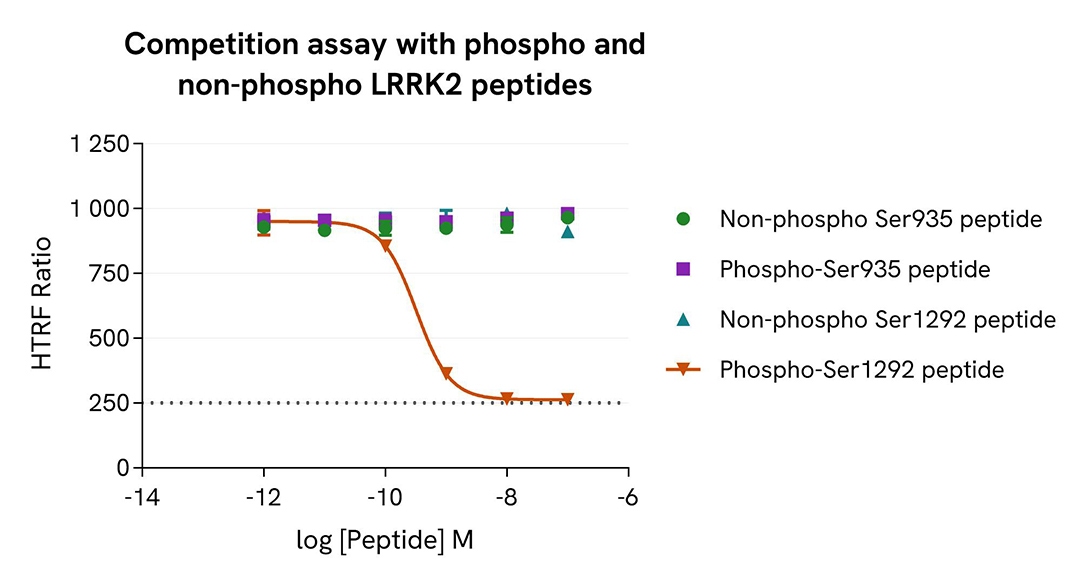
Validation of Phospho-LRRK2 assay specificity for Ser1292 phosphorylation using peptides
HEK293 cells were transfected in a T175 flask with a plasmid encoding the human mutant LRRK2 G2019S protein for 24 hours and then treated with 100 nM Calyculin-A for 30 minutes. The cells were lysed with 3 mL of supplemented lysis buffer #4 for 30 minutes at room temperature under gentle shaking.
The competition assay was carried out in a 384-well low volume white microplate by dispensing 12 µL of cell lysate (previously diluted 20-fold in supplemented lysis buffer), 2 µL of each peptide (serially diluted in supplemented lysis buffer), 2 µL of Activation buffer, and 4 µL of the HTRF Phospho-LRRK2 (Ser1292) detection reagents. The HTRF signal was recorded on an EnVision Nexus reader after an overnight incubation at room temperature.
A dose-dependent inhibition of the HTRF signal was obtained with the LRRK2 phospho-Ser1292 peptide, while the signal remained unchanged in the presence of the other peptides. These results demonstrate that the HTRF assay specifically detects the phosphorylated Ser1292 residue on LRRK2.
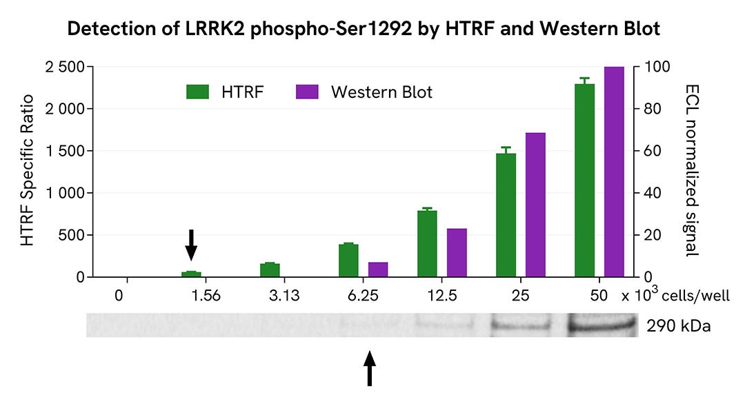
HTRF Phospho-LRRK2 (Ser1292) assay compared to Western Blot
HEK293 cells were transfected in a T175 flask with a plasmid encoding the human mutant LRRK2 G2019S protein for 24 hours, and then treated with 100 nM Calyculin-A for 30 minutes. The cells were lysed with 3 mL of supplemented lysis buffer #4 for 30 minutes at RT under gentle shaking.
Serial dilutions of the cell lysate were performed using supplemented lysis buffer, and 14 µL of each dilution were transferred into a 384-well low volume white microplate before the addition of 2 µL of Activation buffer and 4 µL of HTRF Phospho-LRRK2 (Ser1292) detection reagents. Equal amounts of lysates were used for a side-by-side comparison between HTRF and Western Blot.
Using the HTRF Phospho-LRRK2 (Ser1292) assay, 1560 cells/well were enough to detect a significant signal, while 6250 cells were needed to obtain a minimal chemiluminescent signal using Western Blot. Therefore, in these conditions, the HTRF Phospho-LRRK2 (Ser1292) assay was 4 times more sensitive than the Western Blot technique.
Simplified pathway
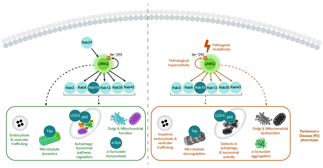
LRRK2 signaling pathway
Under physiological conditions, LRRK2 activation and localization are regulated by the Rab GTPase Rab29. Rab29 recruits LRRK2 to the membranes of different organelles (trans-Golgi network, mitochondria, phagosomes, and damaged lysosomes) under specific stimuli. This recruitment leads to the stimulation of LRRK2 kinase activity and a subsequent increase in LRRK2 autophosphorylation at Ser1292. Activated LRRK2 phosphorylates a subset of membrane-associated Rab GTPases (Rab3, Rab8, Rab10, Rab12, Rab35, Rab43) and interacts with other downstream proteins, which together regulate a variety of cellular processes such as endocytosis and vesicular trafficking, microtubule dynamics, the autophagy-lysosomal pathway (ALP), Golgi and mitochondrial function, as well as α-synuclein homeostasis.
Pathogenic LRRK2 mutations, such as the common G2019S mutation located in the kinase domain, increase LRRK2 kinase activity, resulting in a toxic hyperactive protein that contributes to the Parkinson’s Disease (PD) phenotype.
Specifications
| Application |
Cell Signaling
|
|---|---|
| Automation Compatible |
Yes
|
| Brand |
HTRF
|
| Detection Modality |
HTRF
|
| Lysis Buffer Compatibility |
Lysis Buffer 4
|
| Molecular Modification |
Phosphorylation
|
| Product Group |
Kit
|
| Sample Volume |
16 µL
|
| Shipping Conditions |
Shipped in Dry Ice
|
| Target |
LRRK2
|
| Target Class |
Phosphoproteins
|
| Target Species |
Human
|
| Technology |
TR-FRET
|
| Therapeutic Area |
Neuroscience
|
| Unit Size |
500 assay points
|
Video gallery
HTRF Human Phospho-LRRK2 (Ser1292) Detection Kit, 500 Assay Points
Resources
Are you looking for resources, click on the resource type to explore further.
Application Note
Accelerate Parkinson's disease drug discovery with no-wash LRRK2 assays
Unlock the potential of HTRF™ LRRK2 immunoassays to support and advance Parkinson's disease drug discovery. This comprehensive...
Loading...


How can we help you?
We are here to answer your questions.

