


US
Revvity Sites Globally
Select your location.
*e-commerce not available for this region.

HTRF Phospho-HSP27 (Ser78) Detection Kit, 500 Assay Points

HTRF Phospho-HSP27 (Ser78) Detection Kit, 500 Assay Points

generic HTRF phospho primary image
This HTRF kit allows for the cell-based quantitative detection of HSP27 when phosphorylated at Ser78.
| Feature | Specification |
|---|---|
| Application | Cell Signaling |
| Sample Volume | 16 µL |
This HTRF kit allows for the cell-based quantitative detection of HSP27 when phosphorylated at Ser78.
Product variants
Unit Size: 500 assay points
Part #:
64HSPS78PEG
List price
USD 2,340.00
Your online price:
Unit Size: 10,000 assay points
Part #:
64HSPS78PEH
List price
USD 13,611.00
Your online price:
For research use only. Not for use in diagnostic procedures. All products to be used in accordance with applicable laws and regulations including without limitation, consumption, and disposal requirements under European REACH regulations (EC 1907/2006).
HTRF Phospho-HSP27 (Ser78) Detection Kit, 500 Assay Points
generic HTRF phospho primary image
Loading...
Product information
Overview
Heat shock protein (HSP) 27 (also known as HSPB1) is an ATP-independent, small-sized (approximately 27 kDalton) chaperone molecule. HSP27 is present at basal levels in cells and tissues and is organized as large oligomers up to 1,000 kD. Following phosphorylation (Ser-15, Ser-78, Ser-82, Thr-143), HSP27 reorganizes itself into smaller oligomers, often dimers and tetramers and can interact with other proteins. This simple change in phosphorylation state regulates many of the canonical functions of HSP27. HSP27 is reported to be involved in cell resistance to heat shock and several stress factors, including hypoxia, oxidative stress, infections, and ultraviolet radiation. However, the function of HSP27 is hijacked during disease, and HSP27 helps to promote disease, rather than appropriately regulate cell homeostasis. So, HSP27 is overexpressed in various cancer cell lines and tumor progression, metastatic potential and treatment resistance have been attributed to the increased HSP27 expression or HSP27 phosphorylation, making HSP27, a therapeutic target of interest.
How it works
Phospho-HSP27 (Ser78) assay principle
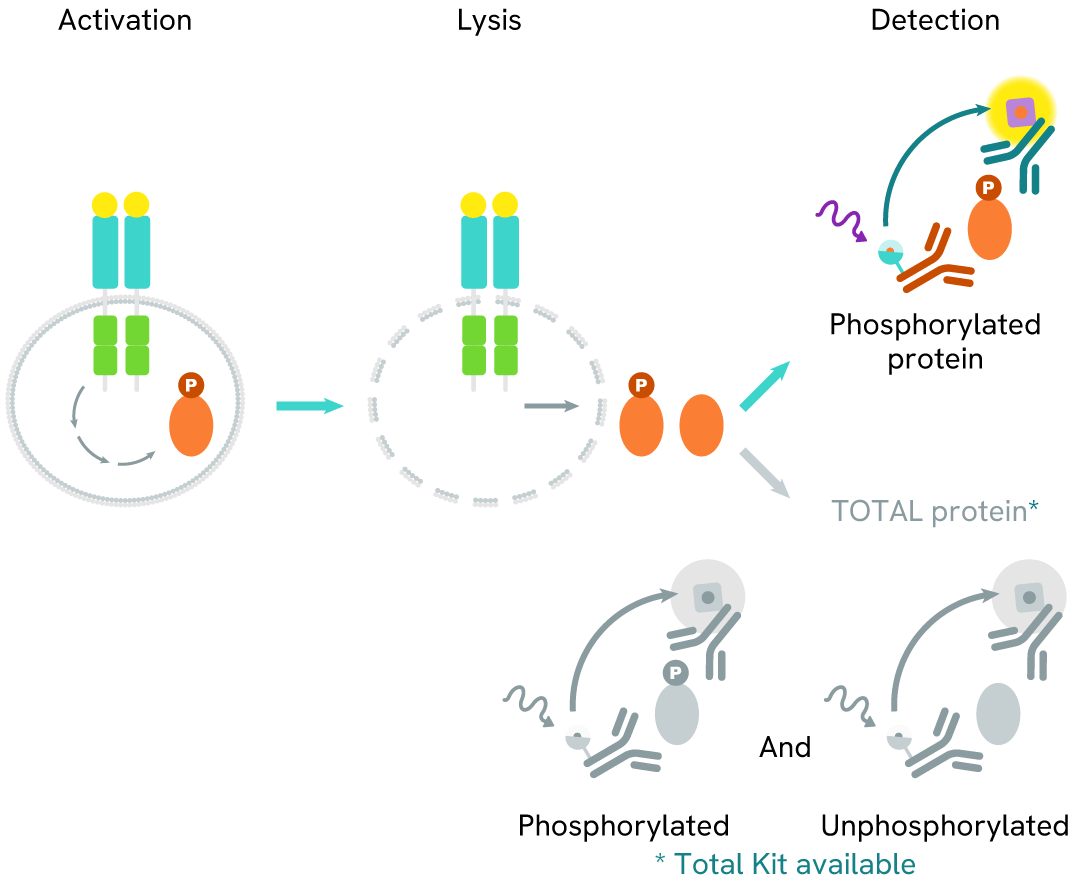
The Phospho-HSP27 (Ser78) assay measures HSP27 when phosphorylated at Ser78. Unlike Western Blot, the assay is entirely plate-based and does not require gels, electrophoresis, or transfer. The assay uses 2 antibodies, one labeled with a donor fluorophore and the other with an acceptor. The first antibody was selected for its specific binding to the phosphorylated motif on the protein, and the second for its ability to recognize the protein independently of its phosphorylation state. Protein phosphorylation enables an immune-complex formation involving both labeled antibodies, and which brings the donor fluorophore into close proximity to the acceptor, thereby generating a FRET signal. Its intensity is directly proportional to the concentration of phosphorylated protein present in the sample and provides a means of assessing the protein's phosphorylation state under a no-wash assay format.
Phospho-HSP27 (Ser78) two-plate assay protocol
The two-plate protocol involves culturing cells in a 96-well plate before lysis, then transferring lysates into a 384-well low volume detection plate before the addition of Phospho-HSP27 (Ser78) HTRF detection reagents. This protocol allows for the cells' viability and confluence to be monitored.

Phospho-HSP27 (Ser78) one-plate assay protocol
Detection of Phosphorylated HSP (Ser78) with HTRF reagents can be performed in a single plate used for culturing, stimulation, and lysis. No washing steps are required. This HTS designed protocol facilitates miniaturization while maintaining robust HTRF quality.

Assay validation
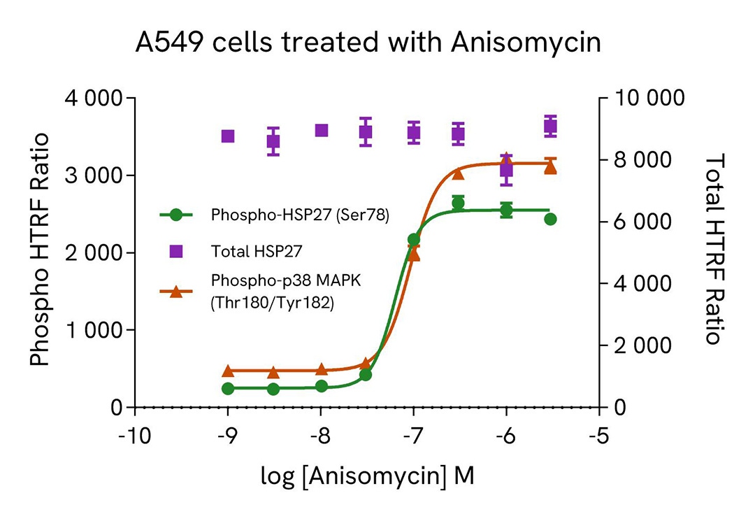
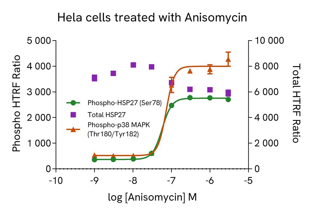
Induction of phospho-HSP27 (Ser78) in A549 and Hela cells
A549 and Hela cells were plated in a 96-well plate (12,500 cells/well) in complete culture medium, and incubated overnight at 37°C, 5% CO2. Then, cells were treated with a dose-response of Anisomycin, a p38 MAPK activator, for 30 min. After culture medium removal, cells were lysed with 50 µl of supplemented Lysis Buffer #1 for 30 min at room temperature under gentle shaking. After cell lysis, 16 µL of cell lysate (dilution 1/2 and 1/8 for Phospho-HSP27 Ser78, dilution 1/4 and 1/8 for Total HSP27 and neat and 1/2 for Phospho-p38 MAPK Thr180/Tyr182 for A549 and Hela samples respectively) were transferred into a HTRF 384-well low volume white microplate then 4 µL of the HTRF Phospho-HSP27 (Ser78) or HTRF Total HSP27 (Revvity, #64HSPTPEG/PEH/PEY) or HTRF Phospho-p38 MAPK Thr180/Tyr182 (Revvity, #64P38PET/PEG/PEH) detection reagents were added. HTRF signal for three kits was recorded using Envision Nexus reader after a 4h-incubation at room temperature. In parallel, cell viability was assessed. To do this, 5 µL of the same cell lysate were transferred into an HTRF 96-well low volume white plate, and 25 µL of ATPlite 1step detection reagent were added (Revvity #6016736/1/9). The luminescence signal was measured after a 10-min incubation in the dark at room temperature.
As expected, Anisomycin, a p38 MAPK activator, induced a dose-dependent increase in p38 MAPK phosphorylation on Threonine180/Tyrosine 182 and HSP27 phosphorylation on Serine 78, without any significant effect on the expression level of the total HSP27 protein. In addition, Anisomycin did not induce cytotoxic effects, as measured by the cell viability indicator ATPLite (data not shown).
Inhibition of phospho-HSP27 (Ser78) in A549 cells
A549 cells were plated in a 96-well plate (12,500 cells/well) in complete culture medium, and incubated overnight at 37°C, 5% CO2. Then, cells were treated with a dose response of PF-3644022, a MAPKAPK2 inhibitor, for 2 h then co-treated with 500 nM Anisomycin, a p38 MAPK activator, for 30 min. After culture medium removal, cells were lysed with 50 µl of supplemented Lysis Buffer #1 for 30 min at room temperature under gentle shaking. After cell lysis, 16 µL of cell lysate (dilution 1/2 for Phospho-HSP27 Ser78 and dilution 1/4 for Total HSP27) were transferred into a HTRF 384-well low volume white microplate then 4 µL of the HTRF Phospho-HSP27 (Ser78) or HTRF Total HSP27 (Revvity, #64HSPTPEG/PEH/PEY) detection reagents were added. HTRF signal for both kits was recorded after a 4h-incubation at room temperature. In parallel, cell viability was assessed. To do this, 5 µL of the same cell lysate were transferred into an HTRF 96-well low volume white plate, and 25 µL of ATPlite 1step detection reagent were added (Revvity, #6016736/1/9). The luminescence signal was measured after a 10-min incubation in the dark at room temperature.
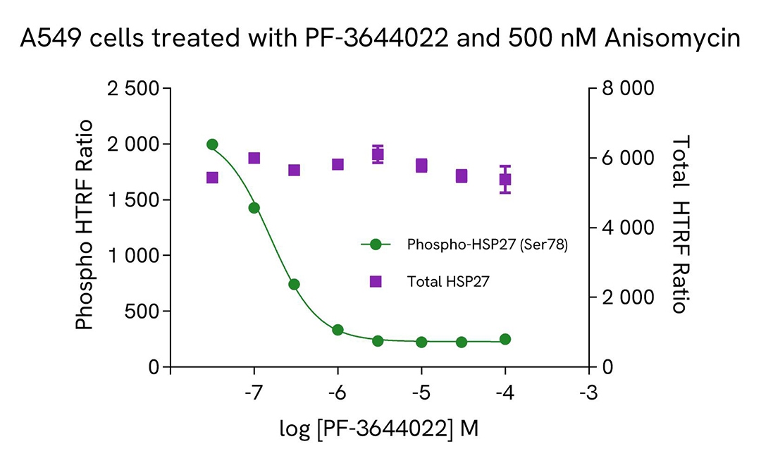
As expected, PF-3644022, a MAPKAPK2 inhibitor, repressed HSP27 phosphorylation on Serine 78 induced by Anisomycin, a p38 MAPK activator, leading to a dose-dependent decrease of Phospho-HSP27 (Ser78) signal, without any significant effect on the expression level of the total HSP27 protein. In addition, these co-treatment did not induce cytotoxic effects, as measured by the cell viability indicator ATPLite (data not shown).
Specificity of phospho-HSP27 (Ser78) assay using HAP1 KO cells
The human phospho-HSP27 Serine 78 expression level was assessed using the HTRF Phospho-HSP27 (Ser78) detection kit in HAP1 Wild Type (WT) cells (Revvity, #C631) and in two HAP1 knock-out (KO) HSP27 cell lines (Revvity, #HZGHC004943c012 and HZGHC004943c001).
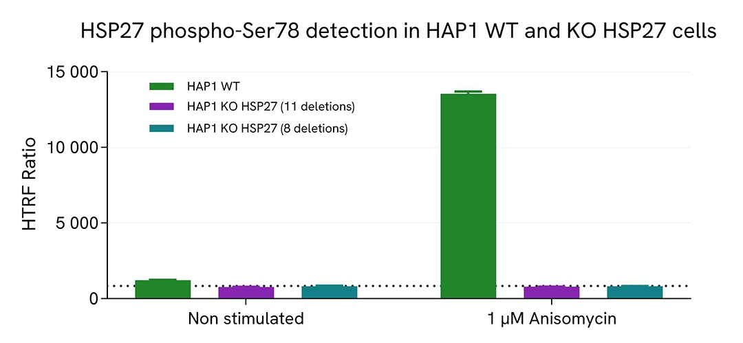
HAP1 WT and KO cells were plated in a 96-well plate (20,000 cells/well) in complete culture medium, and incubated overnight at 37°C, 5% CO2. Then, cells were treated with 1 µM Anisomycin, a p38 MAPK activator, for 30 min. After culture medium removal, cells were lysed with 50 µl of supplemented Lysis Buffer #1 for 30 min at room temperature under gentle shaking. After cell lysis, 16 µL of cell lysate were transferred into a HTRF 384-well low volume white microplate then 4 µL of the HTRF Phospho-HSP27 (Ser78) detection reagents were added. HTRF signal was recorded using HTRF reader after an overnight-incubation at room temperature.
As depicted in figure, HTRF signal was exclusively detected in the HAP1 WT cell line, with basal signal levels increasing following Anisomycin treatment. Conversely, near-background signals were observed in both HAP1 KO HSP27 cell lines, demonstrating the specificity of the HTRF Phospho-HSP27 (Ser78) assay for the detection of the HSP27 protein.
Assessment of phospho-HSP27 (Ser78) level in various human cell lines
A549, Hela and MCF7 cells were plated in a 96-well plate (12,500 cells/well) in complete culture medium, and incubated overnight at 37°C, 5% CO2. Then, cells were treated with 1 µM Anisomycin, a p38 MAPK activator, for 30 min. After culture medium removal, cells were lysed with 50 µl of supplemented Lysis Buffer #1 for 30 min at room temperature under gentle shaking. After cell lysis, 16 µL of cell lysate (dilution 1/2, 1/8 and 1/32 for A549, Hela and MCF7 samples respectively) were transferred into a HTRF 384-well low volume white microplate then 4 µL of the HTRF Phospho-HSP27 (Ser78) detection reagents were added. HTRF signal was recorded using Envision Nexus reader after a 4h-incubation at room temperature.
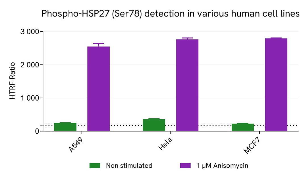
Treatment with Anisomycin induced an increase of HTRF Phospho-HSP27 (Ser78) signal in each tested cell lines.
HTRF Phospho HSP27 (Ser78) assay compared to Western Blot
A549 cells were cultured in flasks and incubated 48 h at 37°C, 5% CO2. Then, cells were treated with 1 µM Anisomycin, a p38 MAPK activator, for 30 min. After culture medium removal, cells were lysed with supplemented Lysis Buffer #1 for 30 min at room temperature under gentle shaking. Equal amounts of cell lysates (16 µL) were used for a side-by-side comparison of Western Blot and HTRF techniques.
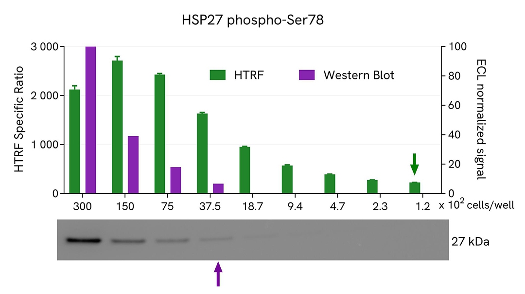
Using the HTRF Phospho-HSP27 (Ser78) assay, 120 cells/well were enough to detect a significant signal, while 3,750 cells were needed to obtain a minimal chemiluminescent signal using Western Blot. Therefore, in these conditions, the HTRF Phospho-HSP27 (Ser78) assay is 32 times more sensitive than Western Blot technique.
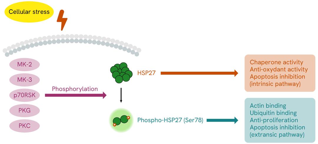
Simplified pathway
HSP27 signaling pathway
HSP27, also known as HSPB1, is an ATP-independent chaperone. It plays a role in cell resistance to stress factors such as heat shock, hypoxia, and oxidative stress. However, during disease, HSP27’s function is hijacked, leading to its overexpression in cancer cells. This increased expression or HSP27 phosphorylation (triggered by MK-2 and MK-3, p70S6K, PKG, and PKC) contributes to tumor progression and treatment resistance, making HSP27 an interesting therapeutic target.
Specifications
| Application |
Cell Signaling
|
|---|---|
| Brand |
HTRF
|
| Buffer/Solvent |
lysis buffer 1
|
| Detection Modality |
HTRF
|
| Host Species |
Human
|
| Molecular Modification |
Phosphorylation
|
| Product Group |
Kit
|
| Sample Volume |
16 µL
|
| Shipping Conditions |
Shipped in Dry Ice
|
| Target |
HSP27
|
| Target Class |
Phosphoproteins
|
| Technology |
TR-FRET
|
| Therapeutic Area |
Inflammation
Oncology
|
| Unit Size |
500 assay points
|
Resources
Are you looking for resources, click on the resource type to explore further.
Brochure
HTRF assays and reagents product list
Discover the versatility and precision of Homogeneous Time-Resolved Fluorescence (HTRF) technology. Our HTRF portfolio offers a...
Infographic
STING, the next candidate for cancer immunotherapy
Discover an introduction to STING in this infographic. Targeting the STING pathway is a strong candidate for cancer therapy, so...
Flyer
The newest AlphaLISA and HTRF kits
Discover the next generation of immunoassay kits with Revvity's newly released AlphaLISA and HTRF assay targets. Our R&D...
Loading...


How can we help you?
We are here to answer your questions.

