


US
Revvity Sites Globally
Select your location.
*e-commerce not available for this region.

HTRF Human and Mouse Total CDK4 Detection Kit, 10,000 Assay Points

HTRF Human and Mouse Total CDK4 Detection Kit, 10,000 Assay Points


generic HTRF total primary image
The Total CDK4 kit is designed to monitor the expression level of cellular CDK4 (Cyclin-Dependent Kinase 4).
| Feature | Specification |
|---|---|
| Application | Cell Signaling |
| Sample Volume | 16 µL |
The Total CDK4 kit is designed to monitor the expression level of cellular CDK4 (Cyclin-Dependent Kinase 4).
Product variants
Unit Size: 500 assay points
Part #:
64CDK4TPEG
List price
USD 2,387.00
Your price:
Unit Size: 10,000 assay points
Part #:
64CDK4TPEH
List price
USD 13,884.00
Your price:
For research use only. Not for use in diagnostic procedures. All products to be used in accordance with applicable laws and regulations including without limitation, consumption, and disposal requirements under European REACH regulations (EC 1907/2006).
HTRF Human and Mouse Total CDK4 Detection Kit, 10,000 Assay Points
generic HTRF total primary image
Loading...
Product information
Overview
The Total CDK4 cellular assay monitors CDK4 protein levels.
CDK4 (Cyclin-Dependent Kinase 4) is a member of the subfamily of CDKs that coordinate cell cycle progression in mammalian cells (including also CDK1, CDK2, and CDK6). In the early G1 phase of the cell cycle, CDK4 is activated by interaction with Cyclin D, and mono-phosphorylates the tumor suppressor RB (protein of retinoblastoma), leading to the transcription of genes required for G1/S phase transition.
Dysregulated activation of CDK4, leading to uncontrolled cell division, is a hallmark of cancers. Inhibition of CDK4 is thus an active research area, especially since the emergence of new approaches involving PROTACs (PROteolysis TArgeting Chimeras).
How it works
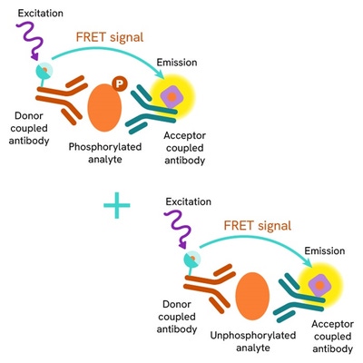
Total CDK4 assay principle
The Total CDK4 assay quantifies the expression level of CDK4 in a cell lysate. Unlike Western Blot, the assay is entirely plate-based and does not require gels, electrophoresis, or transfer. The Total CDK4 assay uses two labeled antibodies, one coupled to a donor fluorophore and the other to an acceptor. Both antibodies are highly specific for a distinct epitope on the protein. In the presence of CDK4 in a cell extract, the addition of these conjugates brings the donor fluorophore into close proximity with the acceptor and thereby generates a FRET signal. Its intensity is directly proportional to the concentration of the protein present in the sample, and provides a means of assessing the protein's expression under a no-wash assay format.
Total CDK4 two-plate assay protocol
The two-plate protocol involves culturing cells in a 96-well plate before lysis, then transferring lysates into a 384-well low volume detection plate before the addition of Total CDK4 HTRF detection reagents. This protocol enables the cells' viability and confluence to be monitored.

Total CDK4 one-plate assay protocol
Detection of Total CDK4 with HTRF reagents can be performed in a single plate used for culturing, stimulation, and lysis. No washing steps are required. This HTS designed protocol enables miniaturization while maintaining robust HTRF quality.

Assay validation
Validation of the specificity of Total CDK2, Total CDK4, and Total CDK6 assays by siRNA experiments
HeLa and HEK293 cells were plated in 96-well plates (40,000 and 50,000 cells/well respectively) and cultured for 24h. The cells were then transfected with siRNAs specific for CDK1, CDK2, CDK4, or CDK6, as well as with a negative control siRNA. After 48h incubation, the cells were lyzed and 16 µL of lysates were transferred into a 384-well low volume white microplate before the addition of 4 µL of the HTRF Total CDK2, Total CDK4, or Total CDK6 detection antibodies. The HTRF signal was recorded after an overnight incubation.
Cell transfection with each specific CDK2, CDK4, or CDK6 siRNA led to a 77 to 97% signal decrease compared to the cells transfected with the negative siRNA. It should be noted that the small signal decrease observed for the total CDK6 assay when cells were transfected with CDK2 siRNA was expected, since CDK2 knockdown leads to down-regulation of CDK6 (Bačević et al., Sci Rep. 2017; 7: 13429). Taken together, these data demonstrate that HTRF Total CDK2, Total CDK4, and Total CDK6 assays are specific for each kinase and do not cross-react with other cell cycle CDK family members.


Assessment of total CDK4 levels in human and mouse cell lines
The human cell lines HEK293, MCF-7, and HeLa, as well as the mouse cell line NIH-3T3, were cultured in T175 flasks for 48h and then lysed with 3 mL of supplemented lysis buffer #1 (1X). Serial dilutions of each cell lysate were analyzed for their total CDK4 content by transferring 16 µL of samples into a 384-well low volume white microplate and adding 4 µL of the HTRF Total CDK4 detection antibodies. The HTRF signal was recorded after an overnight incubation.

HTRF total CDK4 assay compared to Western Blot
HEK293 cells were cultured in a T175 flask in complete culture medium at 37°C-5% CO2. After 48h incubation, the cells were lysed with 3 mL of supplemented lysis buffer #1 (1X) for 30 minutes at RT under gentle shaking.
Serial dilutions of the cell lysate were performed using supplemented lysis buffer, and 16 µL of each dilution were transferred into a low volume white microplate before the addition of 4 µL of HTRF total CDK4 detection reagents. Equal amounts of lysates were used for a side by side comparison between HTRF and Western Blot.
Using the HTRF total CDK4 assay, 400 cells/well were enough to detect a significant signal, and the same number of cells was needed to obtain a minimal chemiluminescent signal using Western Blot. Therefore in these conditions, the HTRF total CDK4 assay was as sensitive as the Western Blot technique.

Simplified pathway
Role of CDK4 in the cell-division cycle
CDK4 (Cyclin-Dependent Kinase 4) is a member of the subfamily of CDKs that coordinate cell cycle progression in mammalian cells (also including CDK1, CDK2, and CDK6).
Mitogenic signals, such as growth factors, trigger cells to enter the G1 phase of the cell cycle by inducing cyclin D synthesis. Cyclin D then interacts with CDK4 and CDK6 to form active complexes. Both activated kinases are then able to mono-phosphorylate the tumor suppressor RB (protein of retinoblastoma), which still binds to transcription factor E2F, but some genes can be transcribed, such as cyclin E. In the late G1 and early S phases, Cyclin E interacts with and activates CDK2, which in turn phosphorylates additional sites on RB resulting in its complete inactivation. The E2F-responsive genes required for S phase progression are thus induced, such as Cyclin A which then interacts with CDK2 to form Cyclin A/CDK2 complexes. CDK2 finally phosphorylates Cdc25B & Cdc25C phosphatases, which in turn activate CDK1, required for progression in the G2 and M phases of the cell-division cycle.

Specifications
| Application |
Cell Signaling
|
|---|---|
| Automation Compatible |
Yes
|
| Brand |
HTRF
|
| Detection Modality |
HTRF
|
| Lysis Buffer Compatibility |
Lysis Buffer 1
Lysis Buffer 4
Lysis Buffer 5
|
| Molecular Modification |
Total
|
| Product Group |
Kit
|
| Sample Volume |
16 µL
|
| Shipping Conditions |
Shipped in Dry Ice
|
| Target |
CDK4
|
| Target Class |
Phosphoproteins
|
| Target Species |
Human
Mouse
|
| Technology |
TR-FRET
|
| Therapeutic Area |
Oncology & Inflammation
|
| Unit Size |
10,000 assay points
|
Video gallery
HTRF Human and Mouse Total CDK4 Detection Kit, 10,000 Assay Points
Resources
Are you looking for resources, click on the resource type to explore further.
Guide
HTRF solutions, guide to major applications
This guide provides you an overview of HTRF applications in several therapeutic areas.
Loading...


How can we help you?
We are here to answer your questions.

