


US
Revvity Sites Globally
Select your location.
*e-commerce not available for this region.

HTRF EPIgeneous Binding Domain B Detection Kit, 500 Assay Points


HTRF EPIgeneous Binding Domain B Detection Kit, 500 Assay Points


Epigeneous binding domain xL665 assay

This biochemical assay format enables the detection of the interaction between a subset of epigenetic binding domains and modified histone sequences.
| Feature | Specification |
|---|---|
| Application | Biochemical Enzymatic Assay |
| Sample Volume | 120 µL |
This biochemical assay format enables the detection of the interaction between a subset of epigenetic binding domains and modified histone sequences.
Product variants
Unit Size: 500 assay points
Part #:
62BDBPEG
List price
USD 784.00
Your online price:
Unit Size: 10,000 assay points
Part #:
62BDBPEH
List price
USD 4,042.00
Your online price:
For research use only. Not for use in diagnostic procedures. All products to be used in accordance with applicable laws and regulations including without limitation, consumption, and disposal requirements under European REACH regulations (EC 1907/2006).
HTRF EPIgeneous Binding Domain B Detection Kit, 500 Assay Points
Epigeneous binding domain xL665 assay
HTRF EPIgeneous Binding Domain B Detection Kit, 500 Assay Points
Product information
Overview
The EPIgeneous Binding Domain kit series provides a simple biochemical approach to study epigenetic reader domain interactions with modified histones. All kits are based on a GST-tagged binding domain / biotin-coupled Histone peptide format, and can be run using the same add-and-read single plate protocol.
Binding Domain kit B has been successfully validated on a wide variety of readers (18), including some important therapeutic epigenetic targets.
HTRF assays offer many advantages over other technologies:
- Homogeneous add-and-read format
- No wash steps
- Low background
- Straightforward miniaturization from 96- or 384-well microplates to high density assay formats such as 384-well low volume and 1536-well plates
- Stable signal, providing flexibility in time of readout or size of assays
How it works
Assay principle
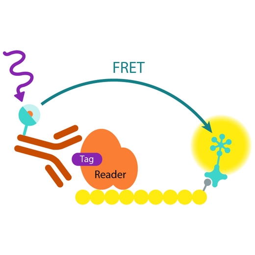
In EPIgeneous Binding Domain kit B, the GST-tagged reader domain protein binding to the biotinylated peptide substrate is detected by a conjugate mix: anti-GST Eu cryptate-labeled antibody conjugate (donor), and XL665-conjugated streptavidin (acceptor). The interaction of the reader domain with the substrate brings the donor and acceptor dyes into close proximity, and allows FRET to occur upon light excitation. The specific signal at 665 nm is inhibited when a specific compound prevents the reader domain protein from binding to its substrate.
Analytical performance
Peptide-biotin titration - UHRF1/ histone H3 peptide interaction
The validation of the EPIgeneous binding Domain B assay was performed using the Tudor Domain UHRF1 binding to [Lys(9)Me3] H3(1-21) peptide. The GST-UHRF1 concentration was fixed at 5 nM while the peptide-biotin was serially diluted.
The HTRF signal was proportional to the specific interaction measured between GST-UHRF1 and peptide-biotin.
The 2nM Kd value was determined from this experiment using a two-site specific binding regression. A slight shift of apparent Kd is observed when DMSO% increases.

DMSO tolerance - peptide-biotin concentration optimization
The assay window slightly decreased as the DMSO percentage increased. The assay window could then be recovered by increasing the peptide-biotin concentration. The optimal peptide-biotin concentration was selected (between real Kd and EC100 obtained on the titration without DMSO) with a compromise between assay window and assay sensitivity for inhibitor studies. Note that the higher the peptide-biotin concentration, the higher the inhibitor IC50. For further study of inhibitors, 1% DMSO and 6nM peptide-biotin conditions were applied.

Inhibitor titration - UHRF1 by reference compound
The EPIgeneous Binding Domain B assay was performed using 4 nM peptide-biotin, 5 nM GST-UHRF1 and 1% DMSO set constant throughout the inhibitor titration. The IC50 of [Lys(9)Me3]-H3(1-21) peptide is in good agreement with published data (Rothbart et al. Nat Struct Mol Biol, 2012 - 2µM)

Assay validation
Validated binding domains
Three kits (A, B and C) have already been validated and fully optimized on a selection of 28 key binding domains. For non-validated reader domains, a fourth one, the Discovery Kit, enables researchers to profile which of the A, B or C kits is the best assay solution.

| BINDING DOMAIN KIT A | BINDING DOMAIN KIT B | BINDING DOMAIN KIT C |
|---|---|---|
| BRD2(1) | CECR2 | BRD4(1/2) |
| BRD3(1) | FALZ (BPTF) | ATAD2A |
| BRD4(1) | BRD2(2) | ATAD2B |
| CBX 1 | BRD2(1/2) | BRD9 |
| BRD3(2) | SMARCA4 (BRG1) | |
| BRD3(1/2) | BAZ2B | |
| BRD4(2) | ||
| BRDT(1) | ||
| BRDT(1/2) | ||
| CREBBP | ||
| BRD1 | ||
| BRPF3 | ||
| TAF1L(2) | ||
| TAF1L(1/2) | ||
| TAF1(2) | ||
| TAF1(1/2) | ||
| L3MBTL1 | ||
| UHRF1 |
Specifications
| Application |
Biochemical Enzymatic Assay
|
|---|---|
| Brand |
EPIgeneous
|
| Detection Modality |
HTRF
|
| Product Group |
Kit
|
| Sample Volume |
120 µL
|
| Shipping Conditions |
Shipped in Dry Ice
|
| Target Class |
Epigenetics
|
| Technology |
TR-FRET
|
| Therapeutic Area |
Metabolism/Diabetes
Neuroscience
Oncology & Inflammation
|
| Unit Size |
500 assay points
|
Video gallery
HTRF EPIgeneous Binding Domain B Detection Kit, 500 Assay Points
HTRF EPIgeneous Binding Domain B Detection Kit, 500 Assay Points
Resources
Are you looking for resources, click on the resource type to explore further.
Brochure
HTRF assays and reagents product list
Discover the versatility and precision of Homogeneous Time-Resolved Fluorescence (HTRF) technology. Our HTRF portfolio offers a...
Guide
HTRF solutions, guide to major applications
This guide provides you an overview of HTRF applications in several therapeutic areas.


Loading...
How can we help you?
We are here to answer your questions.

