


US
Revvity Sites Globally
Select your location.
*e-commerce not available for this region.

HTRF Active GLP1 Detection Kit, 500 Assay Points

HTRF Active GLP1 Detection Kit, 500 Assay Points


Generic HTRF biomarker image
The active GLP1 kit is designed for the accurate quantitative measurement of the glucagon-like peptide-1 active forms on cell supernatants.
| Feature | Specification |
|---|---|
| Application | Protein Quantification |
| Sample Volume | 10 µL |
The active GLP1 kit is designed for the accurate quantitative measurement of the glucagon-like peptide-1 active forms on cell supernatants.
Product variants
Unit Size: 500 assay points
Part #:
62GLPPEG
List price
USD 915.00
Your online price:
Unit Size: 10,000 assay points
Part #:
62GLPPEH
List price
USD 12,278.00
Your online price:
For research use only. Not for use in diagnostic procedures. All products to be used in accordance with applicable laws and regulations including without limitation, consumption, and disposal requirements under European REACH regulations (EC 1907/2006).
HTRF Active GLP1 Detection Kit, 500 Assay Points
Generic HTRF biomarker image
Loading...
Product information
Overview
Active GLP-1, also known as glucagon-like peptide-1 active forms (GLP-1(7-36)NH2, and GLP-1(7-37)), has become a key biomarker in treatment of type 2 diabetes. The main actions of GLP-1 are stimulation of insulin secretion, and inhibition of glucagon secretion and food intake. In vivo, the active forms are rapidly degraded into inactive forms (9-36)NH2 and (9-37) by the dipeptidyl peptidase IV (DPP-IV). The active GLP-1 kit is designed for a rapid detection of GLP-1 in cell supernatants.
HTRF assays offer many advantages over other technologies:
- Homogeneous add-and-read format
- No wash steps
- Low background
- Straightforward miniaturization from 96- or 384-well microplates to high density assay formats such as 384-well low volume and 1536-well plates
- Stable signal, providing flexibility in time of readout or size of assays
How it works
Assay principle
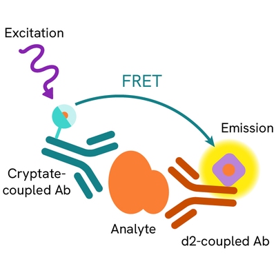
Active GLP-1 is measured using a sandwich immunoassay involving two antibodies, one labelled with Lumi4Tb-Cryptate (Donor) and the second with d2 (Acceptor). The intensity of the signal is proportional to the concentration of GLP-1 active forms present in the sample.
Assay Protocol
The active GLP-1 test can be run on cell supernatants using a simple 'addition and read' procedure described on the right (no wash steps). The HTRF conjugates may be pre-mixed and added in a single dispensing step to further streamline the protocol. An assay can be run in 96- to 384-well plates by simply resizing each addition volume proportionally. The active GLP-1 assay protocol is described here, using a white 384-well small volume plate.

Assay details
Key features
| Active GLP-1 | |
|---|---|
| Incubation | ON at RT |
| Linear range | 43. 8 to 1,400 pg/mL |
| Detection limit | 25 pg/mL* |
| Species reactivity | Rat, mouse, human, porcine |
| Peptide | Cross reactivity |
|---|---|
| GLP-1 (7-36) amide | 100% |
| GLP-1 (7-37) | 98.04% |
| GLP-1 (1-36) amide | 0.39% |
| GLP-1 (1-37) | 0.37% |
| GLP-1 (9-36) amide | <0.01% |
| GLP-1 (7-17) | <0.01% |
| GLP-2 | <0.01% |
| Glucagon | <0.01% |
Standard curve
Active GLP-1 standard curve was performed in a final 20µL assay volume and read on a Pherastar instrument (BMG LABTECH).
* Caution: all HTRF compatible readers but SpectraMax M5e and FlexStation reach this sensitivity criteria.

Analytical performance
Comparaison between HTRF and ELISA assay formats
As shown below, the active GLP-1 assay correlates well with classical ELISA assay while offering handling easiness and labor time saving. Side-by-side comparison of HTRF and ELISA assays for GLP-1 quantification was run in white 384-w small volume plate using crude NCI-H176 cell supernatants. A correlation factor of 0.97 was obtained (n=6).
| HTRF active GLP-1 | Fluorescent ELISA |
|---|---|
| 1 assay step mix and read (no washing) - <1hrs assay time | 6 sequential steps - >4hrs assay time |

Assay validation
Linearity-of-dilution assessment
Dilutions of the NCI-H716 cell supernatants 1 and 2 were performed using the HTRF assay diluent. GLP-1 concentration was then determined in crude supernatants and in diluted samples (1:2, 1:4, 1:8 and 1:16). Figure on the right indicates the means (+/- SD) of 3 independent experiments. The strong correlations obtained between measured and expected GLP-1 concentrations demonstrate the linearity of dilution within the assay range.

Glucose dose-response for active GLP-1 secretion in NCI-H716 cells
A time-course was determined for active GLP-1 secretion in human enteroendocrine NCI-H716 cells after glucose stimulation. Cells (2.105 cells/well) were seeded into 96-well culture plates pre-coated with Matrigel, and cultured for two days with DMEM 10% FBS medium. On the experiment day, cells were washed and incubated for 2 hours with increasing concentration of glucose in 100 µL of KRB supplemented with DPP IV inhibitor . At the end of glucose stimulation, 10 µL of supernatants were transferred to a 384 small volume-well plate, and assayed for active GLP-1 level as described in 'Assay protocol'. As expected, active GLP-1 was induced with Glucose dose dependence. The maximum amplitude of active GLP-1 triggered by 10% glucose versus basal production was 2.1.

Specifications
| Application |
Protein Quantification
|
|---|---|
| Brand |
HTRF
|
| Detection Modality |
HTRF
|
| Product Group |
Kit
|
| Sample Volume |
10 µL
|
| Shipping Conditions |
Shipped in Dry Ice
|
| Target |
GLP1
|
| Target Class |
Biomarkers
|
| Technology |
TR-FRET
|
| Therapeutic Area |
Metabolism/Diabetes
|
| Unit Size |
500 assay points
|
Video gallery
HTRF Active GLP1 Detection Kit, 500 Assay Points
Citations
Resources
Are you looking for resources, click on the resource type to explore further.
Guide
Get your insulin guide
Helping you select an optimal assay for your research
Revvity offers a comprehensive line of insulin quantification assays designed...
Brochure
HTRF assays and reagents product list
Discover the versatility and precision of Homogeneous Time-Resolved Fluorescence (HTRF) technology. Our HTRF portfolio offers a...
Guide
HTRF solutions, guide to major applications
This guide provides you an overview of HTRF applications in several therapeutic areas.
Loading...


How can we help you?
We are here to answer your questions.

