US
Revvity Sites Globally
Select your location.
*e-commerce not available for this region.


PhenoVue 512 Nucleic Acid Stain
 View All
PhenoVue 512 Nucleic Acid Stain

PhenoVue 512 Nucleic Acid Stain
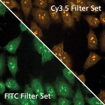
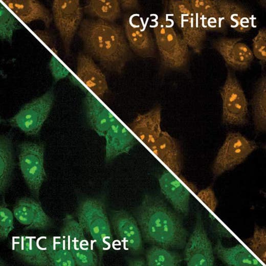
PhenoVue 512 Nucleic acid stain is a cell permeable organic molecule which binds nucleic acids. It displays a higher fluorescence intensity when complexed with RNA compared to DNA and can be used for nucleoli staining.
PhenoVue 512 Nucleic acid stain exhibits bright green or orange fluorescence and is validated for use in imaging microscopy and high-content screening applications.
Part of Revvity's portfolio of cellular imaging reagents, PhenoVue 512 Nucleic acid stain has a maximum excitation wavelength of 525 nm and a maximum emission wavelength of 590 nm, which makes it an alternative to the similar stain SYTO™ 14 green fluorescent nucleic acid stain.
View our extensive validation data in the Product Information Sheet within the Resources tab below.
| Feature | Specification |
|---|---|
| Color | Yellow |
| Filter | Cy3 |
| Organelle and Cell Compartment | Nucleoli |
PhenoVue 512 Nucleic acid stain is a cell permeable organic molecule which binds nucleic acids. It displays a higher fluorescence intensity when complexed with RNA compared to DNA and can be used for nucleoli staining.
PhenoVue 512 Nucleic acid stain exhibits bright green or orange fluorescence and is validated for use in imaging microscopy and high-content screening applications.
Part of Revvity's portfolio of cellular imaging reagents, PhenoVue 512 Nucleic acid stain has a maximum excitation wavelength of 525 nm and a maximum emission wavelength of 590 nm, which makes it an alternative to the similar stain SYTO™ 14 green fluorescent nucleic acid stain.
View our extensive validation data in the Product Information Sheet within the Resources tab below.
Product variant
Quantity: 1 x 250 µL
Part #:
CP61
List price
USD 308.00
Your online price:
For research use only. Not for use in diagnostic procedures.
PhenoVue 512 Nucleic Acid Stain
PhenoVue 512 Nucleic Acid Stain
Loading...
Product information
Overview
PhenoVue 512 Nucleic acid stain is a cell permeable organic molecule which concentrates into RNA enriched organelles, such as nucleoli found in the nucleus of mammalian cells.
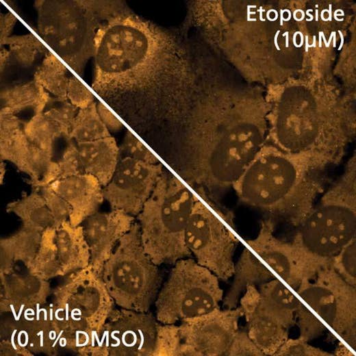
PhenoVue 512 Nucleic acid stain can be used for localization and quantification of nucleoli which are a nuclear sub-compartment that varies in size and number depending on cell type. Nucleoli are involved in the control of many cellular processes that are fundamental to normal cell homeostasis such as synthesis, processing and assembly of ribosomes as well as cell cycle regulation and cellular stress responses. Genetic disorders such as Werner syndrome or fragile X syndrome have been associated with nucleolar proteins. In addition, dysregulations of nucleolus functions have been reported in neurodegenerative diseases, cancer and cardiovascular diseases.
PhenoVue 512 Nucleic acid stain can be used to visualize nucleoli in immunofluorescence, immunohistochemistry and flow cytometry, as well as high-content analysis and screening applications.
Specifications
| Color |
Yellow
|
|---|---|
| Form |
Solution in DMSO
|
| Maximum Emission Wavelength (Emmax) |
590 nm
|
| Maximum Excitation Wavelength (Exmax) |
512/525 nm
|
| Application |
High Content Imaging
Microscopy
|
|---|---|
| Brand |
PhenoVue™
|
| Detection Modality |
Fluorescence
|
| Filter |
Cy3
|
| Organelle and Cell Compartment |
Nucleoli
|
| Quantity |
1 x 250 µL
|
| Sample Type |
Live and fixed samples
|
| Shipping Conditions |
Shipped in Dry Ice
|
| Storage Conditions |
-16 °C or below, protected from light
|
| Type |
Individual reagent
|
Image gallery
PhenoVue 512 Nucleic Acid Stain
Spectra viewer
Resources
Are you looking for resources, click on the resource type to explore further.
Loading...


How can we help you?
We are here to answer your questions.

