


US
Revvity Sites Globally
Select your location.
*e-commerce not available for this region.

HTRF Human and Mouse Total TDP-43 Detection Kit, 500 Assay Points

HTRF Human and Mouse Total TDP-43 Detection Kit, 500 Assay Points


generic HTRF total primary image


This HTRF kit enables the cell-based quantitative detection of total TDP43.
| Feature | Specification |
|---|---|
| Application | Cell Signaling |
| Sample Volume | 16 µL |
This HTRF kit enables the cell-based quantitative detection of total TDP43.
Product variants
Unit Size: 500 assay points
Part #:
64TDPTPEG
List price
USD 2,340.00
Your online price:
Unit Size: 10,000 assay points
Part #:
64TDPTPEH
List price
USD 13,611.00
Your online price:
For research use only. Not for use in diagnostic procedures. All products to be used in accordance with applicable laws and regulations including without limitation, consumption, and disposal requirements under European REACH regulations (EC 1907/2006).
HTRF Human and Mouse Total TDP-43 Detection Kit, 500 Assay Points
generic HTRF total primary image
HTRF Human and Mouse Total TDP-43 Detection Kit, 500 Assay Points
Product information
Overview
TAR DNA binding protein 43 (TDP-43) is a nucleic acid binding protein involved in RNA-related metabolism. Aggregated TDP-43 has been identified as a hallmark of amyotrophic lateral sclerosis (ALS) and frontotemporal lobar dementia (FTLD), and more widely in several neurodegenerative diseases: TDP-43 proteinopathies. In pathological conditions (mutation or dysregulation), TDP-43 forms insoluble inclusion bodies in the cytoplasm of neurons in the brain and spinal cord. The TDP-43 Phospho-Ser409/410 assay detects human TDP-43 phosphorylated in cell lysates.
How it works
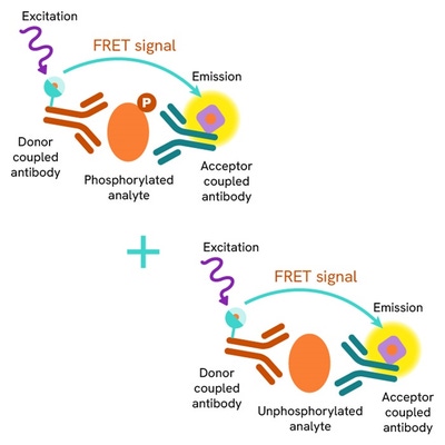
Total TDP-43 assay principle
The HTRF Total TDP-43 assay quantifies the expression level of TDP-43 in a cell lysate. Unlike Western Blot, the assay is entirely plate-based and does not require gels, electrophoresis, or transfer. The Total TDP-43 assay uses two labeled antibodies, one coupled to a donor fluorophore, the other to an acceptor. Both antibodies are highly specific for a distinct epitope on the protein. In presence of TDP-43 in a cell extract, the addition of these conjugates brings the donor fluorophore into close proximity with the acceptor, and thereby generates a FRET signal. Its intensity is directly proportional to the concentration of the protein present in the sample, and provides a means of assessing the protein’s expression under a no-wash assay format.
Total TDP-43 two-plate assay protocol
The two-plate protocol involves culturing cells in a 96-well plate before lysis, then transferring lysates to a 384-well low volume detection plate before the addition of the Total TDP-43 HTRF detection reagents. This protocol enables the cells' viability and confluence to be monitored.

Total TDP-43 one-plate assay protocol
Detection of Total TDP-43 with HTRF reagents can be performed in a single plate used for culturing, stimulation, and lysis. No washing steps are required. This HTS-designed protocol enables miniaturization while maintaining robust HTRF quality.

Assay validation
Phospho-TDP-43 (Ser409/410) modulation using Calyculin A
Neuro2a cells were cultured in a 96-well plate (50,000 cells/well) for 24h and then treated for 30 min with increasing concentrations of Calyculin A (protein phosphatase 1 and 2A inhibitor). After treatment, cells were lysed with 10 µL of supplemented lysis buffer #1 (4X) for 30 min at room temperature under gentle shaking (as per the suspension cell protocol).
After lysis, 16 µL of lysates were transferred into a 384-well low volume white microplate, and 4 µL of the HTRF Phospho-TDP-43 (Ser409/410) or Total TDP-43 detection antibodies were added. The HTRF signal was recorded after an overnight incubation at room temperature.
As expected, Calyculin A triggered a dose-dependent accumulation of phosphorylated TDP-43 at Ser409/410 through inhibition of PP1/2, while the expression level of the protein was not modulated by the treatment.

Inhibition of Phospho-TDP-43 (Ser409/410) / Total TDP-43 with CK1 inhibitors
Neuro2a cells were cultured in a 96-well plate (50,000 cells/well) for 24h, and then treated for 1h30 with the Casein Kinase 1 (CK1) inhibitors IC 261 or PF 670462 (100 µM), followed by Calyculin A activation (30 min, 100 nM).
After cell lysis, 16 µL of lysates were transferred into a 384-well low volume white microplate and 4 µL of the HTRF Phospho-TDP-43 (Ser409/410) or Total TDP-43 detection antibodies were added. The HTRF signal was recorded after an overnight incubation.
As expected, the results obtained showed an inhibition of TDP-43 Ser409/410, triggered by Calyculin A after CK1 inhibitor treatment, while the expression level of the protein was not modulated by the treatment.

Assessment of Total TDP-43 levels in various cell lines
Adherent human & mouse cells Neuro 2A, HeLa, and SH-SY5Y were seeded at 50,000 cells/well in a 96-well microplate. After a 24h incubation, the cells were lysed with 50 µL of supplemented lysis buffer #1 (1X) for 30 minutes at RT under gentle shaking.
Following lysis, 16 µL of lysate were transferred into a 384-well low volume white microplate before the addition of 4 µL of the HTRF Total TDP-43 detection reagents. The HTRF signal was recorded after an overnight incubation.
The HTRF Total TDP-43 assay efficiently detected total TDP-43 in various human and mouse cell models with different expression levels.

HTRF Total TDP-43 assay compared to Western Blot
HeLa cells were cultured in a T175 flask in complete culture medium at 37°C, 5% CO2. After a 48h incubation, the cells were treated with Calyculin A (30 min, 100 nM), then lysed with 3 mL of supplemented lysis buffer #1 (1X) for 30 minutes at RT under gentle shaking.
Serial dilutions of the cell lysate were performed using supplemented lysis buffer, and 16 µL of each dilution were transferred into a low volume white microplate before the addition of 4 µL of HTRF Total TDP-43 detection reagents. Equal amounts of lysates were used for a side-by-side comparison between HTRF and Western Blot.
Using the HTRF Total TDP-43 assay, 1,000 cells/well were enough to detect a significant signal, while 4,000 cells were needed to obtain a minimal chemiluminescent signal using Western Blot. Therefore in these conditions, the HTRF Total TDP-43 assay was 4 times more sensitive than the Western Blot technique.

Simplified pathway
TDP-43 Signaling Pathway
TDP-43 (TAR DNA binding protein 43) is a DNA and RNA-binding protein which plays a crucial role in RNA metabolism. Mainly located in the nucleus, TDP-43 shuttles between the nucleus and the cytoplasm.
The phosphorylation of TDP-43 is regulated, even if it can be an aberrant process in pathological conditions. Several kinases, including Casein Kinases (CK1 & 2), Tau Tubulin Kinases (TTBK 1 & 2) and cell division cycle 7 (CDC7), are known to promote TDP-43 phosphorylation, whereas phosphatases (PP1 &2) and Calcineurin catalyze its dephosphorylation. Dysregulation of these processes by some mutations, oxidative stress, etc. may lead to an increase in TDP-43 phosphorylation. The phosphorylation of TDP-43 impacts cell functions such as RNA binding or alternative splicing, and induces a mislocalization and accumulation in the cytoplasm, triggering aggregate formation.
In pathological conditions, phospho-TDP-43 Ser409/410 is a hallmark of proteinopathies such as Amyotrophic lateral sclerosis (ALS), FrontoTemporal Dementia (FTD), or Alzheimer’s Disease (AD). Aberrant phosphorylation, cytoplasmic accumulation, and aggregation of TDP-43 impair the clearance through the proteasome and autophagy mechanisms, leading to neuron cell toxicity.
TDP-43 phosphorylation and its regulation may provide new therapeutic directions to treat neurodegenenerative diseases.

Specifications
| Application |
Cell Signaling
|
|---|---|
| Brand |
HTRF
|
| Detection Modality |
HTRF
|
| Lysis Buffer Compatibility |
Lysis Buffer 1
Lysis Buffer 2
Lysis Buffer 3
Lysis Buffer 4
|
| Molecular Modification |
Total
|
| Product Group |
Kit
|
| Sample Volume |
16 µL
|
| Shipping Conditions |
Shipped in Dry Ice
|
| Target Class |
Phosphoproteins
|
| Target Species |
Human
Mouse
|
| Technology |
TR-FRET
|
| Unit Size |
500 assay points
|
Video gallery
HTRF Human and Mouse Total TDP-43 Detection Kit, 500 Assay Points
HTRF Human and Mouse Total TDP-43 Detection Kit, 500 Assay Points
Resources
Are you looking for resources, click on the resource type to explore further.
Brochure
HTRF assays and reagents product list
Discover the versatility and precision of Homogeneous Time-Resolved Fluorescence (HTRF) technology. Our HTRF portfolio offers a...


Loading...
How can we help you?
We are here to answer your questions.

