


US
Revvity Sites Globally
Select your location.
*e-commerce not available for this region.

HTRF Human Pro IL-1β Detection Kit, 10,000 Assay Points
HTRF Human Pro IL-1β Detection Kit, 10,000 Assay Points

Generic HTRF cytokine image

The HTRF pro IL-1β detection kit is designed for the simple and rapid quantification of soluble pro IL-1β proteins in cell/tissue lysates.
| Feature | Specification |
|---|---|
| Application | Protein Quantification |
| Dynamic Range | 312.5 - 20,000 pg/mL |
| Limit of Detection | 38.5 pg/mL |
| Limit of Quantification | 130 pg/mL |
| Sample Volume | 16 µL |
The HTRF pro IL-1β detection kit is designed for the simple and rapid quantification of soluble pro IL-1β proteins in cell/tissue lysates.
Product variants
Unit Size: 500 assay points
Part #:
62HPIL1BPEG
List price
USD 1,184.00
Your online price:
Unit Size: 10,000 assay points
Part #:
62HPIL1BPEH
List price
USD 11,044.00
Your online price:
For research use only. Not for use in diagnostic procedures. All products to be used in accordance with applicable laws and regulations including without limitation, consumption and disposal requirements under European REACH regulations (EC 1907/2006).
HTRF Human Pro IL-1β Detection Kit, 10,000 Assay Points
Generic HTRF cytokine image
HTRF Human Pro IL-1β Detection Kit, 10,000 Assay Points
Loading...
Product information
Overview
Pro IL-1β, also known as IL-1β precursor, is a 31 kDa protein precursor of IL-1β. It is produced in response to molecular motifs carried by pathogens called ‘pathogen associated molecular patterns’ (PAMPs). Induction of pro IL-1β expression is referred to as a priming step and is an inefficient secretion stimulus. The processing and cleavage of pro IL-1β into active IL-1β is conditioned by the activation stage involving PAMP, or DAMP (danger associated molecular pattern, endogenous molecules released from dead cells).
Pro IL-1β is cleaved by the pro-inflammatory protease caspase-1, activated via recruitment to a multi-protein complex termed the inflammasome.
HTRF assays offer many advantages over other technologies:
- Homogeneous add-and-read format
- No wash steps
- Low background
- Straightforward miniaturization from 96- or 384-well microplates to high density assay formats such as 384-well low volume and 1536-well plates
- Stable signal, providing flexibility in readout time or size of assays
How it works
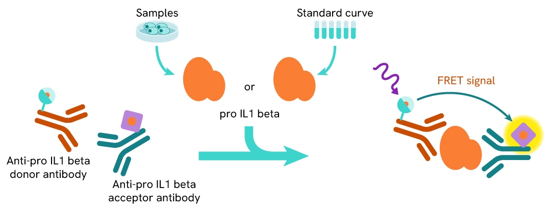
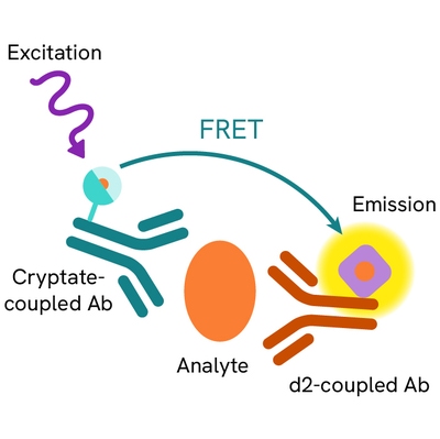
Principle of the HTRF human pro IL-1β assay
The HTRF pro IL-1β assay is based on a TR-FRET sandwich immunoassay involving two specific antibodies, one labeled with Eu3+-cryptate (donor) and the other with d2 (acceptor). One antibody is directed against the IL-1β, and the second recognizes specifically pro IL-1β. When both antibodies bind to pro IL-1β, and the donor-acceptor proximity enables a fluorescent TR-FRET signal. The intensity of the signal is directly proportional to the concentration of pro IL-1β present in the cell lysate sample.
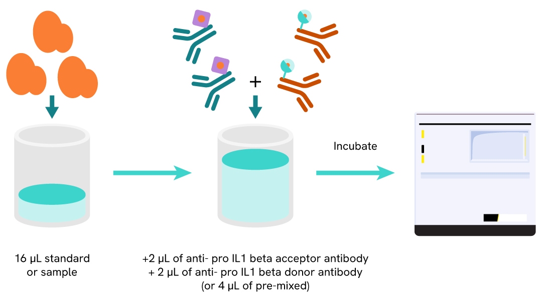
Protocol of the HTRF human pro IL-1β assay
The HTRF pro IL-1β assay can be run in a 96- or 384-well low volume white detection plate (20 µL final). As described here, samples (cell lysates) or standards are dispensed directly into the assay plate for the detection of pro IL-1β by HTRF reagents. The antibodies labelled with HTRF fluorophores may be pre-mixed and added in a single dispensing step. No washing steps are needed. The protocol can be further miniaturized or upscaled by simply resizing each addition volume proportionally.
Assay details
Human pro IL-1β assay details
| Sample size | 16 µL |
|---|---|
| Final assay volume | 20 µL |
| Kit component | Lyophilized standard, frozen detection antibodies & buffers |
| LOD & LOQ (in lysis buffer) | 38.5 & 130 pg/mL |
| Range | 312.5 – 20000 pg/mL |
| Time to result | Overnight at RT |
| Lysis buffer compatibility | LB#1 & LB#4 |
| Species | Human, not tested on mouse samples |
Analytical performance
Intra-assay precision table
All of the 3 samples wwere measured 24 times, and the % CV was calculated for each. The samples were cell lysates from LPS stimulated THP-1.
| Sample | Mean [pro IL-1b] (pg/mL) | CV |
|---|---|---|
| 1 | 333 | 7% |
| 3 | 3728 | 2% |
| 4 | 8626 | 2% |
| Mean CV | 3% |
Inter-assay precision table
All of the samples wwere measured in 3 independent experiments (3 days), and the % CV was calculated for each. The samples were cell lysates from LPS stimulated THP-1.
| Sample | Mean [pro IL-1b] (pg/mL) | CV |
|---|---|---|
| 1 | 490 | 7 % |
| 3 | 4321 | 7 % |
| 5 | 17808 | 5 % |
| Mean CV | 6% |
Dilution linearity
The samples were LPS stimulated THP1 cell lysate serially diluted in LB#1. The excellent % of recovery obtained from these experiments shows the good dilution linearity of the assay.
| Dilution Factor (x) | Expected pro-IL1b concentration (pg/mL) |
Measured pro-IL1b concentration (pg/mL) |
% dilution recovery (%) |
|---|---|---|---|
| Neat | 16004 | 16004 | 100% |
| 2 | 8002 | 8429 | 105% |
| 4 | 4001 | 4205 | 105% |
| 8 | 2001 | 2082 | 104% |
| 16 | 1000 | 1035 | 103% |
| 32 | 500 | 471 | 94% |
| Mean CV | 102% | ||
Spike & recovery
Two known amounts of recombinant pro IL1 beta were spiked into a dilution of native sample from THP1 cell lysates stimulated with LPS. The expected concentrations were compared to those measured, in order to compute antigen recoveries (acceptance criteria: 80-120%). A 100% of recovery indicates similar measurements of cytokine from a sample and the kit standard.
| [pro IL1b] Standard (pg/mL) |
[pro IL1b] Native (pg/mL) |
Expected (pg/mL) | Obtained (pg/mL) | Recovery |
|---|---|---|---|---|
| 3264 | 5600 | 4432 | 5189 | 117% |
| 10083 | 6674 | 7467 | 112% | |
| 8664 | 10083 | 9373 | 10795 | 115% |
| 18541 | 13602 | 15280 | 112% | |
| Mean CV | 114% | |||
Assay validation
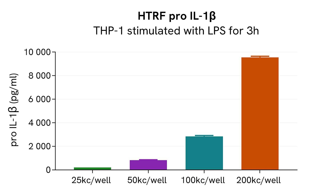
Pro IL-1β expression in LPS stimulated THP1
THP1 cells were plated at 25, 50,100, and 200 kcells/well and were stimulated for 3h with LPS at 1 µg/mL. After treatment, the medium was removed and cells were lysed with 40µL lysis buffer #1 for 30 minutes under gentle shaking. Then 16 µL of cell lysates were transferred into a white detection plate (384 low volume) to be analyzed with the human pro IL-1β assay.
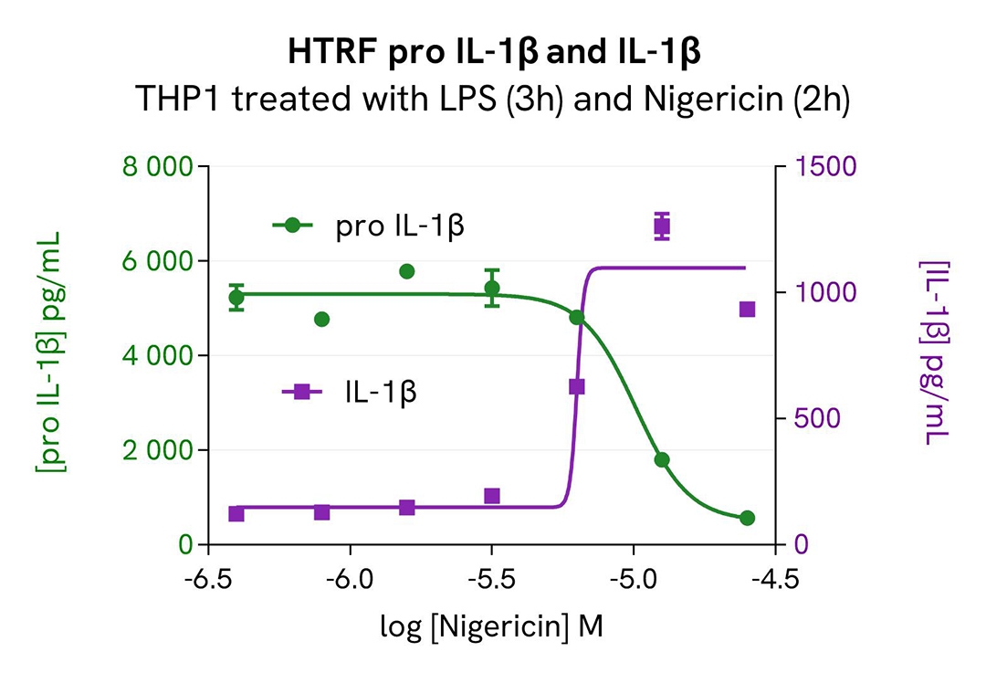
Pro IL-1β modulation using nigericin on LPS stimulated THP1 cells
THP1 cells were seeded in a 96-well culture plate (100,000 cells) and were treated with 1µg/mL LPS for 3 hours. Cells were then treated with increasing concentrations of Nigericin for 2 hours. After treatment, the medium was collected, and cells were lysed with 40µL lysis buffer #1 for 30 minutes under gentle shaking. Next, the cell lysates were tested using the HTRF pro IL-1β kit and the medium was tested using the HTRF IL-1β high performance detection kit (part 62HIL1B2PEG).
As expected, Nigerinicin induced the cleavage of pro IL-1β in LPS stimulated THP1 cells and the secretion of IL-1β in cell medium.
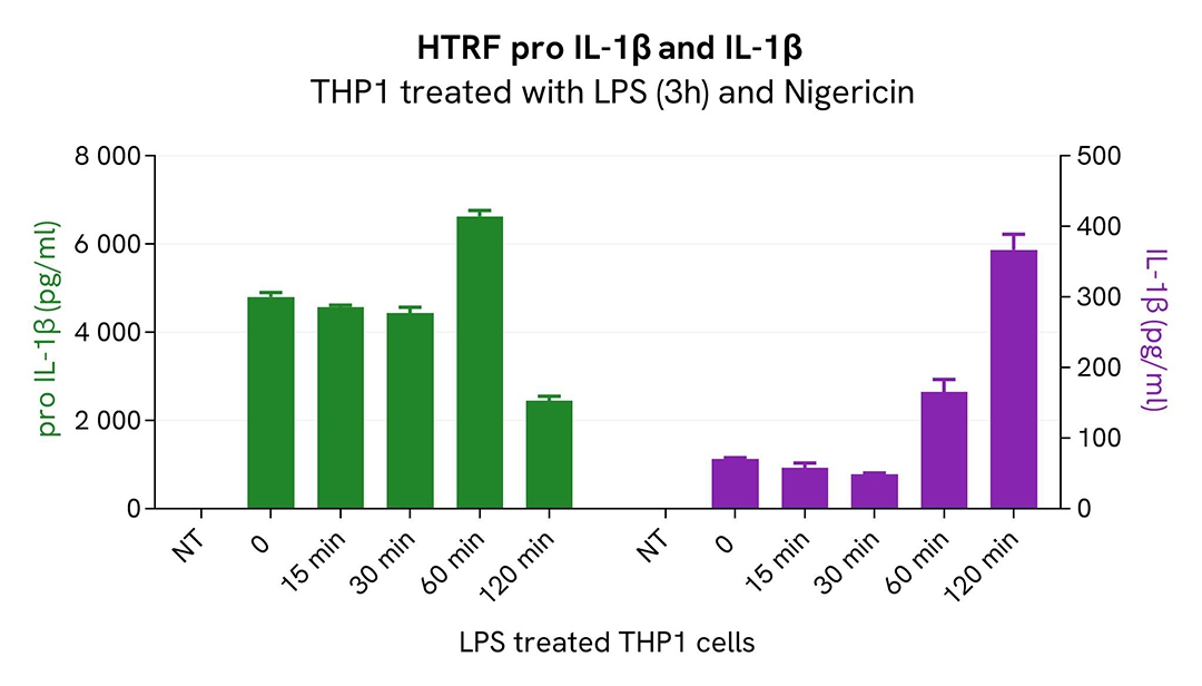
Pro IL-1β modulation kinetics using nigericin
THP1 cells were seeded in a 96-well culture plate (100,000 cells) and were treated with 1µg/mL LPS for 3 hours. Cells were then treated with 12.5µM of Nigericin for 15 to 120 minutes. After treatment, the medium was collected, and cells were lysed with 40µL lysis buffer #1 for 30 minutes under gentle shaking. Next, the cell lysates were tested using the HTRF pro IL-1β kit and the medium was tested using the HTRF IL-1β high performance detection kit (part 62HIL1B2PEG).
Specifications
| Application |
Protein Quantification
|
|---|---|
| Brand |
HTRF
|
| Cellular or Signaling Pathway |
Inflammasome/Pattern Recognition Receptors (PRRs)
|
| Detection Modality |
HTRF
|
| Dynamic Range |
312.5 - 20,000 pg/mL
|
| Limit of Detection |
38.5 pg/mL
|
| Limit of Quantification |
130 pg/mL
|
| Product Group |
Kit
|
| Sample Volume |
16 µL
|
| Shipping Conditions |
Shipped in Dry Ice
|
| Target |
pro-IL1β
|
| Target Class |
Cytokines
|
| Target Species |
Human
|
| Technology |
TR-FRET
|
| Therapeutic Area |
Autoimmunity
Cardiovascular
Immuno-oncology
Infectious Diseases
Inflammation
Neuroscience
Oncology
Virology
|
| Unit Size |
10,000 assay points
|
Resources
Are you looking for resources, click on the resource type to explore further.
Loading...


How can we help you?
We are here to answer your questions.

