
US

Revvity Sites Globally
Select your location.
*e-commerce not available for this region.

HTRF Human TREM2/DAP12 Complex Detection Kit, 10,000 Assay Points
HTRF Human TREM2/DAP12 Complex Detection Kit, 10,000 Assay Points
HTRF PPI primary image
The HTRF Human TREM2/DAP12 complex detection kit supports the cell-based quantitative detection of the TREM2/DAP12 complex, in a homogeneous (no wash steps, no separation steps) format.
| Feature | Specification |
|---|---|
| Application | Cell Signaling |
| Sample Volume | 16 µL |
The HTRF Human TREM2/DAP12 complex detection kit supports the cell-based quantitative detection of the TREM2/DAP12 complex, in a homogeneous (no wash steps, no separation steps) format.
Product variants
Unit Size: 500 Assay Points
Part #:
64TRMDPPEG
List price
USD 2,387.00
Your price:
Unit Size: 10,000 Assay Points
Part #:
64TRMDPPEH
List price
USD 13,884.00
Your price:
For research use only. Not for use in diagnostic procedures. All products to be used in accordance with applicable laws and regulations including without limitation, consumption and disposal requirements under European REACH regulations (EC 1907/2006).
HTRF Human TREM2/DAP12 Complex Detection Kit, 10,000 Assay Points
HTRF PPI primary image
Loading...
Product information
Overview
TREM2 (Triggering Receptor Expressed on Myeloid Cells 2) is a member of the TREM receptor family, which regulates the function of several immune cell types, including neutrophils, monocytes/macrophages, microglia, and dendritic cells.
The kit is designed for the efficient and homogeneous detection of the TREM2/DAP12 complex, a critical early event in TREM2-mediated signaling that plays an important role in the pathogenesis of Alzheimer’s Disease (AD) and has emerged as a promising therapeutic target.
The development of novel small-molecule modulators targeting the TREM2 pathway has gained significant interest as a potential therapeutic strategy for the treatment of neuroinflammatory and neurodegenerative diseases.
HTRF assays offer many advantages over other technologies:
- Homogeneous add-and-read format
- No wash steps
- Low background
- Straightforward miniaturization from 96- or 384-well microplates to high density assay formats such as 384-well low volume and 1536-well plates
- Stable signal, providing flexibility in readout time or assay size
How it works
TREM2/DAP12 Complex assay principle
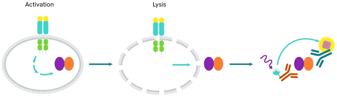
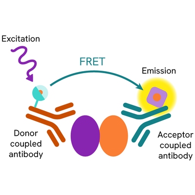
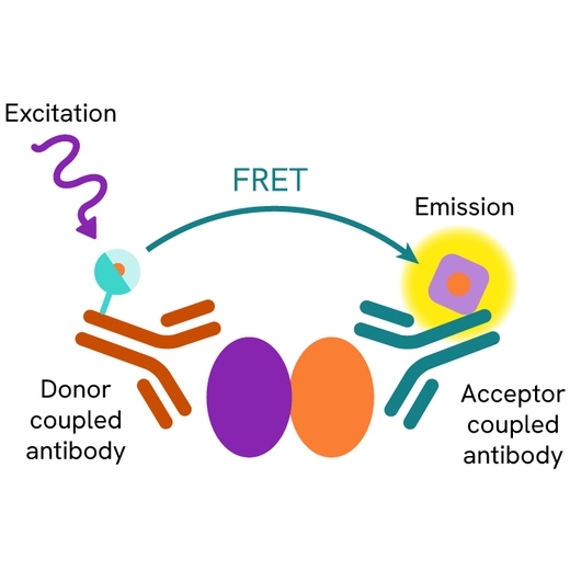
The TREM2/DAP12 Complex assay measures TREM2/DAP12 complex levels in cells. Unlike Western Blot, the assay is entirely plate-based and does not require gel, electrophoresis, or protein transfer. The assay uses two antibodies: one labeled with a donor fluorophore and the other with an acceptor fluorophore. Each antibody specifically recognizes a distinct component of the complex. In the presence of the TREM2/DAP12 complex, the two labeled antibodies bind their respective targets, forming an immunocomplex that brings the donor and acceptor fluorophores into close proximity. This proximity leads to FRET (Fluorescence Resonance Energy Transfer) and generates a signal whose intensity is directly proportional to the amount of TREM2/DAP12 complex present in the sample.
The assay therefore provides a simple and robust method for monitoring TREM2/DAP12 complex formation and assessing protein-protein interactions in a homogeneous, no-wash format.
TREM2/DAP12 Complex two-plate assay protocol
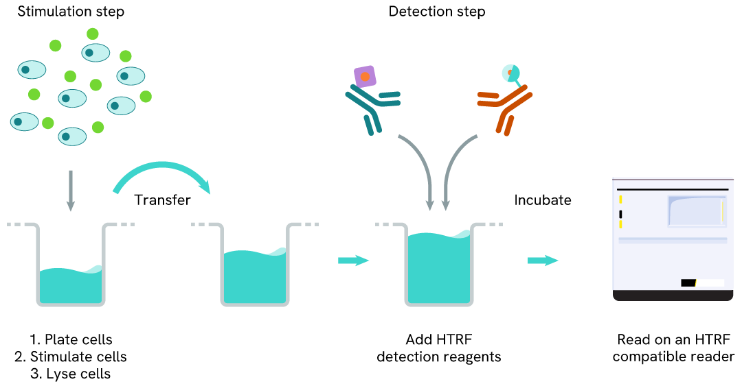
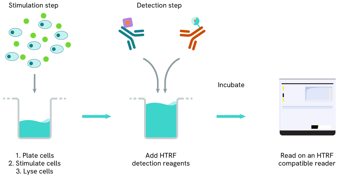
The two-plate protocol involves culturing cells in a 96-well plate before lysis, then transferring lysates into a 384-well low volume detection plate before the addition of TREM2/DAP12 Complex HTRF detection reagents. This protocol allows the cells’ viability and confluence to be monitored.
TREM2/DAP12 Complex one-plate assay protocol
Detection of TREM2/DAP12 Complex with HTRF reagents can be performed in a single plate used for culturing, stimulation, and lysis. No washing steps are required. This HTS designed protocol allows miniaturization while maintaining robust HTRF quality.
Assay validation
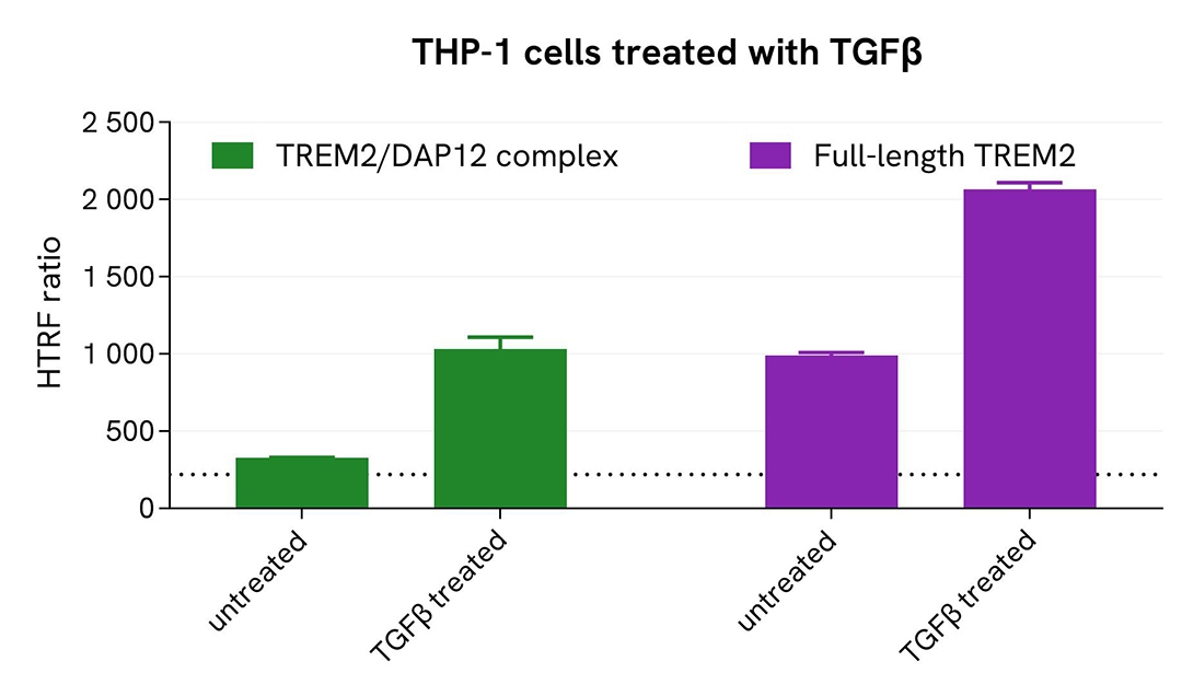
Induction of the TREM2/DAP12 Complex and Full-Length TREM2 in an Endogenous Cellular Model
THP-1 cells were harvested and seeded at a density of 1 × 10⁶ cells/mL in 35 mL of complete RPMI medium in a T175 flask. Cells were then treated with 5 ng/mL TGFβ for 18 hours at 37°C in a 5% CO₂ atmosphere. Following treatment, THP-1 cells were harvested by scraping and centrifuged at 300 × g for 3 minutes. The cells were subsequently seeded into a 96-well tissue culture-treated plate at 300,000 cells per well in complete culture medium, and lysed with 10 µL of supplemented Lysis Buffer #3 (4X) for 30 minutes at room temperature with gentle shaking.
For detection, 16 µL of cell lysate were transferred into a 384-well low-volume white microplate, and 4 µL of HTRF TREM2/DAP12 complex or Full-Length TREM2 detection reagents were added. The HTRF signal was measured following an overnight incubation.
As expected, TGFβ treatment activated the TREM2 pathway, resulting in increased levels of both the TREM2/DAP12 complex and full-length TREM2.
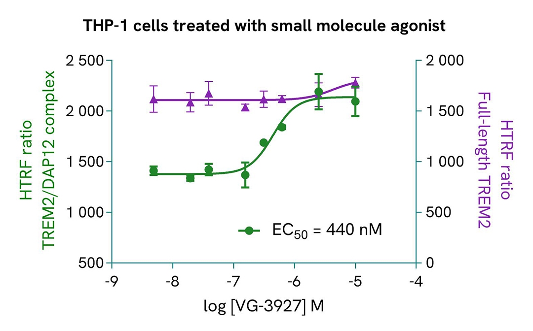
Effect of VG-3927 on TREM2/DAP12 complex formation and Full-Length TREM2 levels in an endogenous cellular model
THP-1 cells were harvested and seeded at a density of 1 × 10⁶ cells/mL in 35 mL of complete RPMI medium in a T175 flask. Cells were then treated with 5 ng/mL TGFβ for 48 hours at 37°C in a 5% CO₂ atmosphere. Following treatment, cells were harvested by scraping and centrifuged at 300 × g for 3 minutes.
The cells were subsequently seeded into a 96-well tissue culture-treated plate at a density of 300,000 cells per well in complete culture medium, and pre-incubated with increasing concentrations of VG-3927 for 20 minutes. After treatment, the cells were lysed with 10 µL of supplemented Lysis Buffer #3 (4X) for 30 minutes at room temperature with gentle shaking.
For detection, 16 µL of cell lysate were transferred into a 384-well low-volume white microplate, and 4 µL of HTRF TREM2/DAP12 Complex or Full-Length TREM2 detection reagents were added. The HTRF signal was measured following an overnight incubation.
As expected, the TREM2 agonist VG-3927 activated the TREM2 pathway and induced a dose-dependent increase in TREM2/DAP12 complex levels, while full-length TREM2 expression remained unchanged.
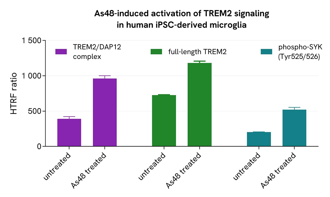
TREM2 pathway activation by As48 in human iPSC-derived microglia
WT microglia differentiated from human iPSCs (iCell® Microglia, FUJIFILM Cellular Dynamics) were seeded at 70,000 cells per well in a 96-well microplate and incubated for 72 hours. Following centrifugation at 200 × g for 3 minutes, the supernatant was removed, and cells were pre-incubated with 30 µM As48 for 20 minutes. After treatment, the culture medium was removed, and cells were lysed with Lysis Buffer #3 (1X) for 30 minutes at room temperature with gentle shaking.
Then, 16 µL of each cell lysate were transferred into a low-volume 384-well white microplate, followed by the addition of 4 µL of premixed HTRF detection reagents. The HTRF signal was measured following an overnight incubation.
These results obtained in human iPSC-derived microglia demonstrate that the dual-function TREM2 modulator As48 activates the TREM2 signaling pathway. As expected, treatment with As48 resulted in a significant increase in TREM2/DAP12 complex formation, full-length TREM2 levels, and SYK phosphorylation at Tyr525/526.
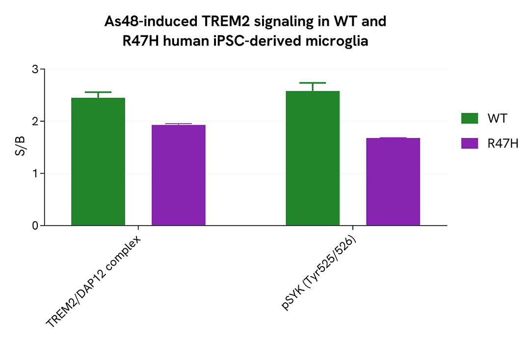
Impaired TREM2/DAP12 complex formation and SYK activation in R47H TREM2 mutant human iPSC-derived microglia
WT and R47H microglia differentiated from human iPSCs (iCell® Microglia, FUJIFILM Cellular Dynamics) were seeded at 70,000 cells per well in a 96-well microplate and incubated for 72 hours. Following centrifugation at 200 × g for 3 minutes, the supernatant was removed, and cells were pre-incubated with 30 µM As48 for 20 minutes. After treatment, the cells were lysed with Lysis Buffer #3 (1X) for 30 minutes at room temperature under gentle shaking.
Then,16 µL of each cell lysate were transferred into a low-volume 384-well white microplate, followed by the addition of 4 µL of premixed HTRF detection reagents. The HTRF signal was measured following an overnight incubation.
The R47H TREM2 variant, a well-characterized Alzheimer’s Disease risk factor, reduces ligand binding and impairs microglial responses, including migration toward and clustering around amyloid plaques, as well as the clearance of cellular debris. Consistent with the impaired signaling associated with this mutation, R47H iPSC-derived microglia exhibited lower levels of TREM2/DAP12 complex formation and reduced SYK phosphorylation (Tyr525/526) compared to WT iPSC-derived microglia.
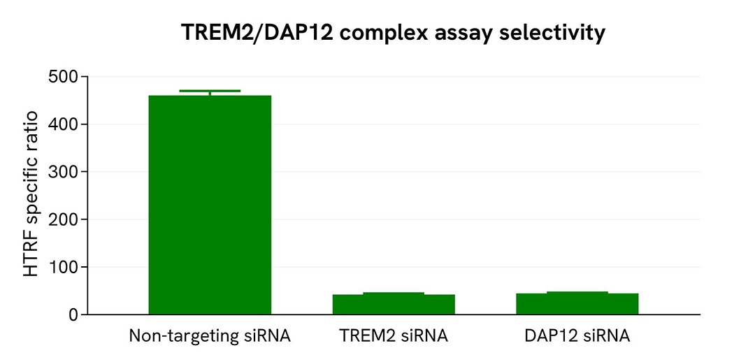
Validation of the specificity of the TREM2/DAP12 Complex assay using Accell siRNA
THP-1 cells were seeded into a 96-well microplate at 25,000 cells per well and transfected with either Accell TREM2 siRNA, Accell DAP12 siRNA, or a non-targeting control siRNA. Following a 72-hour incubation, the cells were treated with 100 nM PMA for 48 hours and then lysed with Lysis Buffer #3 (1X) for 30 minutes at room temperature under gentle shaking.
For the detection step, 16 µL of cell lysate were transferred into a low-volume 384-well white microplate, followed by the addition of 4 µL of premixed HTRF detection reagents. The HTRF signal was measured following an overnight incubation.
Transfection with either TREM2- or DAP12-specific siRNA resulted in an approximately 90% reduction in signal compared to the non-targeting siRNA control, demonstrating the specificity of the HTRF TREM2/DAP12 Complex assay.
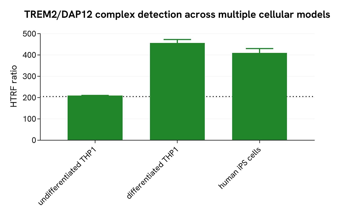
Assessment of the TREM2/DAP12 Complex level in various cell lines
Undifferentiated THP-1 cells, PMA-differentiated THP-1 cells (100 nM PMA for 20 hours), and human iPSC-derived microglia (iCell® Microglia, FUJIFILM Cellular Dynamics) were seeded at 70,000 cells per well in a 96-well microplate and incubated for 20 hours. Following incubation, the supernatant was removed, and the cells were lysed with Lysis Buffer #3 for 30 minutes at room temperature under gentle shaking, according to the appropriate protocol for adherent and suspension cells.
Then, 16 µL of each lysate were transferred into a low-volume 384-well white microplate, followed by the addition of 4 µL of premixed HTRF detection reagents. The HTRF signal was measured following an overnight incubation.
The HTRF TREM2/DAP12 Complex assay successfully detected the TREM2/DAP12 complex across multiple cellular models, revealing distinct levels of TREM2/DAP12 complex formation and demonstrating the assay's broad applicability for studying TREM2 signaling.
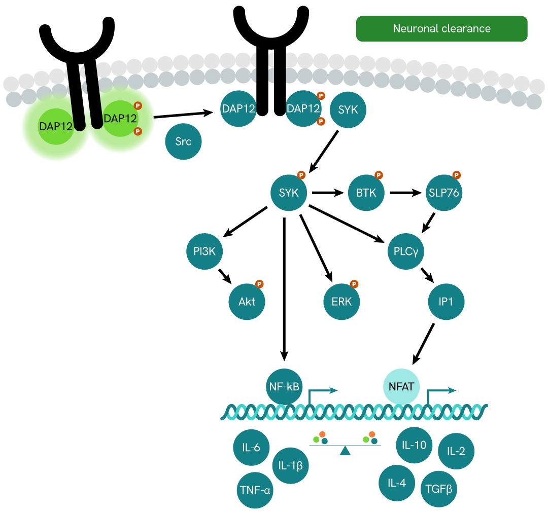
Simplified pathway
TREM2 signaling pathway
TREM2 (Triggering Receptor Expressed on Myeloid Cells 2) is a member of the TREM receptor family, which regulates the activity of various immune cell types, including neutrophils, monocytes/macrophages, microglia, and dendritic cells.TREM2 plays a critical role in modulating innate immune responses, promoting cell survival, phagocytosis, lipid sensing, and tissue homeostasis.
Due to its short cytoplasmic tail, TREM2 lacks intrinsic signaling activity and therefore associates with the adaptor protein DAP12 (TYROBP) to transduce signals into the cell. Upon ligand binding, the TREM2/DAP12 complex is activated, leading to phosphorylation of the two tyrosine residues within the immunoreceptor tyrosine-based activation motif (ITAM) of DAP12 by Src family kinases. These phosphorylated residues recruit and activate SYK kinase, which subsequently triggers downstream signaling pathways including PI3K-AKT, ERK/MAPK, PLCγ, and NF-κB.
In microglia, activation of the TREM2 pathway regulates migration, proliferation, survival, phagocytosis, and the response to pathological stimuli such as amyloid-β (Aβ) aggregates associated with Alzheimer’s Disease. Dysregulation of TREM2 signaling has been linked to impaired microglial function and increased risk of neurodegenerative disorders, highlighting the pathway as an attractive therapeutic target.
Specifications
| Application |
Cell Signaling
|
|---|---|
| Automation Compatible |
Yes
|
| Brand |
HTRF
|
| Detection Modality |
HTRF
|
| Lysis Buffer Compatibility |
Lysis Buffer 3
|
| Product Group |
Kit
|
| Sample Volume |
16 µL
|
| Shipping Conditions |
Shipped in Dry Ice
|
| Target |
TREM2/DAP12
|
| Target Class |
Phosphoproteins
|
| Target Species |
Human
|
| Technology |
TR-FRET
|
| Therapeutic Area |
Alzheimer
Inflammation
Parkinson
|
| Unit Size |
10,000 Assay Points
|
Resources
Are you looking for resources, click on the resource type to explore further.
Loading...


How can we help you?
We are here to answer your questions.