
US

Revvity Sites Globally
Select your location.
*e-commerce not available for this region.

HTRF Human and Mouse Total WDR5 Detection Kit, 10,000 Assay Points
HTRF Human and Mouse Total WDR5 Detection Kit, 10,000 Assay Points
generic HTRF total primary image
This HTRF kit allows for the cell-based quantitative detection of Total WDR5.
| Feature | Specification |
|---|---|
| Application | Cell Signaling |
| Sample Volume | 16 µL |
This HTRF kit allows for the cell-based quantitative detection of Total WDR5.
Product variants
Unit Size: 500 assay points
Part #:
64WDR5TPEG
List price
USD 2,387.00
Your price:
Unit Size: 10,000 assay points
Part #:
64WDR5TPEH
List price
USD 13,884.00
Your price:
For research use only. Not for use in diagnostic procedures. All products to be used in accordance with applicable laws and regulations including without limitation, consumption and disposal requirements under European REACH regulations (EC 1907/2006).
HTRF Human and Mouse Total WDR5 Detection Kit, 10,000 Assay Points
generic HTRF total primary image
Loading...
Product information
Overview
WD repeat-containing protein 5 (WDR5) is a member of the WD40 protein family and plays a key role in various biological processes. It functions as a core component of the mixed lineage leukemia (MLL) histone methyltransferase complexes. WDR5 is essential for the methylation of histone H3 at lysine 4 (H3K4) on chromatin, a reaction catalyzed by the MLL1 complex, and is involved in MLL1-mediated regulation of gene transcription. Overexpression of WDR5 has been linked to a wide range of cancers, making it a prominent target for anti-cancer drug discovery efforts, including the development of PROTAC degraders.
How it works
Total WDR5 assay principle
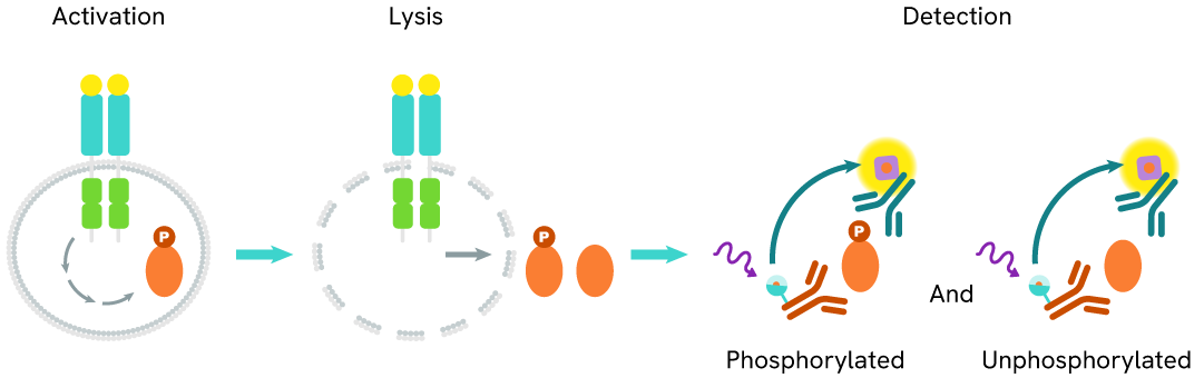
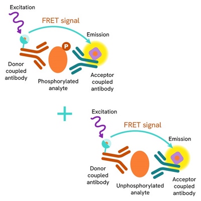
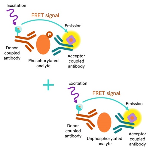
The Total WDR5 assay measures WDR5 levels in cells. Unlike Western Blot, the assay is entirely plate-based and does not require gels, electrophoresis, or transfer. The assay uses 2 antibodies, one labeled with a donor fluorophore and the other with an acceptor. Both antibodies are highly specific for a distinct epitope on the protein.
In the presence of WDR5 this enables an immune-complex formation involving both labeled antibodies, and which brings the donor fluorophore into close proximity to the acceptor, thereby generating a FRET signal. Its intensity is directly proportional to the concentration of total protein present in the sample, and provides a means of assessing the protein's phosphorylation state under a no-wash assay format.
Total WDR5 two-plate assay protocol
The two-plate protocol involves culturing cells in a 96-well plate before lysis, then transferring lysates into a 384-well low volume detection plate before the addition of Total WDR5 HTRF detection reagents. This protocol allows for the cells' viability and confluence to be monitored.

Total WDR5 one-plate assay protocol
Detection of Total WDR5 with HTRF reagents can be performed in a single plate used for culturing, stimulation, and lysis. No washing steps are required. This HTS designed protocol allows miniaturization while maintaining robust HTRF quality.

Assay validation
Total WDR5 modulation using PROTAC on MIA PaCa-2 cells
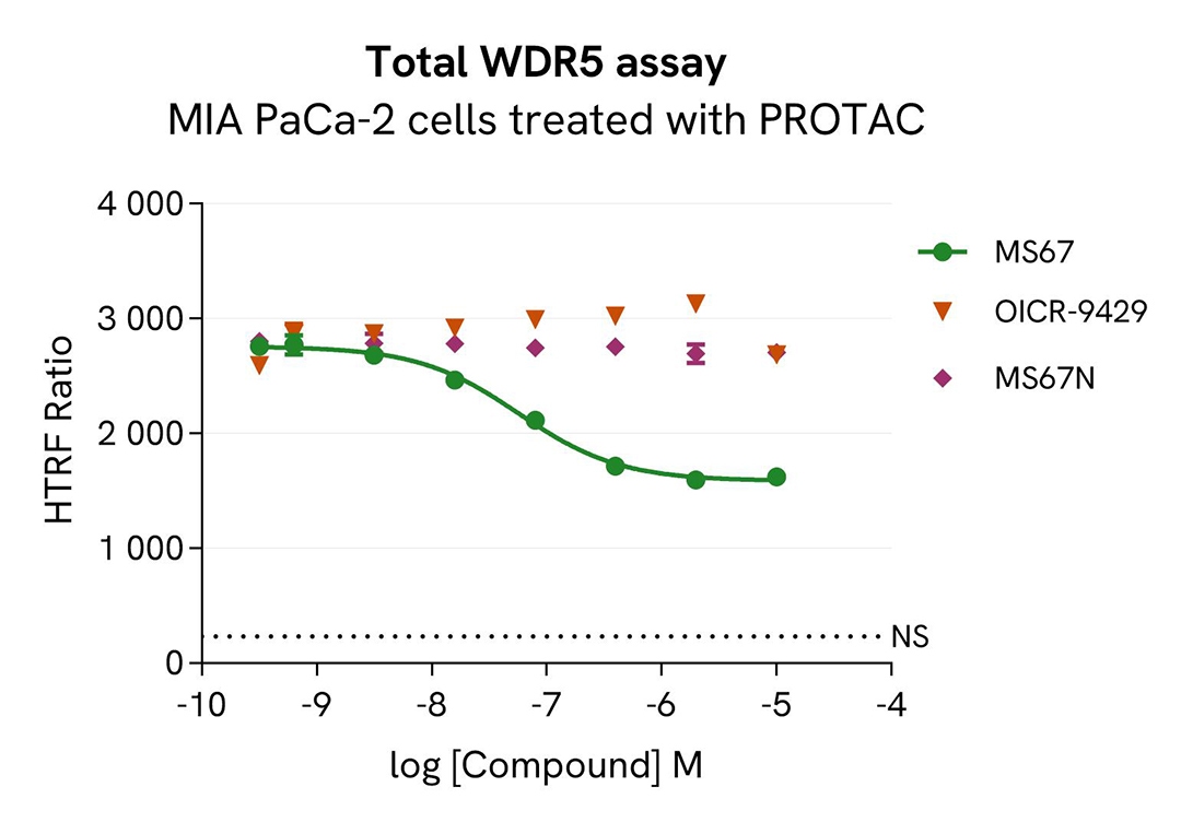
MIA PaCa-2 cells were seeded in 96-well culture plates at a density of 25,000 cells per well, and then incubated for 24 hours at 37°C with 5% CO2. Cells were then treated for 4 hours with increasing concentrations of PROTAC MS67, MS67N, and OICR-9429.
Following treatment, the culture medium was removed, and 50 µL of supplemented lysis buffer #6 (1X) were added to each well. Subsequently, 16 µL of lysate were transferred into a 384-well low-volume white microplate, and 4 µL of HTRF Total WDR5 detection antibodies were added.
An additional volume of lysate was also transferred to the microplate to assess alpha-tubulin levels and cytotoxicity, using the Alpha-Tubulin Housekeeping Cellular Kit (64ATUBPET/G/H) and the ATPlite Luminescence Assay System (#6016736), respectively. HTRF signals were recorded after overnight incubation.
PROTAC MS67 induced a dose-dependent decrease in WDR5 protein levels, with a DC₅₀ of 57 nM after 4 hours. As expected, MS67N and OICR-9429 had no effect on WDR5 protein levels. Alpha-tubulin expression and ATP levels remained stable, as expected (data not shown).
Total WDR5 modulation using PROTAC is mediated by ubiquitin-proteasome system
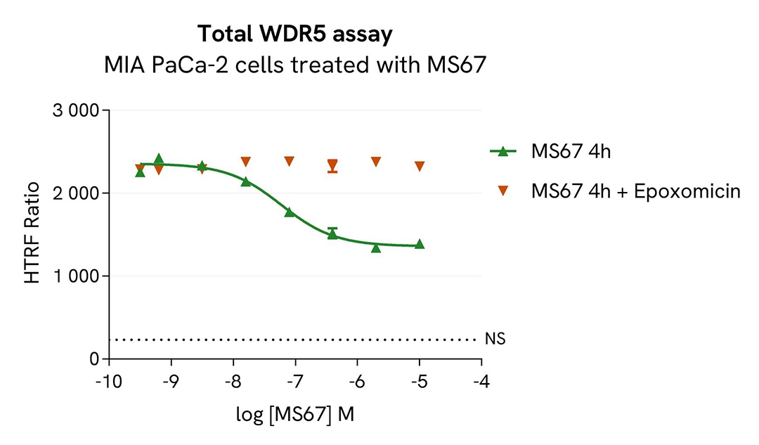
MIA PaCa-2 cells were seeded in 96-well culture plates at a density of 25,000 cells per well, and then incubated for 24 hours at 37°C with 5% CO₂. Cells were next treated with increasing concentrations of PROTAC MS67 for 4 hours. The proteasome inhibitor epoxomicin (1 µM) was either added 1 hour prior to MS67 treatment, or omitted for control conditions.
Following treatment, the culture medium was removed, and 50 µL of supplemented lysis buffer #6 (1X) were added to each well. Then 16 µL of lysate were transferred into a 384-well low-volume white microplate, and 4 µL of HTRF Total WDR5 detection antibodies were added.
An additional volume of lysate was also transferred to assess alpha-tubulin levels and cytotoxicity, using the Alpha-Tubulin Housekeeping Cellular Kit (64ATUBPET/G/H) and the ATPlite Luminescence Assay System (#6016736), respectively. HTRF signals were recorded after overnight incubation.
MS67-induced WDR5 degradation was prevented in the presence of epoxomicin, confirming that this degradation is mediated by the ubiquitin-proteasome system.
Total WDR5 modulation kinetics using PROTAC
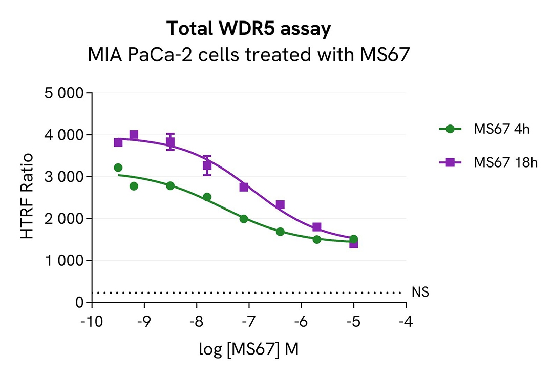
MIA PaCa-2 cells were seeded in 96-well culture plates at a density of 25,000 cells per well and then incubated for 24 hours at 37°C with 5% CO₂. Cells were next treated with increasing concentrations of PROTAC MS67 for either 4 or 18 hours.
Following treatment, the culture medium was removed, and 50 µL of supplemented lysis buffer #6 (1X) were added to each well. Then 16 µL of lysate were transferred to a 384-well low-volume white microplate, and 4 µL of HTRF Total WDR5 detection antibodies were added.
An additional volume of lysate was also transferred to assess alpha-tubulin levels and cytotoxicity, using the Alpha-Tubulin Housekeeping Cellular Kit (64ATUBPET/G/H) and the ATPlite Luminescence Assay System (#6016736), respectively. HTRF signals were recorded after overnight incubation.
PROTAC MS67 induced a dose-dependent decrease in WDR5 protein levels, with a DC₅₀ of 31 nM after 4 hours and 118 nM after 18 hours.
Total WDR5 modulation using PROTAC is mediated by neddylation
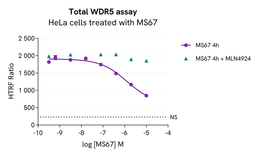
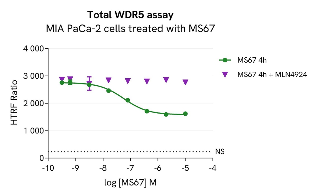
HeLa and MIA PaCa-2 cells were seeded in 96-well culture plates at a density of 25,000 cells per well and incubated for 24 hours at 37 °C with 5% CO2. Cells were then treated with increasing concentrations of PROTAC MS67 for 4 hours. The NEDD8-activating enzyme inhibitor MLN4924 (1 µM) was either added 1 hour prior to MS67 treatment, or omitted for control conditions.
After treatment, the culture medium was removed, and 50 µL of supplemented lysis buffer #6 (1X) were added to each well. Then 16 µL of lysate were transferred into a 384-well low-volume white microplate, and 4 µL of HTRF Total WDR5 detection antibodies were added.
An additional volume of lysate was also transferred to assess alpha-tubulin levels and cytotoxicity, using the Alpha-Tubulin Housekeeping Cellular Kit (64ATUBPET/G/H) and the ATPlite Luminescence Assay System (#6016736), respectively. HTRF signals were recorded after overnight incubation.
MS67-induced WDR5 degradation was prevented in the presence of MLN4924, a protein neddylation inhibitor. This result demonstrated that WDR5 degradation induced by MS67 is mediated by the NEDD8 conjugation pathway.
Validation of the selectivity of Total WDR5 assay using siRNA
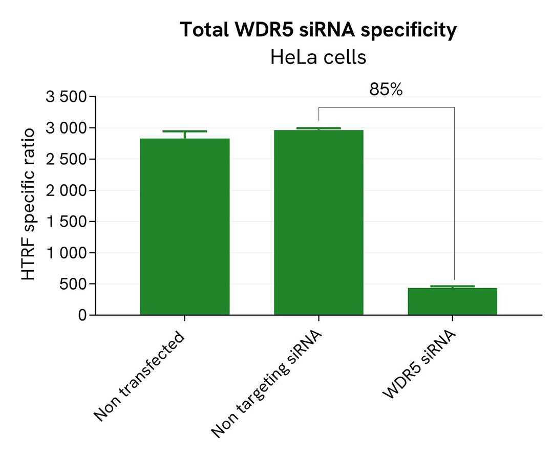
HeLa cells were plated in a 96-well plate at a density of 10,000 cells per well, and then cultured for 24 hours. The cells were next transfected with 25 nM of WDR5-specific siRNA or a negative control siRNA. After 24 hours, the medium was replaced with fresh culture medium, and the cells were incubated for an additional 24 hours.
Following treatment, cells were lysed with 50 µL of supplemented lysis buffer #6 (4X) for 30 minutes at room temperature under gentle shaking. Subsequently, 16 µL of lysate were transferred to a 384-well low-volume white microplate, followed by the addition of 4 µL of HTRF Total WDR5 detection antibodies. HTRF signals were recorded after overnight incubation.
Transfection with WDR5-specific siRNA resulted in an 85% decrease in signal compared to cells transfected with the negative control siRNA. These results demonstrate the selectivity of the HTRF Total WDR5 assay.
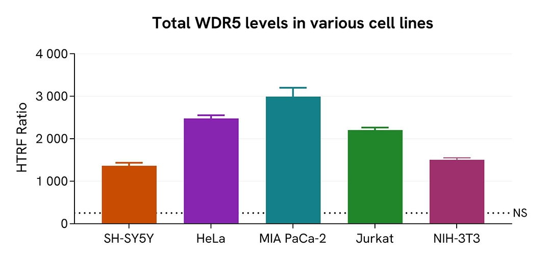
Assessment of Total WDR5 level in various cell lines
Adherent (HeLa, SH-SY5Y, MIA PaCa-2, and NIH3T3) and suspension (Jurkat) cells were seeded at 25,000 cells/well in a 96-well microplate. After 24h of incubation, the cells were lysed for 30 minutes with supplemented lysis buffer #6, following the protocol for adherent or suspended cells at RT under gentle shaking.
16 µL of lysate were transferred into a 384-well low volume white microplate before the addition of 4 µL of the HTRF Total WDR5 detection reagents. The HTRF signal was recorded after overnight incubation.
The HTRF Total WDR5 assay efficiently detected Total WDR5 in various cellular models expressing different levels of the protein.
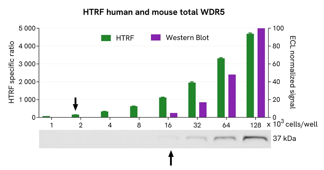
HTRF Total WDR5 assay compared to Western Blot
HeLa cells were grown in a T175 flask in complete culture medium at 37°C, 5% CO2 until 80% confluence. After a 24h incubation, the cells were lysed with 3 mL of supplemented lysis buffer #6 (1X) for 30 minutes at RT under gentle shaking.
Serial dilutions of the cell lysate were performed using supplemented lysis buffer, and 16 µL of each dilution were transferred into a low volume white microplate before the addition of 4 µL of HTRF Total WDR5 detection reagents. Equal amounts of lysates were used for a side-by-side comparison between HTRF and Western Blot.
In these conditions, the HTRF Total WDR5 assay was 8 times more sensitive than the Western Blot technique.
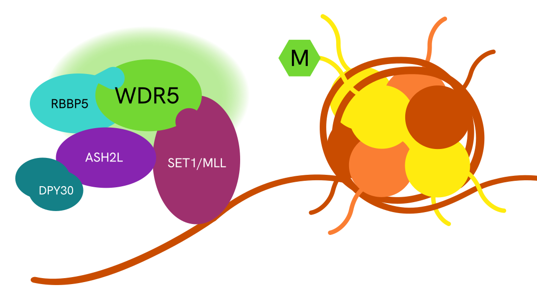
Simplified pathway
WDR5 signaling pathway
WDR5 forms a core component of a multiprotein complex with MLL, RBBP5, ASH2L, and DPY30 at MLL target gene promoters. This complex catalyzes histone H3 lysine 4 (H3K4) trimethylation, promoting transcription of MLL target genes. The resulting gene expression changes drive cell proliferation, block differentiation, support cell survival, and contribute to tumorigenesis and therapy resistance.
Specifications
| Application |
Cell Signaling
|
|---|---|
| Automation Compatible |
Yes
|
| Brand |
HTRF
|
| Detection Modality |
HTRF
|
| Lysis Buffer Compatibility |
Lysis Buffer 6
|
| Molecular Modification |
Total
|
| Product Group |
Kit
|
| Sample Volume |
16 µL
|
| Shipping Conditions |
Shipped in Dry Ice
|
| Target |
WDR5
|
| Target Class |
Phosphoproteins
|
| Target Species |
Human
Mouse
|
| Technology |
TR-FRET
|
| Therapeutic Area |
Oncology
|
| Unit Size |
10,000 assay points
|
Resources
Are you looking for resources, click on the resource type to explore further.

Loading...


How can we help you?
We are here to answer your questions.