
US

Revvity Sites Globally
Select your location.
*e-commerce not available for this region.

HTRF Human Total ITK Detection Kit, 500 Assay Points
HTRF Human Total ITK Detection Kit, 500 Assay Points
HTRF total without phospho primary image
The HTRF human total ITK detection kit supports the cell-based quantitative detection of Total ITK, in a homogeneous (no wash steps, no separation steps) format.
| Feature | Specification |
|---|---|
| Application | Cell Signaling |
| Sample Volume | 16 µL |
The HTRF human total ITK detection kit supports the cell-based quantitative detection of Total ITK, in a homogeneous (no wash steps, no separation steps) format.
Product variants
Unit Size: 500 Assay Points
Part #:
64ITKTPEG
List price
USD 2,387.00
Your price:
Unit Size: 10,000 Assay Points
Part #:
64ITKTPEH
List price
USD 13,884.00
Your price:
For research use only. Not for use in diagnostic procedures. All products to be used in accordance with applicable laws and regulations including without limitation, consumption and disposal requirements under European REACH regulations (EC 1907/2006).
HTRF Human Total ITK Detection Kit, 500 Assay Points
HTRF total without phospho primary image
Loading...
Product information
Overview
Interleukin-2-inducible T-cell kinase (ITK) is a member of the Tec family of cytoplasmic tyrosine kinases. ITK regulates and amplifies T-cell receptor (TCR) signaling cascades and plays a critical role in T-cell development, differentiation, cytokine production, and apoptosis. ITK is frequently overexpressed or constitutively activated in certain T-cell lymphomas, contributing to malignant T-cell proliferation and survival.
ITK is strongly associated with Th2-mediated immune responses through the promotion of IL-4, IL-5, and IL-13 production, which contributes to the development of allergic diseases such as asthma. In addition, aberrant ITK signaling has been implicated in autoimmune disorders, including rheumatoid arthritis and psoriasis, through dysregulation of T-cell activation and function.
Due to its central role in immune regulation, ITK has emerged as an attractive therapeutic target. Several therapeutic strategies, including small-molecule inhibitors and proteolysis-targeting chimeras (PROTACs), are being developed to modulate ITK activity and achieve broad control of T-cell-mediated immune responses.
HTRF assays offer many advantages over other technologies:
- Homogeneous add-and-read format
- No wash steps
- Low background
- Straightforward miniaturization from 96- or 384-well microplates to high density assay formats such as 384-well low volume and 1536-well plates
- Stable signal, providing flexibility in readout time or assay size
How it works
Total ITK assay principle
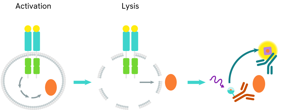
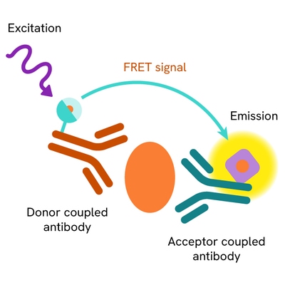
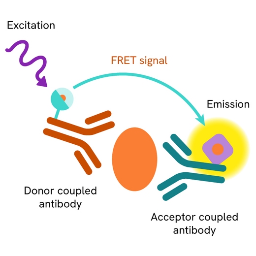
The total ITK assay measures ITK levels in cells. Unlike Western Blot, the assay is entirely plate-based and does not require gels, electrophoresis, or transfer. The assay uses 2 antibodies, one labeled with a donor fluorophore and the other with an acceptor. Both antibodies are highly specific for a distinct epitope on the protein.
The presence of ITK leads to the formation of an immune complex involving both labeled antibodies, bringing the donor fluorophore into close proximity to the acceptor and generating a FRET signal. Its intensity is directly proportional to the concentration of total protein in the sample, and provides a means of assessing the protein's phosphorylation state under a no-wash assay format.
Total ITK two-plate assay protocol
The two-plate protocol involves culturing cells in a 96-well plate before lysis, then transferring lysates into a 384-well low volume detection plate before the addition of total ITK HTRF detection reagents. This protocol allows the cells viability and confluence to be monitored.

Total ITK one-plate assay protocol
Detection of total ITK with HTRF reagents can be performed in a single plate used for culturing, stimulation, and lysis. No washing steps are required. This HTS designed protocol allows miniaturization while maintaining robust HTRF quality.

Assay validation
Validation of HTRF total ITK assay for monitoring PROTAC-induced target degradation
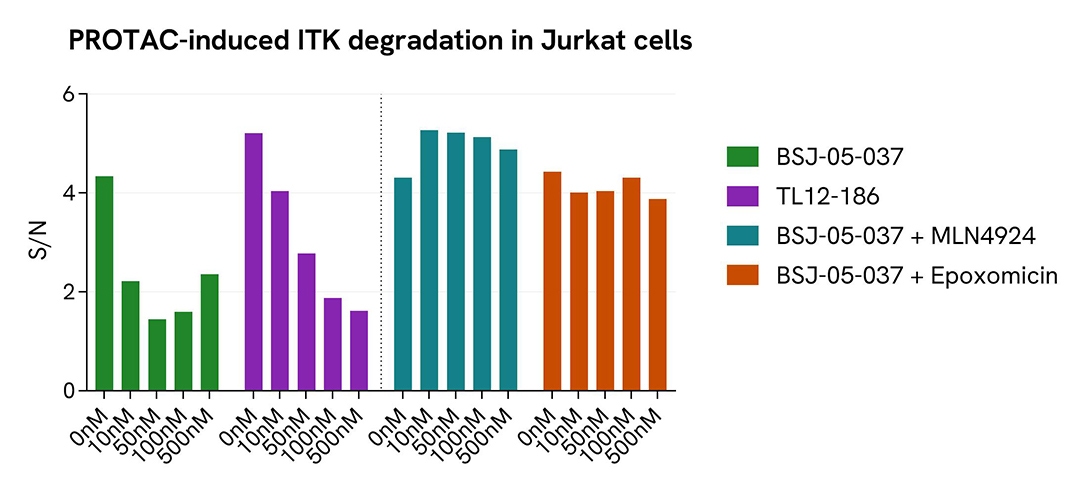
Jurkat cells were seeded in a 96-well tissue culture-treated plate (200,000 cells/well) in complete culture medium and incubated for 24 hours prior to the experiment, at 37°C in a humidified atmosphere containing 5% CO2. Cells were then treated for 4 hours with increasing concentrations of BSJ-05-037 or TL12-186, either alone or in the presence of the proteasome inhibitor epoxomicin (1 µM) or the neddylation inhibitor MLN4924 (3 µM).
BSJ-05-037 is a selective ITK-targeting PROTAC, whereas TL12-186 is a cereblon-dependent multi-kinase degrader.
Following treatment, cells were lysed with supplemented Lysis Buffer #4 for 30 minutes at room temperature under gentle shaking. For detection, 16 µL of cell lysate were transferred into a 384-well low-volume white microplate, and 4 µL of HTRF total ITK detection reagents were added. The HTRF signal was measured on a multimode microplate reader following an overnight incubation at room temperature.
In parallel, cell viability was assessed using the ATPlite™ 1step Kit (Revvity, #6016736/1/9). Five microliters of the same cell lysate were transferred into an ATPlite-compatible 96-well low-volume white microplate, and 25 µL of ATPlite 1step detection reagent were added. Luminescence was measured using an EnVision™ Nexus reader after a 10-minute incubation.
Both PROTACs induced a concentration-dependent reduction in total ITK levels in Jurkat cells after 4 hours of treatment, as measured with the HTRF Total ITK assay, without affecting cell viability. ITK degradation was rescued by co-treatment with either epoxomicin or MLN4924, confirming that the observed effects were dependent on proteasome-mediated degradation and the activity of the Cullin-RING E3 ubiquitin ligase complex.
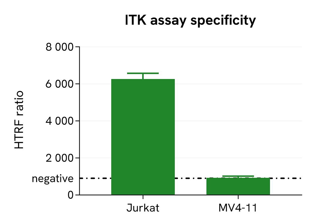
Selective detection of endogenous ITK in Jurkat (ITK+) and MV4-11 (ITK-) cells
Jurkat cells, a human T-lymphocyte cell model, are known to express ITK, whereas MV4-11 cells, a human leukemia cell model, are not expected to express this kinase. Consistent with this observation, publicly available transcriptomic datasets indicate that ITK mRNA expression is undetectable or only present at negligible levels in MV4-11 cells.
Both cell lines were seeded in a 96-well tissue culture-treated plate (400,000 cells/well in 30 µL of complete culture medium) and incubated overnight at 37°C in a humidified atmosphere containing 5% CO2.
Following incubation, cells were lysed with 10 µL of supplemented Lysis Buffer #4 (4X) for 30 minutes at room temperature under gentle shaking. For detection, 16 µL of cell lysate were transferred into a 384-well low-volume white microplate, and 4 µL of HTRF Total ITK detection reagents were added. The HTRF signal was measured on a multimode microplate reader following an overnight incubation at room temperature.
As expected, a robust HTRF signal was detected in ITK-expressing Jurkat cells, whereas no signal above background was observed in MV4-11 cells. These results demonstrate the ability of the HTRF Total ITK assay to specifically detect endogenous ITK expression in cellular lysates.
Specifications
| Application |
Cell Signaling
|
|---|---|
| Automation Compatible |
Yes
|
| Brand |
HTRF
|
| Detection Modality |
HTRF
|
| Lysis Buffer Compatibility |
Lysis Buffer 4
|
| Molecular Modification |
Total
|
| Product Group |
Kit
|
| Sample Volume |
16 µL
|
| Shipping Conditions |
Shipped in Dry Ice
|
| Target |
ITK (Interleukin-2-inducible T-cell kinase)
|
| Target Class |
Phosphoproteins
|
| Target Species |
Human
|
| Technology |
TR-FRET
|
| Therapeutic Area |
Autoimmunity
Inflammation
|
| Unit Size |
500 Assay Points
|
Resources
Are you looking for resources, click on the resource type to explore further.
Loading...


How can we help you?
We are here to answer your questions.