
US

Revvity Sites Globally
Select your location.
*e-commerce not available for this region.

HTRF Human Total BCL-2 Detection Kit, 500 Assay Points
HTRF Human Total BCL-2 Detection Kit, 500 Assay Points
HTRF total without phospho primary image
The HTRF human Total BCL-2 detection kit supports the quantitative detection of total BCL-2 protein levels in cell-based samples. This homogeneous assay requires no wash or separation steps and delivers robust results suitable for drug discovery and signaling studies.
| Feature | Specification |
|---|---|
| Application | Cell Signaling |
| Sample Volume | 16 µL |
The HTRF human Total BCL-2 detection kit supports the quantitative detection of total BCL-2 protein levels in cell-based samples. This homogeneous assay requires no wash or separation steps and delivers robust results suitable for drug discovery and signaling studies.
Product variants
Unit Size: 500 Assay Points
Part #:
64BCL2TPEG
List price
USD 2,387.00
Your price:
Unit Size: 10,000 Assay Points
Part #:
64BCL2TPEH
List price
USD 13,884.00
Your price:
For research use only. Not for use in diagnostic procedures. All products to be used in accordance with applicable laws and regulations including without limitation, consumption and disposal requirements under European REACH regulations (EC 1907/2006).
HTRF Human Total BCL-2 Detection Kit, 500 Assay Points
HTRF total without phospho primary image
Loading...
Product information
Overview
BCL-2 (B-cell lymphoma 2) is a key anti-apoptotic protein belonging to the BCL-2 family and plays a central role in regulating the intrinsic apoptotic pathway. Primarily localized on the outer mitochondrial membrane, BCL-2 preserves mitochondrial integrity by inhibiting the activation of pro-apoptotic proteins such as BAX and BAK, thereby preventing cytochrome c release and subsequent activation of the caspase cascade that leads to programmed cell death.
As a critical regulator of cell fate, BCL-2 contributes to the maintenance of tissue homeostasis across a wide range of physiological processes. However, aberrant overexpression of BCL-2 is frequently observed in both hematological malignancies and solid tumors, where it promotes tumor cell survival and contributes to resistance to chemotherapy, targeted therapies, and other anticancer treatments.
Consequently, BCL-2 has emerged as a highly validated therapeutic target in oncology, highlighted by the development of selective BCL-2 inhibitors such as venetoclax, which has been studied across several leukemia and lymphoma research models.
Beyond cancer, BCL-2 has been implicated in numerous diseases associated with dysregulated apoptosis. In neurodegenerative disorders, including Alzheimer’s Disease and Parkinson’s Disease, modulation of BCL-2 expression is being investigated for its potential role in neuronal survival under stress conditions. In cardiovascular disease, the balance between pro- and anti-apoptotic BCL-2 family members is an active area of research in models of ischemic injury and heart failure. Additionally, BCL-2 is being actively investigated in inflammatory and autoimmune disorders, where prolonged survival of pathogenic immune cells may contribute to disease progression.
Given its central role in regulating cell survival and apoptosis, BCL-2 remains an important biomarker and therapeutic target for understanding disease mechanisms and developing new research approaches.
HTRF assays offer many advantages over other technologies:
- Homogeneous add-and-read format
- No wash steps
- Low background
- Straightforward miniaturization from 96- or 384-well microplates to high density assay formats such as 384-well low volume and 1536-well plates
- Stable signal, providing flexibility in readout time or assay size
How it works
Total BCL-2 assay principle
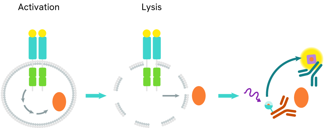
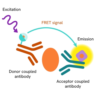
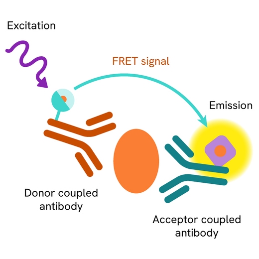
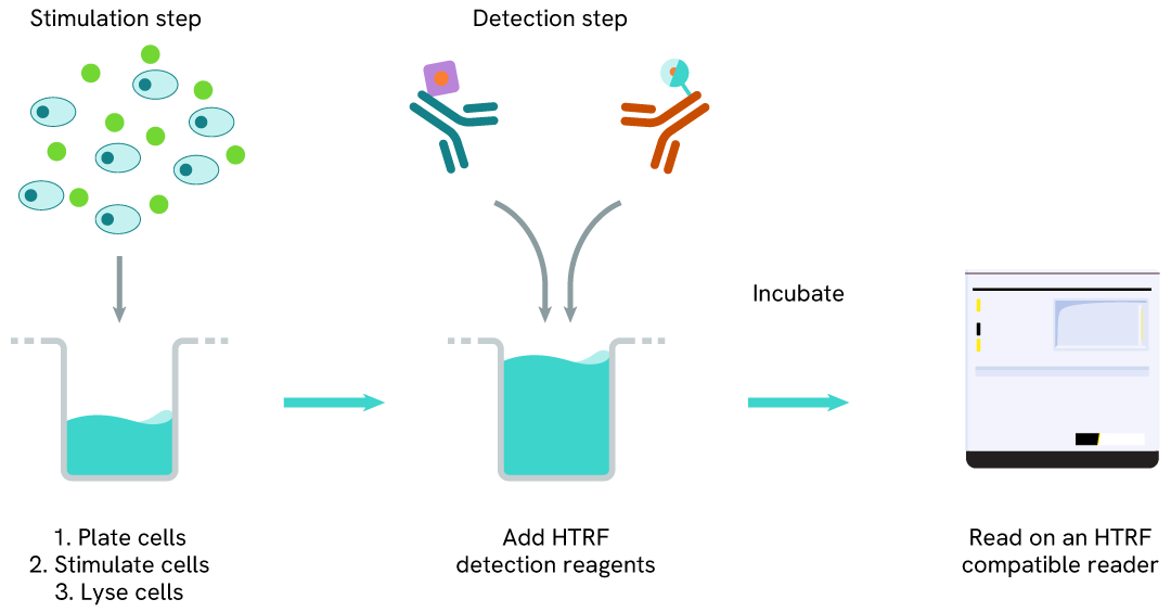
The total BCL-2 assay measures BCL-2 levels in cells. Unlike Western Blot, the assay is entirely plate-based and does not require gels, electrophoresis, or transfer. The assay uses 2 antibodies, one labeled with a donor fluorophore and the other with an acceptor. Both antibodies are highly specific for a distinct epitope on the protein.
The presence of BCL-2 leads to the formation of an immune complex involving both labeled antibodies, bringing the donor fluorophore into close proximity to the acceptor and generating a FRET signal. Its intensity is directly proportional to the concentration of total protein in the sample and provides a means of assessing the protein's phosphorylation state under a no-wash assay format.
Total BCL-2 two-plate assay protocol
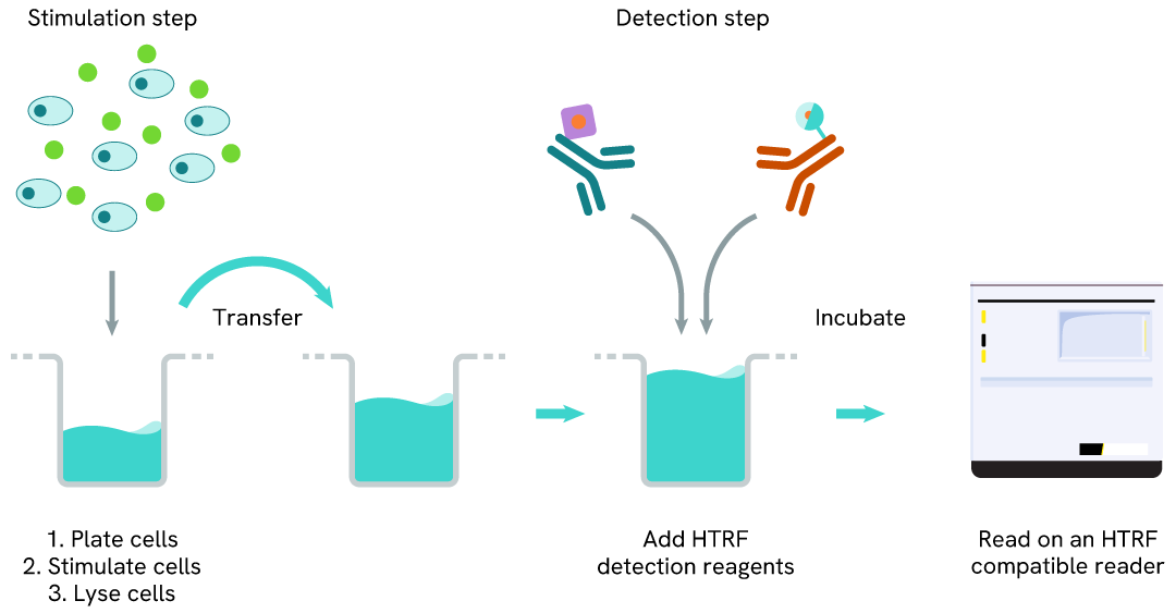
The two-plate protocol involves culturing cells in a 96-well plate before lysis, then transferring lysates into a 384-well low volume detection plate before the addition of total BCL-2 HTRF detection reagents. This protocol allows the cells' viability and confluence to be monitored.
Total BCL-2 one-plate assay protocol
Detection of total BCL-2 with HTRF reagents can be performed in a single plate used for culturing, stimulation, and lysis. No washing steps are required. This HTS designed protocol allows miniaturization while maintaining robust HTRF quality.
Assay validation
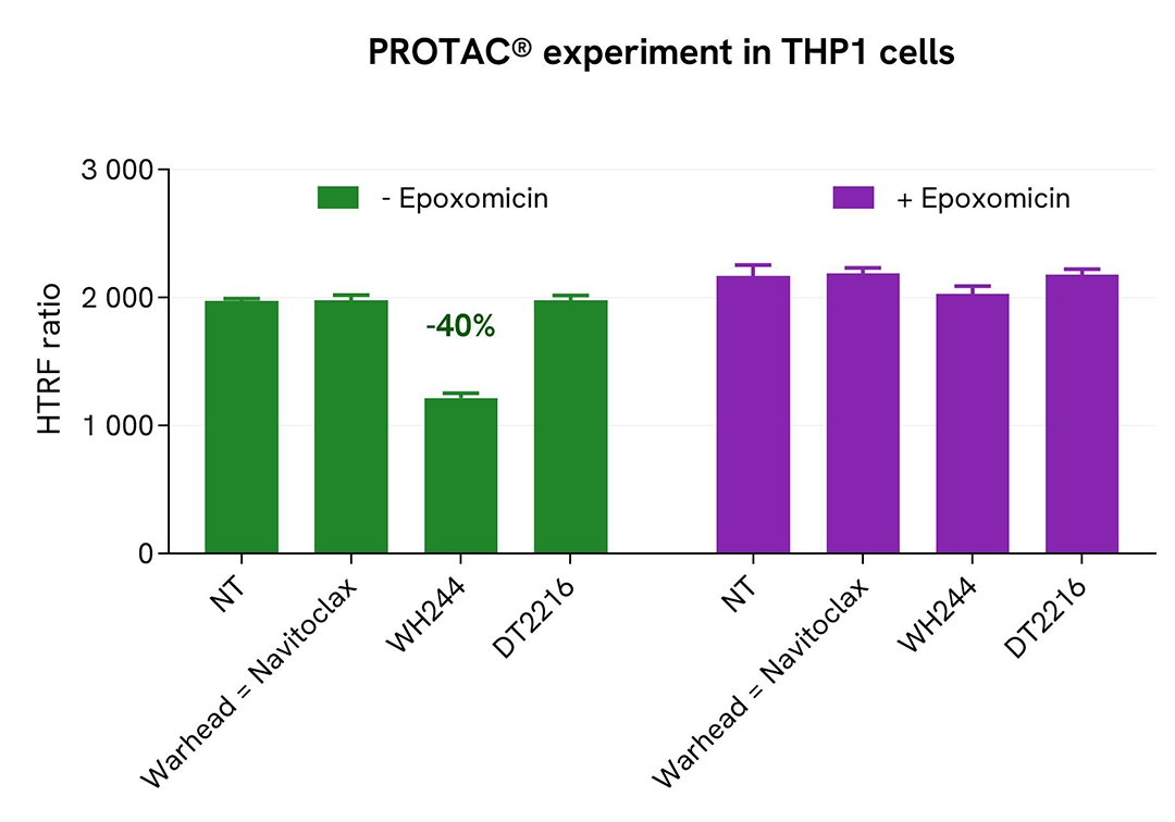
WH244 PROTAC® induces proteasome-dependent BCL-2 degradation in THP-1 cells.
THP-1 cells were cultured in 96-well plates (25,000 cells/well) for 24 h and subsequently treated with 1 µM WH244 (a dual BCL-2/BCL-XL degrader), DT2216 (a selective BCL-XL degrader), or navitoclax (warhead control), in the presence or absence of the proteasome inhibitor epoxomicin, for 4 h at 37°C and 5% CO₂. Following cell lysis, 16 µL of lysate were transferred into a low-volume 384-well white microplate, and 4 µL of HTRF Total BCL-2 detection antibodies were added. The HTRF signal was measured after a 3 h incubation.
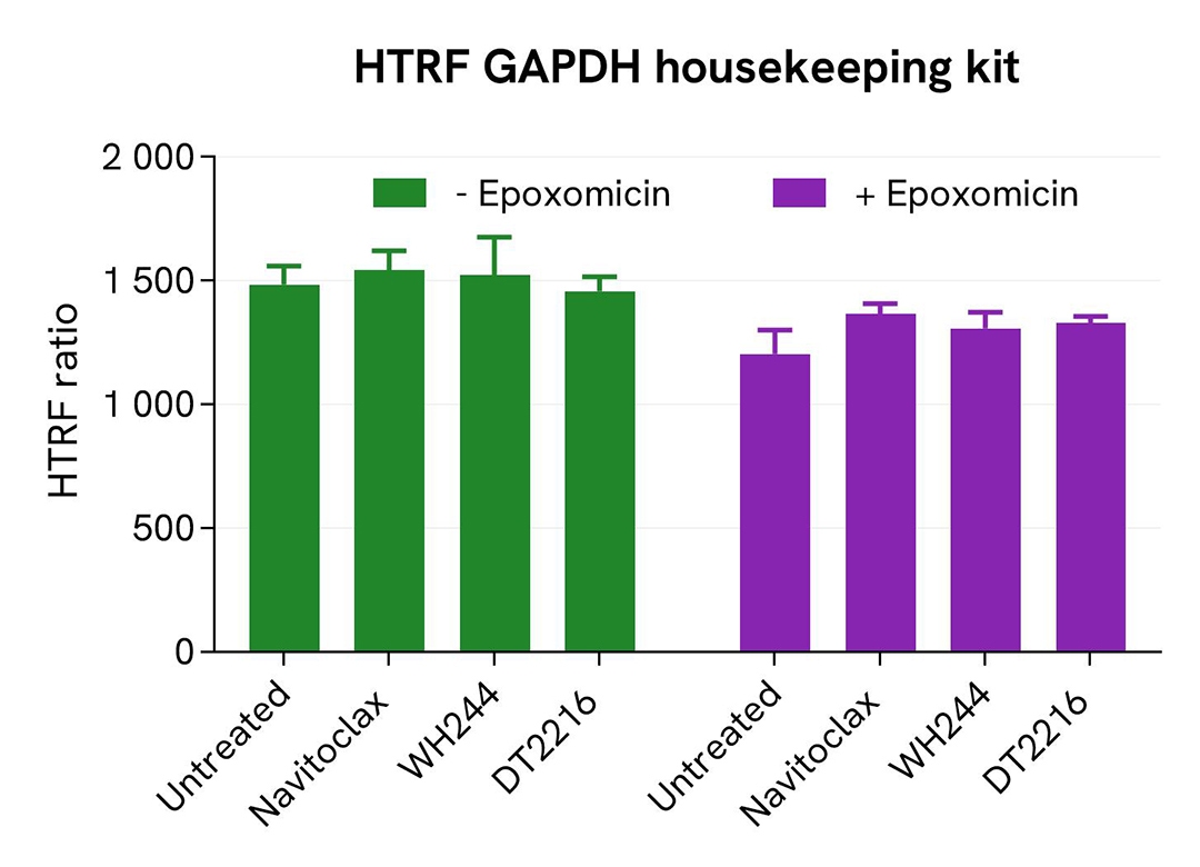
WH244 induced BCL-2 degradation, resulting in a 40% reduction in HTRF signal compared to untreated cells and navitoclax-treated controls. As expected, the selective BCL-XL degrader DT2216 did not affect BCL-2 protein levels. Co-treatment with epoxomicin completely rescued the BCL-2 HTRF signal, confirming that degradation was mediated by the ubiquitin-proteasome pathway. Cell viability was assessed in parallel using the HTRF GAPDH housekeeping cell-based assay and remained largely unaffected under all tested conditions, demonstrating that the observed decrease in BCL-2 levels resulted from specific PROTAC-mediated degradation rather than cytotoxicity.
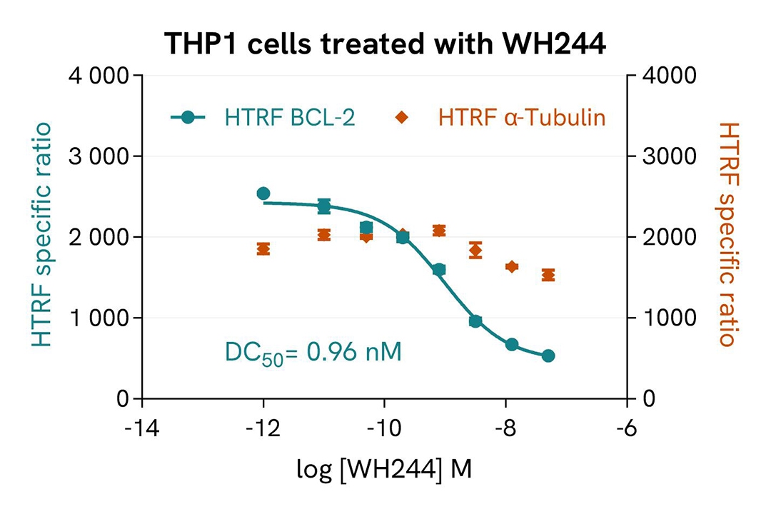
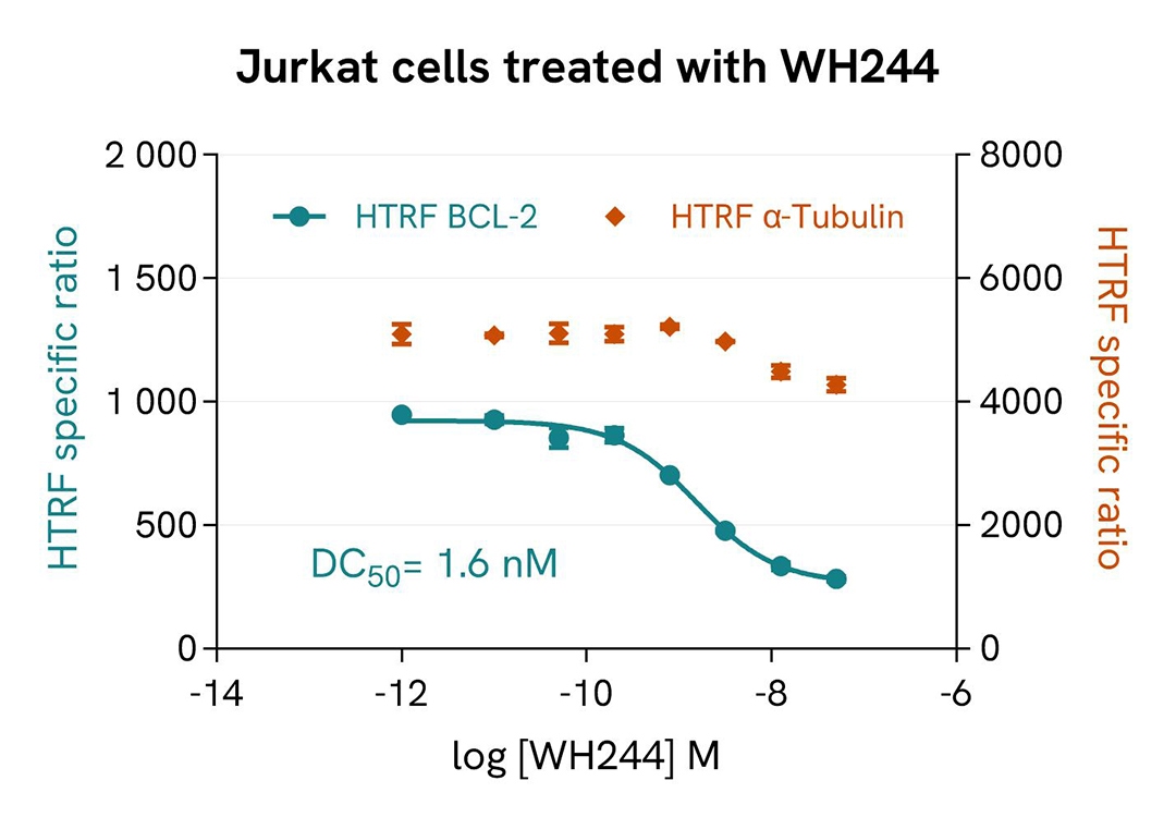
Dose-dependent BCL-2 degradation induced by WH244 PROTAC® in THP-1 and Jurkat cells
THP-1 cells (25,000 cells/well) and Jurkat cells (50,000 cells/well) were seeded in complete culture medium in a 96-well tissue culture-treated plate and incubated for 24 h at 37°C and 5% CO₂. Cells were then treated with increasing concentrations of the WH244 PROTAC® for 16 h and subsequently lysed with supplemented lysis buffer #3 (4X) for 30 min at room temperature under gentle shaking. For detection, 16 µL of cell lysate were transferred into a low-volume white microplate, and 4 µL of total BCL-2 detection reagents were added. The HTRF signal was measured after a 3 h incubation.
In parallel, α-tubulin levels were quantified as a control protein.
Treatment with WH244 induced a dose-dependent reduction in BCL-2 levels in both cell lines, whereas α-tubulin expression remained largely unchanged across the tested concentration range. These results demonstrate the selective degradation of BCL-2 by WH244 while confirming minimal effects on the control protein.
Validation of the specificity of total BCL-2 assay using HAP1 KO cell line
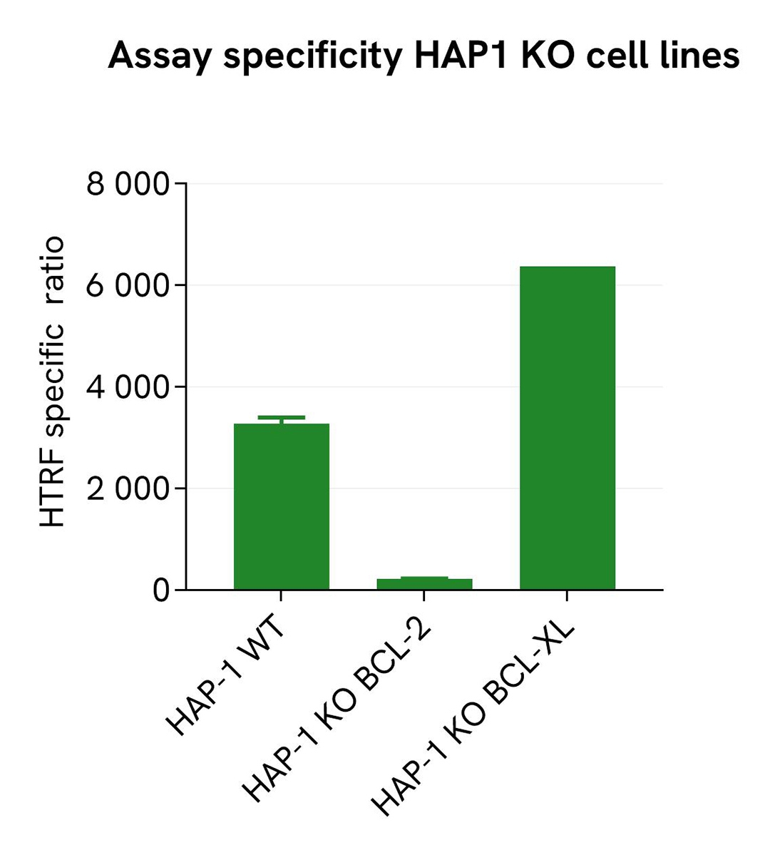
BCL-2 expression levels were assessed using the HTRF total BCL-2 kit in wild-type (WT), BCL-2 knockout (BCL-2 KO), and BCL-XL knockout (BCL-XL KO) HAP1 cell lines (100,000 cells/well).
The different cell lines were cultured in a 96-well plate for 24 h at 37°C and 5% CO₂. Cells were then lysed with supplemented Lysis Buffer #3 (1X), and 16 µL of cell lysate were transferred into a low-volume white microplate. Subsequently, 4 µL of premixed detection reagents were added. The HTRF signal was measured after a 3 h incubation at room temperature.
In BCL-2 KO HAP1 cells, the HTRF signal was reduced to the level of the non-specific signal, indicating a complete loss of BCL-2 expression. In contrast, BCL-2 was readily detected in both WT and BCL-XL KO cell lines.
These results demonstrate the high selectivity of the HTRF total BCL-2 kit, which specifically detects BCL-2 protein with no detectable cross-reactivity to the closely related BCL-XL family member under the tested conditions.
Assessment of total BCL-2 levels in various cell lines
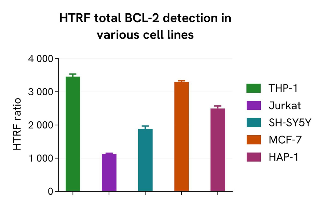
THP-1, Jurkat, SH-SY5Y, MCF-7, and HAP1 cells were seeded at 100,000 cells/well in a 96-well microplate. After overnight incubation, cells were lysed with supplemented Lysis Buffer #3, and 16 µL of lysate were transferred into a low-volume 384-well white microplate. Subsequently, 4 µL of HTRF total BCL-2 detection reagents were added. The HTRF signal was measured after a 3 h incubation at room temperature.
The HTRF total BCL-2 assay supports the robust detection of endogenous BCL-2 across a variety of cellular models expressing different levels of the protein, demonstrating its applicability across multiple cellular models for cell-based studies.
Simplified pathway
BCL-2 signaling pathway
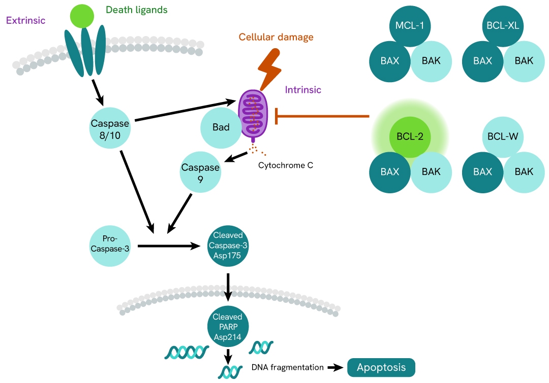
B-cell lymphoma 2 (BCL-2) is a key regulator of cell survival and apoptosis through the mitochondrial (intrinsic) apoptotic pathway. Under physiological conditions, BCL-2 is primarily localized on the outer mitochondrial membrane, endoplasmic reticulum, and nuclear envelope, where it helps maintain cellular homeostasis by preserving mitochondrial integrity and preventing apoptosis.
BCL-2 expression and activity are regulated by several signaling pathways. Activation of receptor tyrosine kinases stimulates the PI3K/AKT pathway, leading to increased BCL-2 expression through transcription factors such as CREB and NF-κB, thereby enhancing cellular resistance to apoptotic stimuli. In addition, activation of the NF-κB pathway by inflammatory cytokines, growth factors, or cellular stress promotes BCL-2 transcription and supports cell survival. Growth factor-mediated activation of the MAPK/ERK pathway can further enhance the anti-apoptotic function of BCL-2 through phosphorylation-dependent stabilization.
As an anti-apoptotic protein, BCL-2 prevents mitochondrial outer membrane permeabilization (MOMP) by binding to and sequestering the pro-apoptotic effector proteins BAX and BAK, thereby preventing their oligomerization within the mitochondrial membrane. Consequently, the release of cytochrome c and other apoptogenic factors into the cytosol is blocked, suppressing caspase activation and apoptosis. BCL-2 also binds to and neutralizes BH3-only proteins such as BIM, BID, PUMA, NOXA, and BAD, which are normally activated in response to cellular stress and promote BAX/BAK activation.
In response to severe cellular damage or prolonged stress, several mechanisms antagonize BCL-2 activity. The tumor suppressor p53 induces the expression of pro-apoptotic genes, including BAX, PUMA, and NOXA, shifting the balance toward apoptosis. Additionally, stress-activated kinases such as JNK can phosphorylate BCL-2, reducing its anti-apoptotic function. When BCL-2-mediated protection is overcome, BAX and BAK oligomerize within the mitochondrial membrane, triggering MOMP, cytochrome c release, apoptosome formation, caspase activation, and ultimately programmed cell death.
Dysregulation of BCL-2 is frequently observed in cancer, where BCL-2 overexpression promotes tumor cell survival and resistance to therapy. Consequently, BCL-2 has become an important therapeutic target, exemplified by the development and clinical investigation of selective BCL-2 inhibitors such as venetoclax in hematological malignancies.
Specifications
| Application |
Cell Signaling
|
|---|---|
| Automation Compatible |
Yes
|
| Brand |
HTRF
|
| Detection Modality |
HTRF
|
| Lysis Buffer Compatibility |
Lysis Buffer 3
|
| Molecular Modification |
Total
|
| Product Group |
Kit
|
| Sample Volume |
16 µL
|
| Shipping Conditions |
Shipped in Dry Ice
|
| Target |
BCL-2
|
| Target Class |
Phosphoproteins
|
| Target Species |
Human
|
| Technology |
TR-FRET
|
| Therapeutic Area |
Autoimmunity
Autophagy
Cancer
Cardiovascular
Central Nervous System
Inflammation
Oncology
|
| Unit Size |
500 Assay Points
|
Resources
Are you looking for resources, click on the resource type to explore further.
Loading...


How can we help you?
We are here to answer your questions.