


US
Revvity Sites Globally
Select your location.
*e-commerce not available for this region.

AlphaLISA Mouse IL-12 p70 Detection Kit, 100 Assay Points
AlphaLISA Mouse IL-12 p70 Detection Kit, 100 Assay Points
AlphaLISA Sandwich Anti-analyte Conjugated Acceptor Bead
The AlphaLISA Mouse IL12p70 kit is designed for the simple and rapid quantification of mouse IL12p70 in cell supernatants and plasma-EDTA, providing a fast no-wash alternative to traditional wash-based ELISA assays.
| Feature | Specification |
|---|---|
| Application | Protein Quantification |
| Dynamic Range | 4.2 - 150,000 pg/mL |
| Limit of Detection | 1.3 pg/mL |
| Limit of Quantification | 6.5 pg/mL |
| Protocol Time | 2step |
| Sample Volume | 5 µL |
The AlphaLISA Mouse IL12p70 kit is designed for the simple and rapid quantification of mouse IL12p70 in cell supernatants and plasma-EDTA, providing a fast no-wash alternative to traditional wash-based ELISA assays.
Product variants
Unit Size: 500 assay points
Part #:
AL595C
List price
USD 2,447.00
Your price:
Unit Size: 5,000 assay points
Part #:
AL595F
List price
USD 16,124.00
Your price:
Unit Size: 100 assay points
Part #:
AL595HV
List price
USD 1,120.00
Your price:
For research use only. Not for use in diagnostic procedures. All products to be used in accordance with applicable laws and regulations including without limitation, consumption and disposal requirements under European REACH regulations (EC 1907/2006).
AlphaLISA Mouse IL-12 p70 Detection Kit, 100 Assay Points
AlphaLISA Sandwich Anti-analyte Conjugated Acceptor Bead
Loading...
Product information
Overview
Mouse IL-12p70 is a heterodimeric pro-inflammatory cytokine composed of the p35 and p40 subunits, and belongs to the IL-12 cytokine family, which also includes IL-23, IL-27, and IL-35. The p35 subunit also associates with EBI3 to form IL-35, whereas the p40 subunit can form IL-23 (in combination with p19) or exist as a p40 homodimer (IL-12p80) or monomer. These different combinations confer distinct and sometimes opposing biological functions within the IL-12 family.
IL-12p70 is primarily produced by antigen-presenting cells such as dendritic cells, macrophages, and neutrophils, and can also be secreted by activated B cells and helper T cells in response to antigenic stimulation and inflammatory signals. Functionally, IL-12 plays a central role in bridging innate and adaptive immunity. It promotes the differentiation of naive CD4⁺ T cells into Th1 cells, enhances interferon-γ (IFN-γ) production, and stimulates the cytotoxic activity of natural killer (NK) cells and CD8⁺ T lymphocytes.
These effects are mediated through binding to the heterodimeric IL-12 receptor (IL-12Rβ1 and IL-12Rβ2), leading to activation of the JAK/STAT signaling pathway, particularly STAT4.
Studying the production and regulation of IL-12p70 during infection is therefore critical for understanding immune defense mechanisms and the orchestration of inflammatory responses.
Formats
- Our 100 assay point kit allows you to run 100 wells in 96-well format, using a 100 µL reaction volume (10 µL of sample).
- Our 500 assay point kit allows you to run 500 wells in 96-well or 384-well format, using a 50 µL reaction volume (5 µL of sample).
- Our 5,000 assay point kit allows you to run 5,000 wells in 96-well or 384-well format, using a 50 µL reaction volume (5 µL of sample).
Features
- No wash steps, no separation steps
- Alternative to ELISA technology
- Sensitive detection
- Broad sample compatibility
- Small sample volume
- Results in less than 4 hours
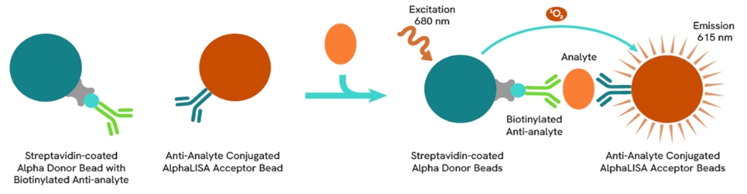
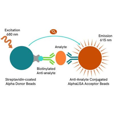
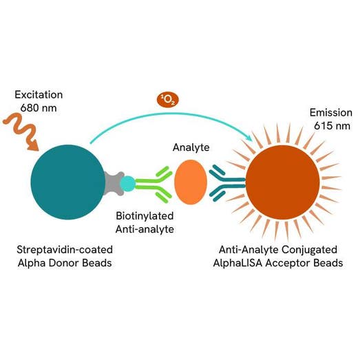
AlphaLISA technology allows the detection of molecules of interest in a no-wash, highly sensitive, quantitative assay. In an AlphaLISA assay, a biotinylated anti-analyte antibody binds to the Streptavidin-coated Donor beads while another anti-analyte antibody is conjugated to AlphaLISA Acceptor beads. In the presence of the analyte, the beads come into close proximity. The excitation of the Donor beads causes the release of singlet oxygen molecules, triggering a cascade of energy transfer in the Acceptor beads, and resulting in a sharp peak of light emission at 615 nm.
How it works
Principle of the AlphaLISA Mouse IL12p70 assay
The AlphaLISA Mouse IL12p70 assay is based on an AlphaLISA sandwich immunoassay involving a biotinylated anti-mIL12p70 antibody bound to Streptavidin-coated AlphaLISA Donor beads and an anti-mIL12p70 antibody conjugated to AlphaLISA Acceptor beads. One antibody is directed against the mouse p35 subunit protein, and the second recognizes specifically the p40 subunit forming mIL12p70. In the presence of the target, both antibodies bind to mouse IL12p70, and the beads come into proximity. The excitation of the Donor beads provokes the release of singlet oxygen molecules, triggering a cascade of energy transfer within the Acceptor beads, and resulting in emission with λmax at 615 nm. The intensity of the signal is directly proportional to the concentration of mIL12p70 present in the sample (cell supernatant or plasma EDTA).
Protocol of the AlphaLISA Mouse IL12p70 assay
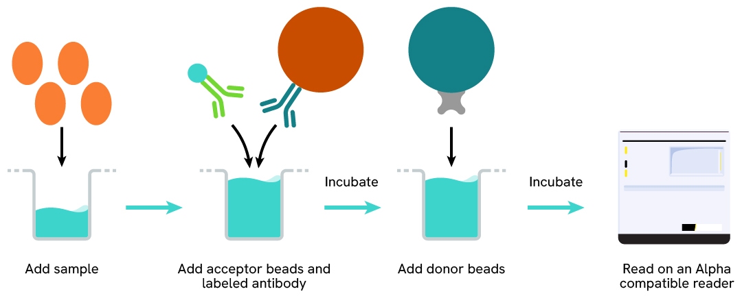
The AlphaLISA Mouse IL12p70 assay can be run in a 96- or 384-well detection plate (50 µL final). As described here, samples (cell supernatants or plasma EDTA) or standards are dispensed directly into the assay plate for the detection of Mouse IL12p70 by AlphaLISA reagents. No washing steps are needed. The protocol can be further miniaturized or upscaled by simply resizing each addition volume proportionally.
Assay details
Mouse IL12p70 assay details
| Kit Components | Lyophilized mIL12p70, SA-Donor Beads, Biotinylated anti-analyte, Anti-analyte conjugated Acceptor Beads, AlphaLISA BSA Assay Buffer |
|---|---|
| LDL & LLOQ (in Assay Buffer) | 1.3 pg/ml & 6.5 pg/mL |
| LDL & LLOQ (in DMEM) | 1.8 pg/ml & 6.7 pg/mL |
| LDL & LLOQ (in RPMI) | 3.4 pg/ml & 8.4 pg/mL |
| LDL & LLOQ (in Plasma EDTA) | 3.1 pg/ml & 10.9 pg/mL |
| Species | Mouse |
Analytical performance
Intra-assay precision table
Each of the 3 samples was measured 24 times, and their % CV were calculated. Samples were diluted in RPMI + 10% FBS.
| Sample | [mIL12p70] (pg/mL) | CV |
|---|---|---|
| 1 | 113 | 4% |
| 2 | 1169 | 4% |
| 3 | 11225 | 4% |
| Mean CV | 4% |
Inter-assay precision table
Each of the samples was measured in 3 independent experiments (3 days), and their % CV were calculated. Samples were diluted in RPMI + 10% FBS.
| Sample | [mIL12p70] (pg/mL) | CV |
|---|---|---|
| 1 | 133 | 7% |
| 2 | 1160 | 4% |
| 3 | 11243 | 4% |
| Mean CV | 5% |
Dilutional linearity table
Samples containing a known concentration of analyte were serially diluted in either RPMI + 10% FBS or mouse plasma EDTA. The assay was performed on serially diluted samples, along with a standard curve prepared in the same matrix. The recovery % obtained from these experiments show the good dilutional linearity of the assay.
| Standard in RPMI + 10% FBS | Expected mIL12p70 (pg/mL) | Observed mIL12p70 (pg/mL) | Dilution Recovery (%) |
|---|---|---|---|
| Neat | - | 2305 | - |
| 1/2 | 1153 | 1236 | 107 |
| 1/4 | 576 | 639 | 111 |
| 1/8 | 288 | 341 | 118 |
| 1/16 | 144 | 173 | 120 |
| 1/32 | 72 | 87 | 121 |
| Standard in Plasma EDTA | Expected mIL12p70 (pg/mL) | Observed mIL12p70 (pg/mL) | Dilution Recovery (%) |
|---|---|---|---|
| Neat | - | 9772 | - |
| 1/2 | 4886 | 5302 | 104 |
| 1/4 | 2443 | 2538 | 108 |
| 1/8 | 1221 | 1318 | 111 |
| 1/16 | 611 | 680 | 111 |
| 1/32 | 305 | 340 | 110 |
| 1/64 | 153 | 168 | 117 |
Specificity table
Cross reactivities were assessed using recombinant proteins from the mouse IL12 family (mIL23, mIL35, and p40) or human and rat IL12p70. Proteins were tested up to 1,500,000 pg/mL and standard curves were generated for each protein diluted in the kit diluent for recombinant proteins.
Percentage recoveries were computed by comparing the measured Interpolated concentrations versus the theoretical ones. The assay is mouse specific for IL12p70 targeting p35 and p40 subunits.
| Proteins | Cross reactivity (%) |
|---|---|
| Mouse IL-23 (p19+p40) | 0 |
| Mouse IL-35 (p35+EBI3) | 0 |
| Mouse IL-12p40 | 0 |
| Human IL12p70 | 0 |
| Rat IL12p70 | 1.3% |
Specifications
| Application |
Protein Quantification
|
|---|---|
| Automation Compatible |
Yes
|
| Brand |
AlphaLISA
|
| Detection Modality |
Alpha
|
| Dynamic Range |
4.2 - 150,000 pg/mL
|
| Limit of Detection |
1.3 pg/mL
|
| Limit of Quantification |
6.5 pg/mL
|
| Lysis Buffer Compatibility |
BSA Assay Buffer
|
| Product Group |
Kit
|
| Protocol Time |
2step
|
| Sample Volume |
5 µL
|
| Shipping Conditions |
Shipped in Blue Ice
|
| Target |
IL12p70
|
| Target Class |
Cytokines
|
| Target Species |
Mouse
|
| Technology |
Alpha
|
| Therapeutic Area |
Infectious Diseases
Inflammation
|
| Unit Size |
100 assay points
|
Resources
Are you looking for resources, click on the resource type to explore further.
Loading...


How can we help you?
We are here to answer your questions.

