
US

Revvity Sites Globally
Select your location.
*e-commerce not available for this region.

AlphaLISA Mouse CXCL1 Detection Kit, 5,000 Assay Points
AlphaLISA Mouse CXCL1 Detection Kit, 5,000 Assay Points
AlphaLISA Sandwich Anti-analyte Conjugated Acceptor Bead
The AlphaLISA™ Mouse CXCL1 kit is designed for the simple and rapid quantification of soluble mouse CXCL1 in cell supernatants, providing a fast no-wash alternative to traditional wash-based ELISA assays.
| Feature | Specification |
|---|---|
| Application | Protein Quantification |
| Dynamic Range | 2 - 15,000 pg/mL |
| Limit of Detection | 0.62 pg/mL |
| Limit of Quantification | 2.18 pg/mL |
| Protocol Time | 2-step |
| Sample Volume | 5 µL |
The AlphaLISA™ Mouse CXCL1 kit is designed for the simple and rapid quantification of soluble mouse CXCL1 in cell supernatants, providing a fast no-wash alternative to traditional wash-based ELISA assays.
Product variants
Unit Size: 500 assay points
Part #:
AL594C
List price
USD 2,447.00
Your price:
Unit Size: 5,000 assay points
Part #:
AL594F
List price
USD 16,124.00
Your price:
Unit Size: 100 assay points
Part #:
AL594HV
List price
USD 1,120.00
Your price:
For research use only. Not for use in diagnostic procedures. All products to be used in accordance with applicable laws and regulations including without limitation, consumption and disposal requirements under European REACH regulations (EC 1907/2006).
AlphaLISA Mouse CXCL1 Detection Kit, 5,000 Assay Points
AlphaLISA Sandwich Anti-analyte Conjugated Acceptor Bead
Loading...
Product information
Overview
Mouse CXCL1 is a small peptide chemokine of 8 kDa that belongs to the CXC subfamily. It is also called growth-regulated oncogene-α (GRO-α) and keratinocyte-derived chemokine (KC). It is expressed in neutrophils, macrophages, and epithelial cells. CXCL1 produces its effect via its receptor CXCR2, mainly expressed on neutrophils. Although CXCL1 plays an important role in inflammatory responses by recruiting and activating neutrophils, aberrant expression of CXCL1 has been reported as being associated with the growth and progression of certain tumors such as breast, lung, and gastric cancers.
Formats
- Our 100 assay point kit allows you to run 100 wells in 96-well format, using a 100 µL reaction volume (10 µL of sample).
- Our 500 assay point kit allows you to run 500 wells in 96-well or 384-well format, using a 50 µL reaction volume (5 µL of sample).
- Our 5,000 assay point kit allows you to run 5,000 wells in 96-well or 384-well format, using a 50 µL reaction volume (5 µL of sample).
Features
- No-wash steps, no separation steps
- ELISA alternative technology
- Reliable detection
- Broad sample compatibility
- Small sample volume
- Results in less than 3 hours
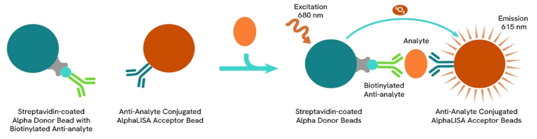
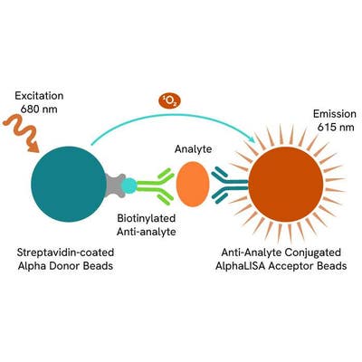
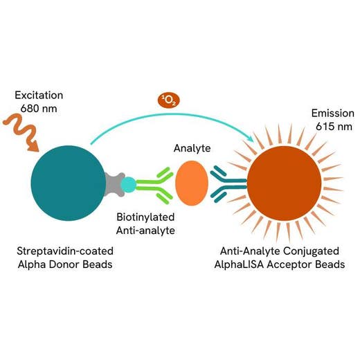
AlphaLISA technology allows the detection of molecules of interest in a no-wash, highly reliable, quantitative assay. In an AlphaLISA assay, a biotinylated anti-analyte antibody binds to the Streptavidin-coated Donor beads while another anti-analyte antibody is conjugated to AlphaLISA Acceptor beads. In the presence of the analyte, the beads come into close proximity. The excitation of the Donor beads causes the release of singlet oxygen molecules, triggering a cascade of energy transfer in the Acceptor beads. This results in a sharp peak of light emission at 615 nm.
How it works
Principle of the AlphaLISA Mouse CXCL1 assay
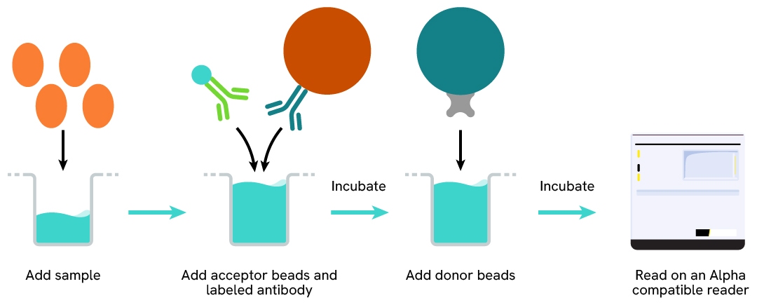
The AlphaLISA Mouse CXCL1 assay is based on an AlphaLISA sandwich immunoassay involving a biotinylated anti-target antibody bound to Streptavidin-coated AlphaLISA Donor beads and an anti-target antibody conjugated to AlphaLISA Acceptor beads. In the presence of the target, both antibodies bind to CXCL1 and the beads come into proximity. The excitation of the Donor beads provokes the release of singlet oxygen molecules, triggering a cascade of energy transfer within the Acceptor beads. This results in emission with λmax at 615 nm. The intensity of the signal is directly proportional to the concentration of CXCL1 present in the sample.
Protocol of the AlphaLISA Mouse CXCL1 assay
The AlphaLISA CXCL1 assay can be run in a 96- or 384-well detection plate (50 µL final). As described here, samples or standards are dispensed directly into the assay plate for the detection of mCXCL1 by AlphaLISA reagents. No washing steps are necessary. The protocol can be further miniaturized or upscaled by simply resizing each addition volume proportionally.
Assay details
Mouse CXCL1 details
| Kit components | Lyophilized analyte, SA-Donor Beads, Biotinylated anti-analyte, Anti-analyte conjugated Acceptor Beads, Assay Buffer |
|---|---|
| LDL & LLOQ (in Assay Buffer) | 0.62 pg/ml & 2.18 pg/mL |
| LDL & LLOQ (in DMEM) | 1.4 pg/ml & 4 pg/mL |
| LDL & LLOQ (in RPMI) | 33 pg/ml & 115 pg/mL |
| Species | Mouse, Rat (not tested on rat samples) |
| Time to result | 3 hours at RT |
Analytical performance
Intra-assay precision table
The 3 samples were all measured 24 times, and the % CV was calculated for each sample. Samples were mouse recombinant CXCL1.
| Sample | [mCXCL1] (pg/mL) | CV |
|---|---|---|
| 1 | 12772 | 3% |
| 2 | 1577 | 2% |
| 3 | 166 | 2% |
| Mean CV | 2,3% |
Inter-assay precision table
The three samples were measured in 3 independent experiments (3 days), and the % CV was calculated for each sample. Samples were mouse recombinant CXCL1.
| Sample | [mCXCL1] (pg/mL) | CV |
|---|---|---|
| 1 | 13445 | 6% |
| 2 | 1584 | 1% |
| 3 | 165 | 2% |
| Mean CV | 3% |
Spike and recovery table
Known concentrations of a standard (exogenous analyte) were spiked into NIH-3T3 cell supernatant medium containing the endogenous analyte. Non-spiked and spiked samples were measured in the assay.
| Sample type | [Endogenous mCXCL1] in sample (pg/mL) | [Exogenous mCXCL1] spiked into sample (pg/mL) | Expected [mCXCL1] in spiked sample (pg/mL) | Measured [mCXCL1] in spiked sample (pg/mL) | Recovery (%) |
|---|---|---|---|---|---|
| Sample 1 | 869 | 459 | 1328 | 1130 | 85 |
| Sample 2 | 869 | 1571 | 2439 | 1968 | 81 |
| Sample 3 | 869 | 4422 | 5291 | 5117 | 97 |
Dilutional linearity table
A mouse CXCL1 sample collected from NIH-3T3 supernatant was serially diluted. Concentrations of CXCL1 in neat and diluted samples were determined.
| Sample dilution factor | Expected [mCXCL1] (pg/mL) | Measured [mCXCL1] (pg/mL) | Dilution Recovery (%) |
|---|---|---|---|
| neat | - | 903 | - |
| 2 | 452 | 465 | 103% |
| 4 | 226 | 239 | 106% |
| 8 | 113 | 118 | 105% |
| 16 | 53 | 63 | 112% |
| 32 | 28 | 31 | 110% |
| 64 | 14 | 13,7 | 97% |
Specificity table
Cross reactivity was assessed using recombinant proteins and tested at 18 000 pg/mL. The assay is compatible with rat CXCL1.
| Target | Cross reactivity |
|---|---|
| Rat CXCL1 | 100% |
| Human CXCL1 | 0 |
| Mouse CXCL2 | 0 |
| Human CXCL2 | 0 |
| Mouse CXCL3 | 0 |
| Human CXCL3 | 0 |
Assay validation
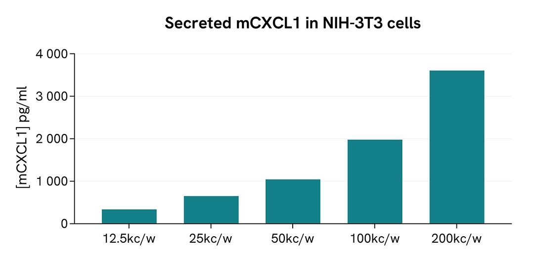
Validation of AlphaLISA mouse CXCL1 detection kit on NIH-3T3 cell supernatant
NIH-3T3 murine fibroblast cells were plated in a 96-well culture plate at serial densities ranging from 200k cells/well to 12.5k cells/well in DMEM medium with 10% FBS for 24h at 37°C. Then 5 µL of supernatant were transferred into an AlphaPlate-384 to measure the concentration of secreted CXCL1. The standard curve was prepared in DMEM medium + 10% FBS. All other assay components were diluted in AlphaLISA Assay Buffer 1 and added to the wells using the kit protocol. AlphaLISA counts were measured, and the CXCL1 concentration in each supernatant sample was interpolated from the standard curve.
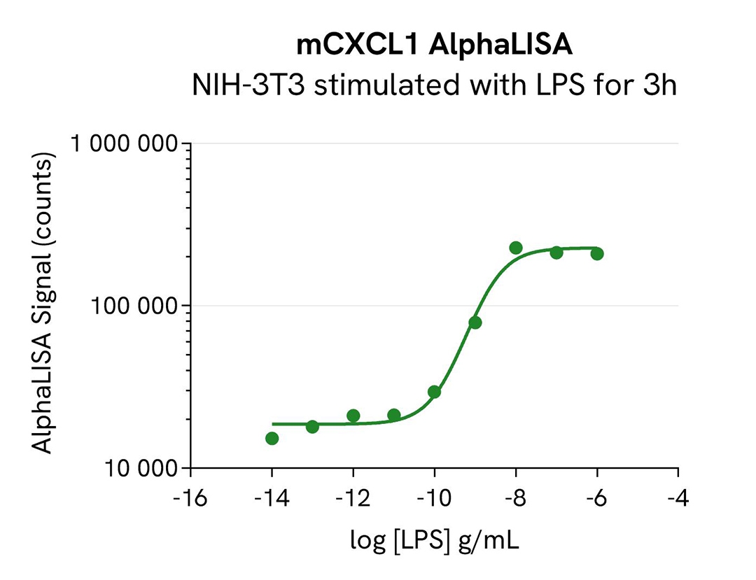
Validation of AlphaLISA mouse CXCL1 detection kit on LPS treated NIH-3T3 cells
NIH-3T3 cells were plated in a 96-well culture-treated plate at a density of 12.5k cells per well and allowed to adhere in complete DMEM+10%FBS culture medium for 24h at 37°C. Subsequently, the cells were exposed to increasing concentrations of LPS for 3h at 37°C, 5% CO2. Following treatment, 5 µL of supernatant were transferred into an AlphaPlate-384 to measure the concentration of secreted CXCL1. The standard curve was prepared in DMEM +10% FBS. All other assay components were diluted in AlphaLISA Assay Buffer 1 and added to the wells using the kit protocol. AlphaLISA counts were measured, and the CXCL1 concentration in each supernatant sample was interpolated from the standard curve.
Specifications
| Application |
Protein Quantification
|
|---|---|
| Automation Compatible |
Yes
|
| Brand |
AlphaLISA
|
| Cellular or Signaling Pathway |
GPCR signaling
|
| Detection Modality |
Alpha
|
| Dynamic Range |
2 - 15,000 pg/mL
|
| Limit of Detection |
0.62 pg/mL
|
| Limit of Quantification |
2.18 pg/mL
|
| Lysis Buffer Compatibility |
AlphaLISA Assay Buffer 1
|
| Product Group |
Kit
|
| Protocol Time |
2-step
|
| Sample Volume |
5 µL
|
| Shipping Conditions |
Shipped in Dry Ice
|
| Target |
CXCL1
|
| Target Class |
Cytokines
|
| Target Species |
Mouse
|
| Technology |
Alpha
|
| Therapeutic Area |
Immuno-oncology
Inflammation
Oncology
|
| Unit Size |
5,000 assay points
|
Resources
Are you looking for resources, click on the resource type to explore further.

Brochure
Alpha assays and reagents catalog
Alpha technolgy enables the rapid and straightforward mesaure of virtually any target. This includes enzymes, receptor-ligand...
Loading...


How can we help you?
We are here to answer your questions.