


US
Revvity Sites Globally
Select your location.
*e-commerce not available for this region.

AlphaLISA Human and Mouse LC3B-II Detection Kit, 100 assay points
AlphaLISA Human and Mouse LC3B-II Detection Kit, 100 assay points
AlphaLISA Sandwich Anti-analyte Conjugated Acceptor Bead
The AlphaLISA™ Human and Mouse LC3B-II kit is designed for the simple and rapid detection of LC3B-II protein in cell or tissue lysates, providing a fast no-wash alternative to traditional wash-based ELISA assays.
| Feature | Specification |
|---|---|
| Application | Protein Quantification |
| Protocol Time | Overnight at RT |
| Sample Volume | 5 µL |
The AlphaLISA™ Human and Mouse LC3B-II kit is designed for the simple and rapid detection of LC3B-II protein in cell or tissue lysates, providing a fast no-wash alternative to traditional wash-based ELISA assays.
Product variants
Unit Size: 100 assay points
Part #:
AL3210HV
List price
USD 1,171.00
Your online price:
Unit Size: 500 assay points
Part #:
AL3210C
List price
USD 2,712.00
Your online price:
Unit Size: 5,000 assay points
Part #:
AL3210F
List price
USD 15,807.00
Your online price:
For research use only. Not for use in diagnostic procedures. All products to be used in accordance with applicable laws and regulations including without limitation, consumption and disposal requirements under European REACH regulations (EC 1907/2006).
AlphaLISA Human and Mouse LC3B-II Detection Kit, 100 assay points
AlphaLISA Sandwich Anti-analyte Conjugated Acceptor Bead
AlphaLISA Human and Mouse LC3B-II Detection Kit, 100 assay points
Loading...
Product information
Overview
LC3 is a mammalian homolog of the yeast ATG8 protein and is distributed throughout mammalian tissues and cultured cells. LC3 is a key component of the autophagy process - a degradative program that maintains cellular homeostasis. During autophagy, cytoplasmic components such as cytosolic proteins and organelles are engulfed by autophagosomes Concurrently, a cytosolic form of LC3 (LC3-I) is conjugated to phosphatidylethanolamine to form LC3-phosphatidylethanolamine conjugate (LC3-II). This newly formed conjugate is recruited to autophagosomal membranes. The subsequent fusion of autophagosomes with lysosomes results in the formation of autolysosomes, and intra-autophagosomal components are degraded by lysosomal hydrolases. At the same time, LC3-II within the autolysosomal lumen is degraded. The LC3-II level has been found to be proportional to autophagosome abundance making LC3-II the most popular marker of autophagy.
Formats:
- Our 100 assay point kit allows you to run 100 wells in 96-well format, using a 100 µL reaction volume (10 µL of sample).
- Our 500 assay point kit allows you to run 500 wells in 96-well or 384-well format, using a 50 µL reaction volume (5 µL of sample).
- Our 5,000 assay point kit allows you to run 5,000 wells in 96-well or 384-well format, using a 50 µL reaction volume (5 µL of sample).
Features:
- No-wash steps, no separation steps
- ELISA alternative technology
- Sensitive detection
- Broad sample compatibility
- Small sample volume
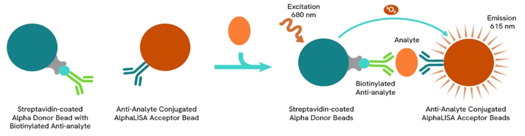
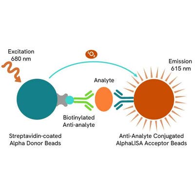
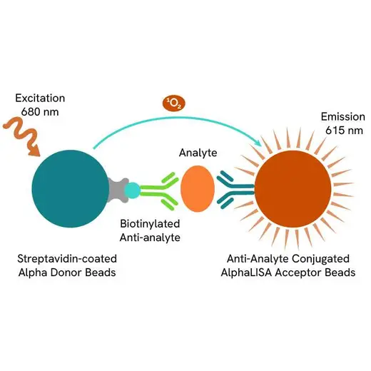
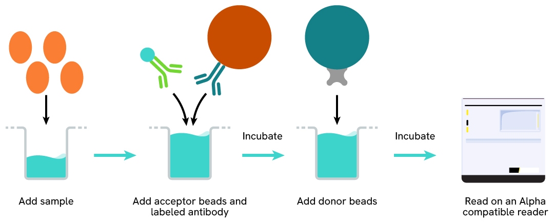
AlphaLISA technology allows the detection of molecules of interest in a no-wash, highly sensitive, quantitative assay. In an AlphaLISA assay, a biotinylated anti-analyte antibody binds to the Streptavidin-coated Donor beads while another anti-analyte antibody is conjugated to AlphaLISA Acceptor beads. In the presence of the analyte, the beads come into close proximity. The excitation of the Donor beads causes the release of singlet oxygen molecules that triggers a cascade of energy transfer in the Acceptor beads, resulting in a sharp peak of light emission at 615 nm.
How it works
Principle of the AlphaLISA human and mouse LC3B-II detection kit
The AlphaLISA Human and Mouse LC3B-II assay is based on an AlphaLISA sandwich immunoassay. A biotinylated anti-LC3B-II antibody binds to the streptavidin coated AlphaLISA Donor beads, while an anti- LC3B-II antibody is conjugated to AlphaLISA Acceptor beads.
In the presence of LC3B-II, the beads come into proximity. The excitation of the Donor beads provokes the release of singlet oxygen molecules that triggers a cascade of energy transfer within the Acceptor beads, resulting in emission with λmax at 615 nm.
Protocol of the AlphaLISA human and mouse LC3B-II assay
The AlphaLISA Human and Mouse LC3B-II assay can be run in a 96- or 384-well detection plate (50 µL final). As described here, samples (cell lysates or tissue lysates) or control lysate are dispensed directly into the assay plate for the detection of LC3B-II by AlphaLISA reagents. No washing steps are needed. The protocol can be further miniaturized or upscaled by simply resizing each addition volume proportionally.
Assay details
AlphaLISA human and mouse LC3B-II assay details
| Sample size | 5 µL |
|---|---|
| Final assay volume | 50 µL |
| Time to result | Overnight (18-22h) at RT |
| Kit components | AlphaLISA Acceptor beads coated with Anti-LC3B-II Antibody, Streptavidin-coated Donor beads, Biotinylated Anti-LC3B-II Antibody, Lyophilized LC3B-II Control Lysate, 4X AlphaLISA Human and Mouse LC3B-II Lysis Buffer, 100X AlphaLISA LC3B-II Blocking Reagent and 10X AlphaLISA Immunoassay Buffer |
| Species | Human and mouse (rat predicted) |
Assay validation
Strong correlation between AlphaLISA, HTRF and Western Blot for detection of LC3B-II modulation
U-87 MG cells were plated in a 96-well culture-treated plate (55,000 cells per well) in complete culture medium, and incubated overnight at 37°C, 5% CO2. The cells were then treated with increasing concentrations of Hydroxycholoroquine (HCQ), a late-stage autophagic inhibitor, for 4 h at 37 °C, 5% CO2. Following culture medium removal, cells were lysed with 50 µL of 1X Supplemented AlphaLISA Human and Mouse LC3B-II Lysis Buffer for 30 min at room temperature under gentle shaking.
After cell lysis, samples were used for a side-by-side comparison of AlphaLISA LC3B-II, HTRF Human and Mouse LC3B II Detection Kit (#64LC3B2PEG) and Western Blot techniques.
AlphaLISA, HTRF and chemiluminescent signals were normalized with GAPDH Housekeeping protein (HTRF kit: #64GAPDHPEG).
Fold of change corresponds to LC3B-II normalized signal treated over untreated condition.
As evidenced, AlphaLISA LC3B-II, HTRF LC3B-II and Western Blot results are comparable demonstrating that these three methods are correlated.

Induction of autophagy with AZD2014, a mTOR inhibitor
U-87 MG cells were plated in a 96-well culture-treated plate (50,000 cells per well) in complete culture medium, and incubated overnight at 37°C, 5% CO2. Then, cells were then treated with increasing concentrations of AZD2014 (Vistusertib) for 4 h at 37°C, 5%CO2. Following culture medium removal, cells were lysed with 50 µl of 1X Supplemented AlphaLISA Human and Mouse LC3B-II Lysis Buffer for 30 min at room temperature under gentle shaking. After cell lysis, samples were then tested with the AlphaLISA LC3B-II.
As expected, AZD2014, a potent mTOR inhibitor, induced autophagy activation, leading to a dose-dependent increase of LC3B-II signal. In addition, no significant cytotoxic effects were observed with ATPlite Luminescence Assay System (#6016736) in the same experimental conditions.

Autophagic flux inhibition with Hydroxychloroquine, a late-stage autophagy inhibitor
U-87 MG cells were plated in a 96-well culture-treated plate (50,000 cells per well) in complete culture medium, and incubated overnight at 37°C, 5% CO2. Then, cells were then treated with increasing concentrations of Hydroxycholoroquine (HCQ), a late-stage autophagic inhibitor, for 4 h at 37°C, 5%CO2. Following culture medium removal, cells were lysed with 50 µl of 1X Supplemented AlphaLISA Human and Mouse LC3B-II Lysis Buffer for 30 min at room temperature under gentle shaking. After cell lysis, samples were then tested with the AlphaLISA LC3B-II.
As expected, HCQ, a late-stage autophagic inhibitor, inhibited autophagic flux, leading to a dose-dependent increase of LC3B-II signal. In addition, no significant cytotoxic effects were observed with ATPlite Luminescence Assay System (#6016736) in the same experimental conditions (data not shown).

Autophagy inhibition with ULK-101, a potent and selective ULK1/2 inhibitor
U-87 MG cells were plated in a 96-well culture-treated plate (50,000 cells per well) in complete culture medium, and incubated overnight at 37°C, 5% CO2. Then, cells then co-treated with 2.5 µM of AZD2014, a mTOR inhibitor, and 10 µM of ULK-101, a potent and selective ULK1/2 inhibitor, for 4 h at 37°C, 5%CO2. Following culture medium removal, cells were lysed with 50 µl of 1X Supplemented AlphaLISA Human and Mouse LC3B-II Lysis Buffer for 30 min at room temperature under gentle shaking. After cell lysis, samples were then tested with the AlphaLISA LC3B-II.
As expected, ULK-101, a potent and dual autophagy kinase ULK1/2 inhibitor, repressed autophagy induced by AZD2014, leading to a significant decrease of LC3B-II signal. In addition, no significant cytotoxic effects were observed with ATPlite Luminescence Assay System (#6016736) in the same experimental conditions (data not shown).

Specificity of AlphaLISA LC3B-II assay using siRNA
NCI-H1272 cells were plated in a 96-well culture plate (10,000 cells/well) in complete culture medium, and incubated overnight at 37°C, 5% CO2. The day after, the cells were transfected with ON-TARGETplus SMARTPool siRNAs (Horizon Discovery/Revvity) LC3A, LC3B and LC3C isoforms, as well as with a non-targeting siRNA used as negative control using DharmaFECT1 transfection reagent. After a 24h-incubation, the medium was replaced by fresh culture medium, and cells were incubated for an additional 24h-incubation. Then, cells were then treated with 300 µM Hydroxycholoroquine (HCQ), a late-stage autophagic inhibitor, for 4 h at 37°C, 5% CO2. Following culture medium removal, cells were lysed with 50 µl of 1X Supplemented AlphaLISA Human and Mouse LC3B-II Lysis Buffer for 30 min at room temperature under gentle shaking. After cell lysis, samples were then tested with the AlphaLISA LC3B-II.
The siRNA experiments demonstrate that AlphaLISA detection antibodies specifically measure LC3B isoform and do not recognize the other isoforms. It was found that treatment with the LC3B siRNA induced almost complete AlphaLISA signal loss, while the knockdown of LC3A and LC3C genes did not lead to any significant signal modulation in this assay.

Versatility of AlphaLISA LC3B-II assay using various human and mouse cell lines
U-87 MG (human) and NIH3T3 (mouse) cells were plated in a 96-well culture-treated plate (50,000 cells/well) in complete culture medium, and incubated 48 h at 37°C, 5% CO2. Then, cells were then treated with 300 µM Hydroxycholoroquine (HCQ), a late-stage autophagic inhibitor, for 4 h at 37°C, 5% CO2. Following culture medium removal, cells were lysed with 50 µl of 1X Supplemented AlphaLISA Human and Mouse LC3B-II Lysis Buffer for 30 min at room temperature under gentle shaking. After cell lysis, samples were then tested with the AlphaLISA LC3B-II.
The AlphaLISA LC3B-II assay efficiently displayed significant positive signals in each tested cell lines, demonstrating its capability to detect LC3II-B protein in human and mouse and its suitability for translational research.

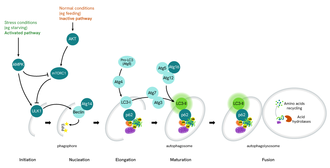
Simplified pathway
LC3B-II in autophagy
Autophagy is an evolutionary conserved cellular process that occurs in virtually all eukaryotic cells, ranging from yeast to mammals. Autophagy is crucial in the maintenance of homeostasis, being involved in numerous physiological processes including stress responses (e.g. starvation, hypoxia, high temperature), cell growth, and aging. Conversely, dysfunctions in autophagic mechanisms have been associated with diseases such as cancer, neurodegenerative diseases, infectious diseases, and cardiac and metabolic diseases.
During autophagy, cytoplasmic components, including cytosolic proteins and organelles, are engulfed by autophagosomes. Concomitantly, a cytosolic form of LC3 (LC3-I) is conjugated to phosphatidylethanolamine to form LC3-phosphatidylethanolamine conjugate (LC3-II). This conjugate is recruited to autophagosomal membranes. Then, autophagosomes fuse with lysosomes to form autolysosomes, and intra-autophagosomal components are degraded by lysosomal hydrolases. At the same time, LC3-II in autolysosomal lumen is degraded. LC3-II level has been found to be proportional to the amount of autophagosomes and thus LC3-II detection has become the most reliable method for monitoring autophagy and autophagy-related processes.
Specifications
| Application |
Protein Quantification
|
|---|---|
| Automation Compatible |
Yes
|
| Brand |
AlphaLISA
|
| Detection Modality |
Alpha
|
| Product Group |
Kit
|
| Protocol Time |
Overnight at RT
|
| Sample Volume |
5 µL
|
| Shipping Conditions |
Shipped in Blue Ice
|
| Target |
LC3B-II
|
| Target Class |
Biomarkers
|
| Target Species |
Human
Mouse
|
| Technology |
Alpha
|
| Unit Size |
100 assay points
|
Resources
Are you looking for resources, click on the resource type to explore further.
Brochure
Alpha assays and reagents catalog
Alpha technolgy enables the rapid and straightforward mesaure of virtually any target. This includes enzymes, receptor-ligand...
Brochure
Species compatibility for HTRF, AlphaLISA SureFire Ultra and Alpha SureFire Ultra Multiplex assays
This document includes detailed tables listing HTRF™, AlphaLISA™ SureFire® Ultra™, and Alpha SureFire® Ultra™ Multiplex assays...


How can we help you?
We are here to answer your questions.

