


US
Revvity Sites Globally
Select your location.
*e-commerce not available for this region.

AlphaLISA SureFire Ultra Human and Mouse Total ARID1A Detection Kit, 500 Assay Points
AlphaLISA SureFire Ultra Human and Mouse Total ARID1A Detection Kit, 500 Assay Points

AlphaLISA Surefire Ultra Total Protein
The AlphaLISA™ SureFire® Ultra™ Human and Mouse Total ARID1A assay is a sandwich immunoassay for quantitative detection of total ARID1A in cellular lysates using Alpha Technology.
| Feature | Specification |
|---|---|
| Application | Cell Signaling |
| Protocol Time | 2h at RT |
| Sample Volume | 10 µL |
The AlphaLISA™ SureFire® Ultra™ Human and Mouse Total ARID1A assay is a sandwich immunoassay for quantitative detection of total ARID1A in cellular lysates using Alpha Technology.
Product variants
Unit Size: 100 assay points
Part #:
ALSU-TARID-A-HV
List price
USD 737.00
Your price:
Unit Size: 500 assay points
Part #:
ALSU-TARID-A500
List price
USD 2,490.00
Your price:
Unit Size: 10,000 assay points
Part #:
ALSU-TARID-A10K
List price
USD 14,982.00
Your price:
Unit Size: 50,000 assay points
Part #:
ALSU-TARID-A50K
List price
USD 47,624.00
Your price:
For research use only. Not for use in diagnostic procedures. All products to be used in accordance with applicable laws and regulations including without limitation, consumption and disposal requirements under European REACH regulations (EC 1907/2006).
AlphaLISA SureFire Ultra Human and Mouse Total ARID1A Detection Kit, 500 Assay Points
AlphaLISA Surefire Ultra Total Protein
Loading...
Product information
Overview
AT-rich interactive domain-containing protein 1A (ARID1A) is a critical component of the SWI/SNF chromatin remodeling complex, which regulates gene expression by modulating chromatin structure and accessibility. ARID1A plays essential roles in transcriptional regulation, DNA damage repair, and the maintenance of genomic stability. It acts as a tumor suppressor, with frequent inactivating mutations observed in a variety of cancers, including ovarian clear cell carcinoma, endometrial cancer, and gastric cancer. Loss of ARID1A disrupts chromatin remodeling, leading to altered transcriptional programs, impaired DNA damage responses, and increased sensitivity to epigenetic and DNA repair inhibitors. Notably, synthetic lethality with EZH2 and ATR inhibitors has emerged as a promising therapeutic strategy, positioning ARID1A as a key biomarker and vulnerability in precision oncology.
The AlphaLISA SureFire Ultra Human and Mouse Total ARID1A Detection Kit is a sandwich immunoassay for the quantitative detection of total ARID1A in cellular lysates, using Alpha Technology.
Formats:
- The HV (high volume) kit contains reagents to run 100 wells in 96-well format, using a 60 μL reaction volume.
- The 500-point kit contains enough reagents to run 500 wells in 384-well format, using a 20 μL reaction volume.
- The 10,000-point kit contains enough reagents to run 10,000 wells in 384-well format, using a 20 μL reaction volume.
- The 50,000-point kit contains enough reagents to run 50,000 wells in 384-well format, using a 20 μL reaction volume.
AlphaLISA SureFire Ultra kits are compatible with:
- Cell and tissue lysates
- Antibody modulators
- Biotherapeutic antibodies
AlphaLISA SureFire Ultra kits can be used for:
- Cellular kinase assays
- Receptor activation studies
- High-throughput screening for preclinical studies
How it works
Total-AlphaLISA SureFire Ultra assay principle
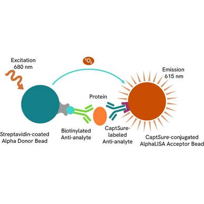
The Total-AlphaLISA SureFire Ultra assay measures the expression level of a target protein in a biological sample (e.g. cell lysate).
The Total-AlphaLISA SureFire Ultra assay uses two antibodies which recognize two different distal epitopes on the target protein. AlphaLISA assays require two bead types: Acceptor and Donor Beads. Acceptor Beads are coated with a proprietary CaptSure™ agent to specifically immobilize the assay specific antibody, labeled with a CaptSure tag. Donor Beads are coated with streptavidin to capture one of the detection antibodies, which is biotinylated. In the presence of target protein, the two antibodies bring the Donor and Acceptor Beads in close proximity whereby the singlet oxygen transfers energy to excite the Acceptor Bead, allowing for the generation of a luminescent Alpha signal. The amount of light emission is directly proportional to the quantity of protein present in the sample.
Total-AlphaLISA SureFire Ultra two-plate assay protocol
The two-plate protocol involves culturing and treating the cells in a 96-well plate before lysis, then transferring lysates into a 384-well OptiPlate™ plate before the addition of Total-AlphaLISA SureFire Ultra detection reagents. This protocol enables cell viability and confluence to be monitored. In addition, lysates from a single well can be used to measure multiple targets.

Total-AlphaLISA SureFire Ultra one-plate assay protocol
Detection of Total target protein with AlphaLISA SureFire Ultra reagents can be performed in a single plate used for culturing, treatment, and lysis. No washing steps are required. This HTS designed protocol allows for miniaturization while maintaining robust AlphaLISA SureFire Ultra quality.

Assay specificity/selectivity
Selectivity of ARID1A Total assay
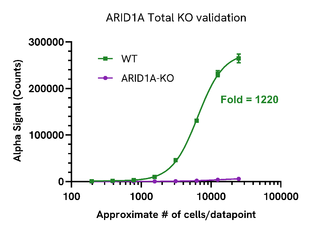
Total ARID1A levels were assessed in wild type (WT) HEK293T cells and ARID1A knockout (KO) HEK293T cells (Abcam ab266189) cultured to confluency in T175 flasks at 37°C, 5% CO2.
Each flask was lysed with 4 mL of Lysis Buffer B for 10 minutes at RT with shaking. Lysates were then evaluated for ARID1A expression using the AlphaLISA SureFire Ultra assay kit.
For the detection step, 10 µL of cell lysate was transferred into a 384-well white OptiPlate, followed by 5 µL of Acceptor mix and incubated for 1 hour at RT. Finally, 5 µL of Donor mix was then added to each well and incubated for 1 hour at RT in the dark. The plate was read on an Envision using standard AlphaLISA settings.
ARID1A was detected only in the HEK293T-WT cells demonstrating assay selectivity.
Assay versatility
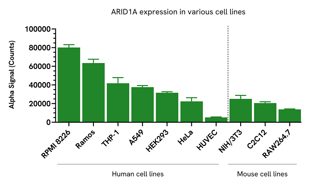
Versatility of Total ARID1A assay in various cell lines
Adherent cells were seeded at 40,000 cells/well in a 96-well culture plate in complete medium and incubated overnight at 37°C, 5% CO2. Cells were lysed with 100 µL of Lysis Buffer.
Suspension cells were seeded at 400,000 cells/well in a 96-well culture plate in HBSS + 0.1% BSA and lysed with 100 µL of Lysis Buffer.
ARID1A levels were evaluated by AlphaLISA SureFire Ultra. For the detection step, 10 µL of cell lysate (approximately 4,000 adherent cells or 40,000 suspension cells) was transferred into a 384-well white OptiPlate, followed by 5 µL of Acceptor Mix and incubated for 1 hour at RT. Finally, 5 µL of Donor Mix was then added to each well and incubated for 1 hour at RT in the dark. The plate was read on an Envision using standard AlphaLISA settings.
Specifications
| Application |
Cell Signaling
|
|---|---|
| Automation Compatible |
Yes
|
| Brand |
AlphaLISA SureFire Ultra
|
| Detection Modality |
Alpha
|
| Molecular Modification |
Total
|
| Product Group |
Kit
|
| Protocol Time |
2h at RT
|
| Sample Volume |
10 µL
|
| Shipping Conditions |
Shipped in Blue Ice
|
| Target |
ARID1A
|
| Target Class |
Phosphoproteins
|
| Target Species |
Human
Mouse
|
| Technology |
Alpha
|
| Therapeutic Area |
Oncology
|
| Unit Size |
500 assay points
|
Resources
Are you looking for resources, click on the resource type to explore further.
Guide
AlphaLISA SureFire Ultra: the ultimate guide for successful experiments
The definitive guide for setting up a successful AlphaLISA SureFire Ultra assay
Several biological processes are regulated by...
Loading...


How can we help you?
We are here to answer your questions.

