


US
Revvity Sites Globally
Select your location.
*e-commerce not available for this region.

AlphaLISA SureFire Ultra Human Total TNF-α Induced Protein 3 (TNFAIP3) Detection Kit, 100 Assay Points

AlphaLISA SureFire Ultra Human Total TNF-α Induced Protein 3 (TNFAIP3) Detection Kit, 100 Assay Points
AlphaLISA Surefire Ultra Total Protein

The AlphaLISA™ SureFire® Ultra™ Human Total TNFAIP3 assay is a sandwich immunoassay for quantitative detection of total TNFAIP3 in cellular lysates using Alpha Technology.
| Feature | Specification |
|---|---|
| Application | Cell Signaling |
| Protocol Time | 2h at RT |
| Sample Volume | 30 µL |
The AlphaLISA™ SureFire® Ultra™ Human Total TNFAIP3 assay is a sandwich immunoassay for quantitative detection of total TNFAIP3 in cellular lysates using Alpha Technology.
Product variants
Unit Size: 100 assay points
Part #:
ALSU-TTNFA-A-HV
List price
USD 722.00
Your online price:
Unit Size: 500 assay points
Part #:
ALSU-TTNFA-A500
List price
USD 2,441.00
Your online price:
Unit Size: 10,000 assay points
Part #:
ALSU-TTNFA-A10K
List price
USD 14,688.00
Your online price:
Unit Size: 50,000 assay points
Part #:
ALSU-TTNFA-A50K
List price
USD 46,690.00
Your online price:
For research use only. Not for use in diagnostic procedures. All products to be used in accordance with applicable laws and regulations including without limitation, consumption and disposal requirements under European REACH regulations (EC 1907/2006).
AlphaLISA SureFire Ultra Human Total TNF-α Induced Protein 3 (TNFAIP3) Detection Kit, 100 Assay Points
AlphaLISA Surefire Ultra Total Protein
AlphaLISA SureFire Ultra Human Total TNF-α Induced Protein 3 (TNFAIP3) Detection Kit, 100 Assay Points
Loading...
Product information
Overview
Tumor necrosis factor alpha-induced protein 3 (TNFAIP3), also known as A20, is a ubiquitin-editing enzyme that negatively regulates NF-κB signaling and inflammation. A20 exhibits both deubiquitinase and E3 ligase activities, allowing it to disassemble K63-linked polyubiquitin chains and add K48-linked chains for proteasomal degradation. This dual function is essential for maintaining immune homeostasis and preventing excessive inflammation. Loss-of-function mutations or reduced expression of A20 are associated with autoimmune diseases, lymphomas, and inflammatory conditions. A20 is a key checkpoint in immune signaling and a potential target for modulating chronic inflammation and enhancing anti-tumor responses.
The AlphaLISA SureFire Ultra Human Total TNFAIP3 Detection Kit is a sandwich immunoassay for the quantitative detection of total TNFAIP3 in cellular lysates, using Alpha Technology.
Formats:
- The HV (high volume) kit contains reagents to run 100 wells in 96-well format, using a 60 μL reaction volume.
- The 500-point kit contains enough reagents to run 500 wells in 384-well format, using a 20 μL reaction volume.
- The 10,000-point kit contains enough reagents to run 10,000 wells in 384-well format, using a 20 μL reaction volume.
- The 50,000-point kit contains enough reagents to run 50,000 wells in 384-well format, using a 20 μL reaction volume.
AlphaLISA SureFire Ultra kits are compatible with:
- Cell and tissue lysates
- Antibody modulators
- Biotherapeutic antibodies
AlphaLISA SureFire Ultra kits can be used for:
- Cellular kinase assays
- Receptor activation studies
- High-throughput screening for preclinical studies
How it works
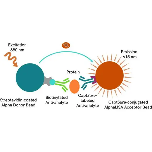
Total-AlphaLISA SureFire Ultra assay principle
The Total-AlphaLISA SureFire Ultra assay measures the expression level of a protein target in a cell lysate.
The Total-AlphaLISA SureFire Ultra assay uses two antibodies which recognize two different distal epitopes on the targeted protein. AlphaLISA assays require two bead types: Acceptor and Donor beads. Acceptor beads are coated with a proprietary CaptSure™ agent to specifically immobilize the assay specific antibody, labeled with a CaptSure tag. Donor beads are coated with streptavidin to capture one of the detection antibodies, which is biotinylated. In the presence of targeted protein, the two antibodies bring the Donor and Acceptor beads in close proximity whereby the singlet oxygen transfers energy to excite the Acceptor bead, allowing the generation of a luminescent Alpha signal. The amount of light emission is directly proportional to the quantity of protein present in the sample.
Total-AlphaLISA SureFire Ultra two-plate assay protocol
The two-plate protocol involves culturing and treating the cells in a 96-well plate before lysis, then transferring lysates into a 384-well OptiPlate™ plate before the addition of Total-AlphaLISA SureFire Ultra detection reagents. This protocol permits the cells viability and confluence to be monitored. In addition, lysates from a single well can be used to measure multiple targets.

Total-AlphaLISA SureFire Ultra one-plate assay protocol
Detection of Total target protein with AlphaLISA SureFire Ultra reagents can be performed in a single plate used for culturing, treatment, and lysis. No washing steps are required. This HTS designed protocol allows for miniaturization while maintaining AlphaLISA SureFire Ultra quality.

Assay validation
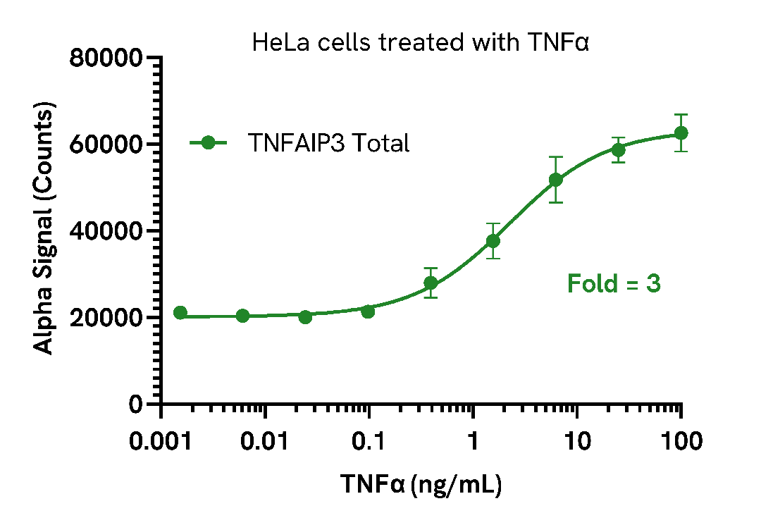
Induction of TNFAIP3 in TNFα treated cells
HeLa cells were seeded in a 96-well plate (40,000 cells/well) in complete medium and incubated overnight at 37°C, 5% CO2. The cells were then treated with increasing concentrations of TNFα for 5 hours.
After treatment, the cells were lysed with 200 µL of Lysis Buffer for 10 minutes at RT with shaking (350 rpm). TNFAIP3 levels were evaluated using the AlphaLISA SureFire Ultra assay. For the detection step, 10 µL of cell lysate (approximately 2,000 cells) was transferred into a 384-well white OptiPlate, followed by 5 µL of Acceptor mix and incubated for 1 hour at RT. Finally, 5 µL of Donor mix was then added to each well and incubated for 1 hour at RT in the dark. The plate was read on an Envision using standard AlphaLISA settings.
As expected, TNFα triggered a dose-dependent increase in the levels of TNFAIP3, while Cofilin levels were unchanged (data not shown).
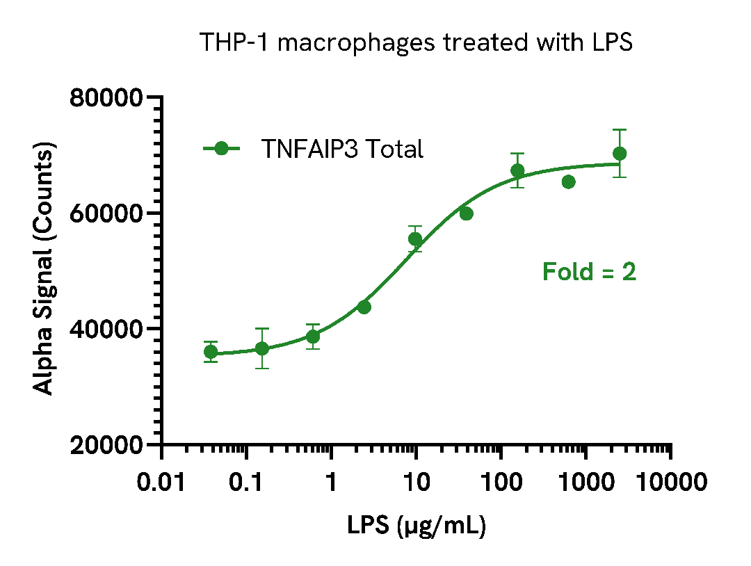
Induction of TNFAIP3 in LPS treated cells
THP-1 cells were seeded in a 96-well plate (50,000 cells/well) in complete medium containing 100 nM of PMA for 24 hours at 37°C, 5% CO2. The THP-1 derived macrophages were then treated with increasing concentrations of LPS for 24 hours.
After treatment, the cells were washed with HBSS and lysed with 200 µL of Lysis Buffer for 10 minutes at RT with shaking (350 rpm). TNFAIP3 levels were evaluated using the AlphaLISA SureFire Ultra assay. For the detection step, 10 µL of cell lysate (approximately 2,500 cells) was transferred into a 384-well white OptiPlate, followed by 5 µL of Acceptor mix and incubated for 1 hour at RT. Finally, 5 µL of Donor mix was then added to each well and incubated for 1 hour at RT in the dark. The plate was read on an Envision using standard AlphaLISA settings.
As expected, LPS triggered a dose-dependent increase in the levels of Total TNFAIP3, while Cofilin levels were unchanged (data not shown).
Assay specificity/selectivity
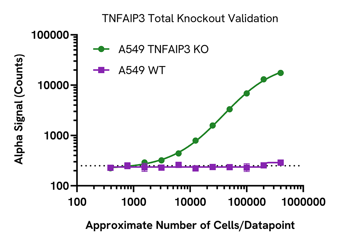
Selectivity of TNFAIP3 Total assay
Total TNFAIP3 protein levels were assessed in A549 wild type (WT) and A549 TNFAIP3 KO (Abcam ab266946) cell lines cultured to confluency in T175 flasks at 37°C, 5% CO2.
Each flask was lysed in 4 mL of Lysis Buffer for 10 minutes at RT with shaking. Lysates were serially diluted in Lysis Buffer and evaluated for Total TNFAIP3 using the AlphaLISA SureFire Ultra assay kit. For the detection step, 10 µL of cell lysate was transferred into a 384-well white OptiPlate, followed by 5 µL Acceptor Mix and incubated for 1 hour at RT. Finally, 5 µL of Donor Mix was added to each well and incubated for 1 hour at RT in the dark. The plate was read on an Envision using standard AlphaLISA settings.
As expected TNFAIP3 was only detected in WT cells, demonstrating assay selectivity.
Assay versatility
Expression of TNFAIP3 in various cell lines
Adherent cell lines were seeded in a 96-well plate (40,000 cells/well) and incubated overnight at 37°C, 5% CO2. Cells were lysed with 100 µL of Lysis Buffer for 10 minutes at RT with shaking (350 rpm).
Suspension cell lines were seeded in a 96-well plate (400,000 cells/well) in HBSS + 0.1% BSA and then lysed with 100 µL of Lysis Buffer for 10 minutes at RT with shaking (350 rpm).
TNFAIP3 levels were evaluated by AlphaLISA SureFire Ultra. For the detection step, 10 µL of cell lysate were transferred into a 384-well white OptiPlate, followed by 5 µL of Acceptor Mix and incubated for 1 hour at RT. Finally, 5 µL of Donor Mix was then added to each well and incubated for 1 hour at RT in the dark. The plate was read on an Envision using standard AlphaLISA settings.

Specifications
| Application |
Cell Signaling
|
|---|---|
| Automation Compatible |
Yes
|
| Brand |
AlphaLISA SureFire Ultra
|
| Detection Modality |
Alpha
|
| Product Group |
Kit
|
| Protocol Time |
2h at RT
|
| Sample Volume |
30 µL
|
| Shipping Conditions |
Shipped in Blue Ice
|
| Target |
TNFAIP3
|
| Target Class |
Phosphoproteins
|
| Target Species |
Human
|
| Technology |
Alpha
|
| Therapeutic Area |
Inflammation
Oncology
|
| Unit Size |
100 assay points
|
Resources
Are you looking for resources, click on the resource type to explore further.
Guide
AlphaLISA SureFire Ultra: the ultimate guide for successful experiments
The definitive guide for setting up a successful AlphaLISA SureFire Ultra assay
Several biological processes are regulated by...
Loading...


How can we help you?
We are here to answer your questions.

