
US

Revvity Sites Globally
Select your location.
*e-commerce not available for this region.

AlphaLISA SureFire Biotin-Free Human Total STING Detection Kit, 100 Assay Points
AlphaLISA SureFire Biotin-Free Human Total STING Detection Kit, 100 Assay Points
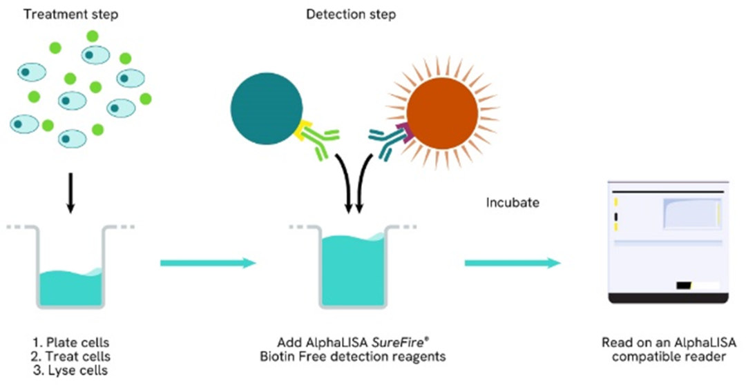
AlphaLISA SureFire Biotin Free Total Kit Schematic
The AlphaLISA™ SureFire® Biotin-Free Human Total STING assay is a sandwich immunoassay for quantitative detection of total STING in cellular lysates using Alpha Technology.
| Feature | Specification |
|---|---|
| Application | Cell Signaling |
| Protocol Time | 2h at RT |
| Sample Volume | 30 µL |
The AlphaLISA™ SureFire® Biotin-Free Human Total STING assay is a sandwich immunoassay for quantitative detection of total STING in cellular lysates using Alpha Technology.
Product variants
Unit Size: 100 Assay Points
Part #:
ASBF-TSTNG-A-HV
List price
USD 737.00
Your price:
Unit Size: 500 Assay Points
Part #:
ASBF-TSTNG-A500
List price
USD 2,490.00
Your price:
Unit Size: 10,000 Assay Points
Part #:
ASBF-TSTNG-A10K
List price
USD 14,982.00
Your price:
Unit Size: 50,000 Assay Points
Part #:
ASBF-TSTNG-A50K
List price
USD 47,624.00
Your price:
For research use only. Not for use in diagnostic procedures. All products to be used in accordance with applicable laws and regulations including without limitation, consumption and disposal requirements under European REACH regulations (EC 1907/2006).
AlphaLISA SureFire Biotin-Free Human Total STING Detection Kit, 100 Assay Points
AlphaLISA SureFire Biotin Free Total Kit Schematic
Loading...
Product information
Overview
Stimulator of Interferon Genes (STING, also known as TMEM173/MITA/MPYS) is an endoplasmic reticulum-resident transmembrane adaptor protein that serves as the central signaling hub of the cGAS-STING innate immune pathway, which detects cytosolic double-stranded DNA as a danger signal. Upon binding cyclic GMP-AMP (cGAMP) produced by the DNA sensor cGAS, STING undergoes conformational change, palmitoylation, and trafficking from the ER through the Golgi to perinuclear compartments, where it recruits and activates TBK1. TBK1 phosphorylates IRF3, which dimerizes and translocates to the nucleus to induce type I interferon and interferon-stimulated gene expression, while STING also activates NF-κB to drive pro-inflammatory cytokine production. STING plays critical roles in antiviral immunity, anti-tumor immune surveillance, and the detection of self-DNA released during cellular stress, senescence, and mitotic errors. In cancer, STING pathway activation in dendritic cells and macrophages promotes anti-tumor immunity, and STING agonists are under clinical investigation as cancer immunotherapy agents and vaccine adjuvants. Conversely, chronic STING activation by self-DNA contributes to autoinflammatory diseases including STING-associated vasculopathy with onset in infancy (SAVI), and STING inhibitors are being explored for autoimmune indications.
The AlphaLISA SureFire Biotin-Free Human Total STING Detection Kit is a sandwich immunoassay for the quantitative detection of total STING in cellular lysates, using Alpha Technology.
Formats:
- The HV (high volume) kit contains reagents to run 100 wells in 96-well format, using a 60 μL reaction volume.
- The 500-point kit contains enough reagents to run 500 wells in 384-well format, using a 20 μL reaction volume.
- The 10,000-point kit contains enough reagents to run 10,000 wells in 384-well format, using a 20 μL reaction volume.
- The 50,000-point kit contains enough reagents to run 50,000 wells in 384-well format, using a 20 μL reaction volume.
AlphaLISA SureFire Biotin Free kits are compatible with:
- Cell and tissue lysates
- Antibody modulators
- Biotherapeutic antibodies
- Biotin rich samples
AlphaLISA SureFire Biotin Free kits can be used for:
- Cellular kinase assays
- Receptor activation studies
- High-throughput screening for preclinical studies
How it works
Total-AlphaLISA SureFire Biotin Free assay principle
The Total-AlphaLISA SureFire Biotin Free assay measures the expression level of a target protein in a biological sample (e.g. cell lysate).
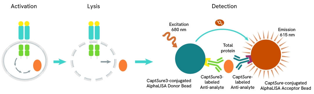
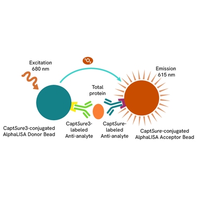
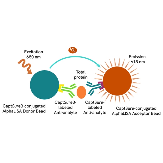
The Total-AlphaLISA SureFire Biotin Free assay uses two antibodies which recognize two different distal epitopes on the target protein. AlphaLISA assays require two bead types: Acceptor and Donor Beads. Acceptor Beads are coated with a proprietary CaptSure™ agent to specifically immobilize the assay specific antibody, labeled with a CaptSure tag. Donor Beads are coated with a proprietary CaptSure3 agent to capture one of the detection antibodies, which is labeled with the CaptSure3 tag. In the presence of target protein, the two antibodies bring the Donor and Acceptor Beads in close proximity whereby the singlet oxygen transfers energy to excite the Acceptor Bead, allowing for the generation of a luminescent Alpha signal. The amount of light emission is directly proportional to the quantity of protein present in the sample.
Total-AlphaLISA SureFire Biotin Free two-plate assay protocol
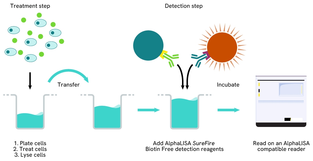
The two-plate protocol involves culturing and treating the cells in a 96-well plate before lysis, then transferring lysates into a 384-well OptiPlate™ plate before the addition of Total-AlphaLISA SureFire Biotin Free detection reagents. This protocol enables cell viability and confluence to be monitored. In addition, lysates from a single well can be used to measure multiple targets.
Total-AlphaLISA SureFire Biotin Free one-plate assay protocol
Detection of Total target protein with AlphaLISA SureFire Biotin Free reagents can be performed in a single plate used for culturing, treatment, and lysis. No washing steps are required. This HTS designed protocol allows for miniaturization while maintaining robust AlphaLISA SureFire quality.
Assay validation
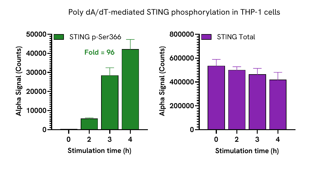
Induction of STING Phosphorylation in THP-1 Cells
THP-1 cells were seeded in a 96-well plate (400,000 cells/mL) in complete medium and transfected with 20 µg/mL Poly dA/dT at the indicated time points.
After treatment, the cells were spun down and lysed with 100 µL of Lysis Buffer B for 30 minutes at RT with shaking (350 rpm). STING Phospho (Ser366)* and Total levels were evaluated using respective AlphaLISA SureFire Biotin Free assays. For the detection step, 10 µL of cell lysate (approximately 40,000 cells) was transferred into a 384-well white OptiPlate, followed by 5 µL of Acceptor mix and incubated for 1 hour at RT. Finally, 5 µL of Donor mix was then added to each well and incubated for 1 hour at RT in the dark. The plate was read on an Envision using standard AlphaLISA settings.
As expected, Poly dA/dT induced STING phosphorylation, peaking at 4 hours, with no significant changes to STING Total levels.
*The highly specific and sensitive Phospho STING monoclonal antibody used in this assay was developed by Cell Signaling Technology (clone E9A9K, #72650).
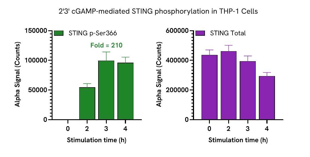
THP-1 cells were seeded in a 96-well plate (400,000 cells/mL) in complete medium and transfected with 200 µg/mL of 2’3’cGAMP at the indicated time points.
After treatment, the cells were spun down and lysed with 100 µL of Lysis Buffer B for 30 minutes at RT with shaking (350 rpm). STING Phospho (Ser366) and Total levels were evaluated using respective AlphaLISA SureFire Biotin Free assays. For the detection step, 10 µL of cell lysate (approximately 40,000 cells) was transferred into a 384-well white OptiPlate, followed by 5 µL of Acceptor mix and incubated for 1 hour at RT. Finally, 5 µL of Donor mix was then added to each well and incubated for 1 hour at RT in the dark. The plate was read on an Envision using standard AlphaLISA settings.
As expected, 2’3’cGAMP induced STING phosphorylation, peaking at 3-4 hours with a modest decrease in STING total levels.
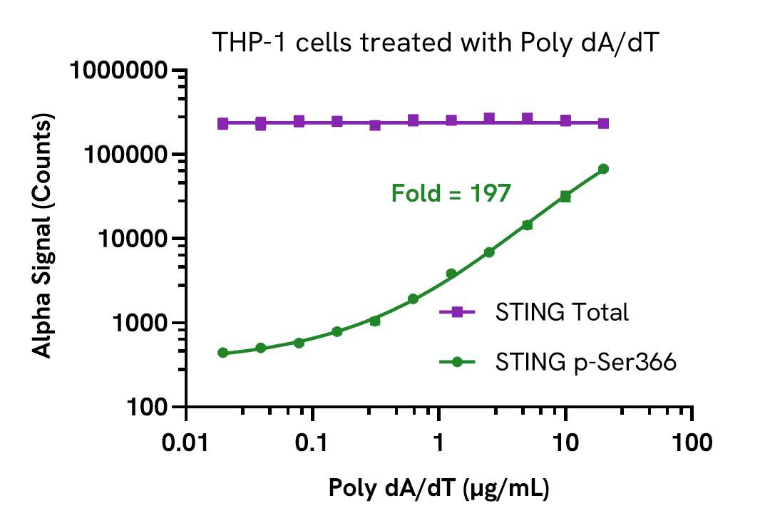
Dose-dependent activation of STING phosphorylation
THP-1 cells were seeded in a 96-well plate (400,000 cells/well) in complete medium and transfected with 20 µg/mL Poly dA/dT at the indicated concentrations for 4 hours.
After treatment, the cells were spun down and lysed with 100µL of Lysis Buffer B for 30 minutes at RT with shaking (350 rpm). STING Phospho (Ser366) and Total levels were evaluated using respective AlphaLISA SureFire Biotin Free assays. For the detection step, 10 µL of diluted cell lysate (approximately 8,000 cells) was transferred into a 384-well white OptiPlate, followed by 5 µL of Acceptor mix and incubated for 1 hour at RT. Finally, 5 µL of Donor mix was then added to each well and incubated for 1 hour at RT in the dark. The plate was read on an Envision using standard AlphaLISA settings.
As expected, Poly dA/dT triggered a dose-dependent increase in the levels of Phospho STING (Ser366) while Total levels remained unchanged.
Assay versatility
STING expression in various cell lines
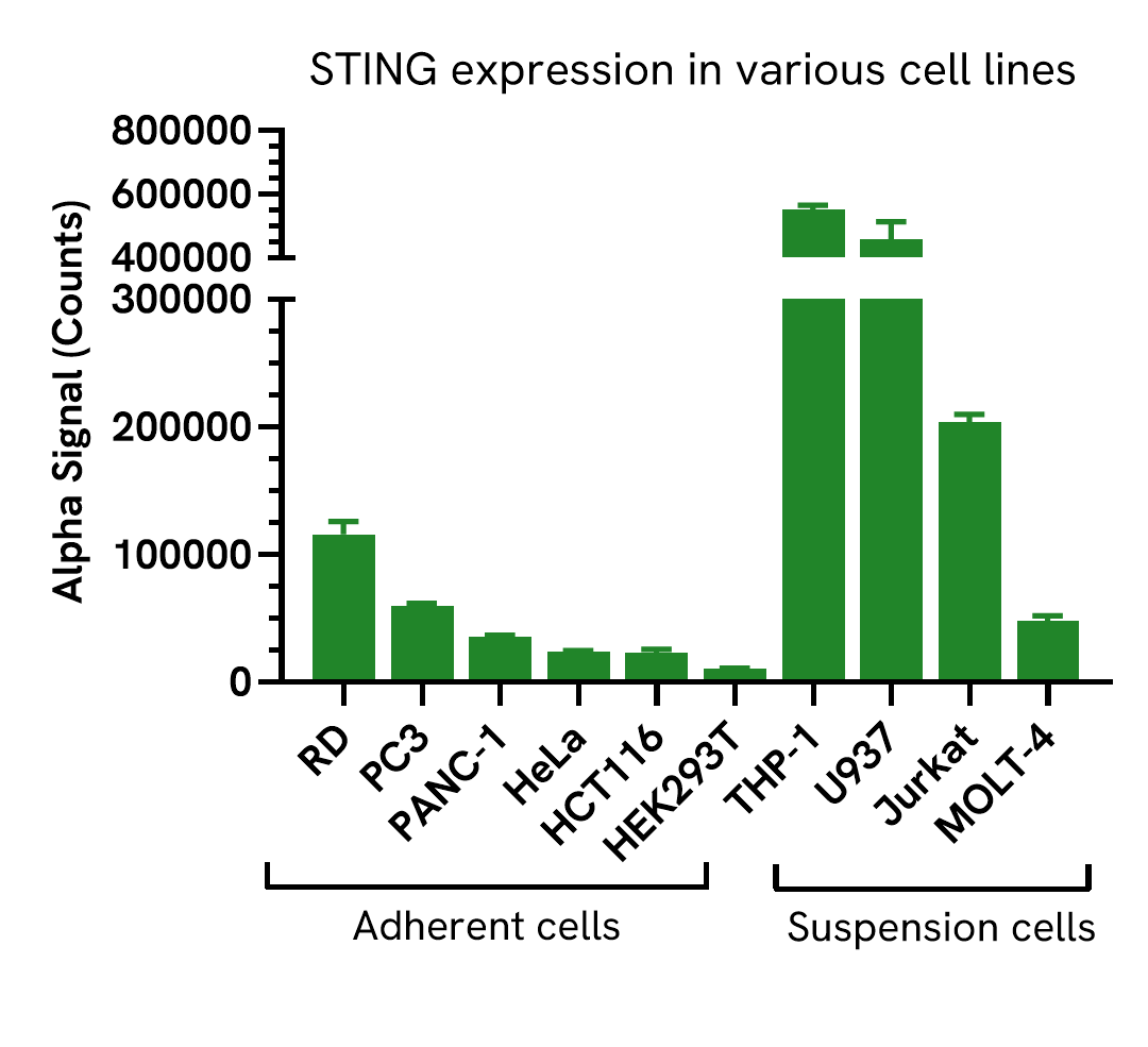
Adherent cells were seeded in a 96-well plates (40,000 cells/well) in complete medium and incubated overnight at 37°C, 5% CO2. Cells were lysed with 100 µL Lysis buffer. Suspension cells were harvested and seeded in a 96-well plate (400,000 cells/well). Cells were lysed with 100µL of Lysis Buffer for 10 minutes at RT with shaking (350 rpm). STING levels were evaluated using the AlphaLISA SureFire Ultra Biotin Free assay. For the detection step, 10 µL of cell lysate (4,000 adherent cells or 40,000 suspension cells) were transferred into a 384-well white OptiPlate, followed by 5 µL of Acceptor Mix and incubated for 1 hour at RT. Finally, 5 µL of Donor Mix was then added to each well and incubated for 1 hour at RT in the dark. The plate was read on an Envision using standard AlphaLISA settings.
STING expression was detected in a wide range of human cell lines with higher levels observed in THP-1 and U937 cells.
Assay sensitivity
Assay sensitivity - cell lysate dilution
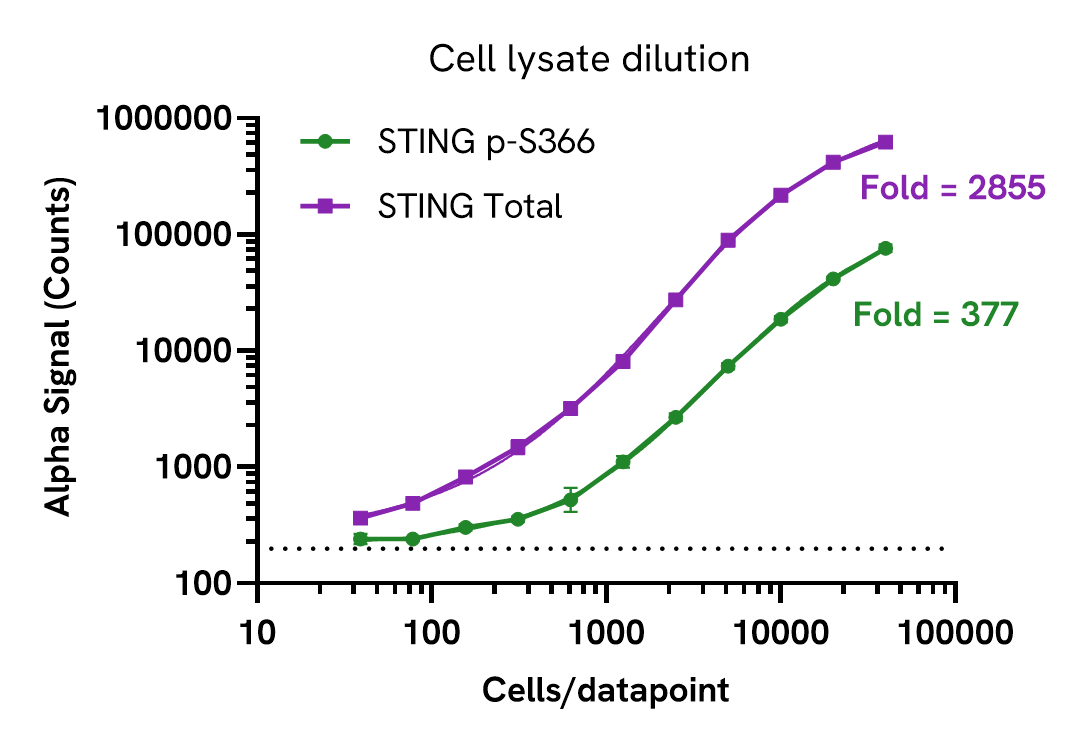
Cell lysate was prepared from THP-1 cells treated with 20 µg/mL of Calyculin A for 2 hours and lysed at 4 x 10⁶ cells/mL with Lysis Buffer B for 30 minutes at RT with shaking. Lysate was serially diluted and Phospho (Ser366) and Total STING levels were evaluated using the AlphaLISA SureFire Biotin Free assay. For the detection step, 10 µL of lysate was transferred into a 384-well white OptiPlate, followed by 5 µL of Acceptor mix and incubated for 1 hour at room temperature. Finally, 5 µL of Donor mix was then added to each well and incubated for 1 hour at RT in the dark. The plate was read on an Envision using standard AlphaLISA settings.
Approximate number of cells per datapoint is indicated. Dotted line represents assay background. This assay can detect STING expression in less than 500 cells/datapoint.
Specifications
| Application |
Cell Signaling
|
|---|---|
| Automation Compatible |
Yes
|
| Brand |
AlphaLISA SureFire Biotin-Free
|
| Detection Modality |
Alpha
|
| Product Group |
Kit
|
| Protocol Time |
2h at RT
|
| Sample Volume |
30 µL
|
| Shipping Conditions |
Shipped in Blue Ice
|
| Target |
STING
|
| Target Class |
Phosphoproteins
|
| Target Species |
Human
|
| Technology |
Alpha
|
| Therapeutic Area |
Autoimmunity
Oncology
Virology
|
| Unit Size |
100 Assay Points
|
Resources
Are you looking for resources, click on the resource type to explore further.

Brochure
Alpha assays and reagents catalog
Alpha technolgy enables the rapid and straightforward mesaure of virtually any target. This includes enzymes, receptor-ligand...
Brochure
Alpha SureFire Ultra no-wash immunoassay catalog
Discover Alpha SureFire® Ultra™ assays, the no-wash cellular kinase assays leveraging Revvity's exclusive bead-based technology...
Brochure
Species compatibility for HTRF, AlphaLISA SureFire Ultra and Alpha SureFire Ultra Multiplex assays
This document includes detailed tables listing HTRF™, AlphaLISA™ SureFire® Ultra™, and Alpha SureFire® Ultra™ Multiplex assays...
Loading...


How can we help you?
We are here to answer your questions.