- The host cells are seeded in a 24-well microplate and incubated for 100% confluence
- The cells are then infected by GFP-RSV and incubated with low, medium, and high hamster sera concentrations
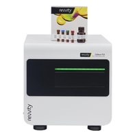
- Finally, the plate is scanned and analyzed with the Celigo™ image cytometer
 Celigo Image Cytometer
to count the number of foci in each well

Zoomed in bright and fluorescent images of GFP-RSV infected foci

Plate-view of the 24-wells showing reduction in number of GFP foci at high concentration of serum
You may also be interested in these products
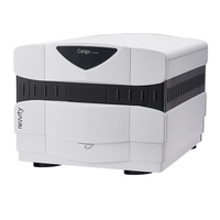
Part number:
200-BFFL-5C
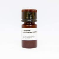
Part number:
CS2-0106-5ML,
CS2-0106-25ML
List price:
USD 133.00 - USD 594.00
Log in to view
your online price
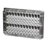
List price:
USD 286.00 - USD 5,260.00
Log in to view
your online price
List price:
USD 286.00 - USD 5,260.00
Log in to view
your online price
of 5
For research use only. Not for use in diagnostic procedures.


