US
Revvity Sites Globally
Select your location.
*e-commerce not available for this region.


IVIS Lumina XRMS In Vivo Imaging System
Industry leading fluorescence, bioluminescence and x-ray imaging - All-In-One benchtop instrument! The IVIS™ Lumina XRMS (x-ray multi-species) adds multimodal versatility for imaging large animals up to 500 grams with optical and x-ray overlay.
For research use only. Not for use in diagnostic procedures.
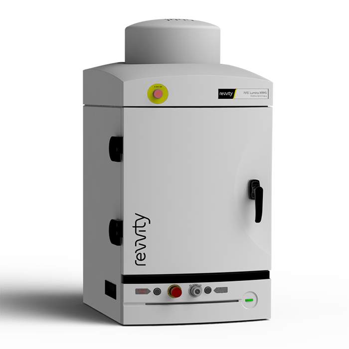
IVIS Lumina XRMS In Vivo Imaging System
IVIS Lumina XRMS 2D Optical Imaging System with Integrated X-ray
IVIS Lumina XRMS In Vivo Imaging System
Part #:
CLS136340
Imaging Modality:
2D Bioluminescence, 2D Fluorescence, X-ray
Loading...
Product information
Overview
The Lumina XRMS includes state of the art spectral unmixing for sensitive multispectral imaging to monitor multiple biological events in the same animal. Use our Living Image® software to automate all the controls and settings required for seamless image acquisition and processing. Typical X-ray image acquisitions take only 10 seconds and can be overlaid with both optical and photographic images.
Superior Optical Imaging with Spectral Unmixing
The IVIS Lumina XRMS Series III is capable of imaging all common fluorescent and bioluminescent reporters or dyes. The system is equipped with up to 21 filter sets to image reporters that emit from green to near-infrared. High resolution, sharp cut-off filters are simply interchangeable to achieve the highest performance, sensitivity and spectral unmixing. The Lumina XRMS imaging system also accommodates Petri dishes or micro-titer plates for in vitro imaging.
The system can incorporate premium animal handling features such as a heated stage, gas anesthesia connections and a syringe injection system for simultaneous compound administration. Living Image software yields high-quality, reproducible, quantitative results incorporating instrument calibration, background subtraction and the image algorithms. Simple user guided spectral unmixing allows detection and separation of multiple reporters, and Living Image provides the precise overlay to see your optical reporters together with anatomical surface or X-ray features.
Features and Benefits:
- Multispecies optical and X-ray imaging
- Image mice, rats and other large animals
- High resolution, low dose digital X-Ray
- Exquisite sensitivity in bioluminescence
- Compute Pure Spectrum (CPS) spectral unmixing for ultimate fluorescence sensitivity
- Full fluorescence tunability through the NIR Spectrum
- Complimentary Living Image™ software licenses are provided with the IVIS systems and upon request.
Specifications
| Dimensions | 48.0 cm (W) x 104.0 cm (H) |
|---|---|
| Weight |
73.0 kg
|
| Brand |
IVIS
|
|---|---|
| Imaging Modality |
2D Bioluminescence
2D Fluorescence
X-ray
|
| Unit Size |
1 unit
|
References
- Zhang et al (2021). One-step and facile synthesis of peptide-like poly(melphalan) nanodrug for cancer therapy. Nanotoday. 37; 101098. https://doi.org/10.1016/j.nantod.2021.101098
- Zou et al (2020). A ZIF-90 nanoplatform loaded with an enzyme-responsive organic small-molecule probe for imaging the hypoxia status of tumor cells. Nanoscale. 12, 14870-14881. https://doi.org/10.1039/D0NR02580A
- Parkins et al (2020). Engineering Circulating Tumor Cells as Novel Cancer Theranostics. Theranostics. 10(17): 7925–7937. https://dx.doi.org/10.7150%2Fthno.44259
- Son et al (2019). Gelatin–chlorin e6 conjugate for in vivo photodynamic therapy. J Nanobiotechnology. 17(50). https://doi.org/10.1186/s12951-019-0475-1
- Wang et al (2019). A Y1 receptor ligand synergized with a P-glycoprotein inhibitor improves the therapeutic efficacy of multidrug resistant breast cancer. Biomater. Sci., 7, 4748-4757. https://doi.org/10.1039/C9BM00337A
- Gao et al (2018). A unique off-on near-infrared cyanine-based probe for imaging of endogenous alkaline phosphatase activity in cells and in vivo. Sensors & Actuators: Chemical. 265: 565-574. https://doi.org/10.1016/j.snb.2018.03.078
- Shen et al (2018). Versatile hyaluronic acid modified AQ4N-Cu(II)-gossypol infinite coordination polymer nanoparticles: Multiple tumor targeting, highly efficient synergistic chemotherapy, and real-time self-monitoring. Biomaterials. 154: 197-212. https://doi.org/10.1016/j.biomaterials.2017.11.001
- Gong et al (2018). A Ultrasensitive Near‐Infrared Fluorescent Probe Reveals Pyroglutamate Aminopeptidase 1 Can Be a New Inflammatory Cytokine. Adv Sci. https://doi.org/10.1002/advs.201700664
- Lai et al (2016). Fluorescent gold nanoclusters for in vivo target imaging of Alzheimer's disease. RSC Advances. 6, 30081-30088. https://doi.org/10.1039/C6RA01027J
- Zhao et al (2016). In vivo target bio-imaging of cerebral ischemic stroke by real-time labeling of zinc. RSC Adv., 2016,6, 110525-110534. https://doi.org/10.1039/C6RA23507G
Citations
Resources
Are you looking for resources, click on the resource type to explore further.
Case Study
A novel non-Invasive in vivo tool for the assessment of NASH
Non-alcoholic fatty liver disease (NAFLD) describes a progressive pathology that affects the liver. Fat accumulation causes fatty...
Technical Note
Adaptive Fluorescence Background Subtraction for IVIS systems
Instrument background occurs when excitation light leaks through the emission filter. This occurs more frequently when the...
Application Note
AI-assisted high-throughput analysis of multimodal in vivo imaging data for cancer research
Preclinical cancer research faces significant workflow bottlenecks due to the manual, time-intensive nature of analyzing...
Literature - Publication Review
Applications of In Vivo bioluminescence imaging for SARS-CoV-2
Scientists continue to explore several options to treat SARS-CoV-2 infection with hundreds of therapeutics at various phases of...
Technical Note
AutoExposure
This tech note outlines procedures on using auto-exposure on the IVIS® preclinical optical imaging platform using Living Image®...
Loading...


How can we help you?
We are here to answer your questions.

